Review
. 2017 Dec 1;144(23):4271-4283.
doi: 10.1242/dev.151266.

Mechanical regulation of musculoskeletal system development

Affiliations
Review

Mechanical regulation of musculoskeletal system development

Neta Felsenthal et al. Development. .

Abstract

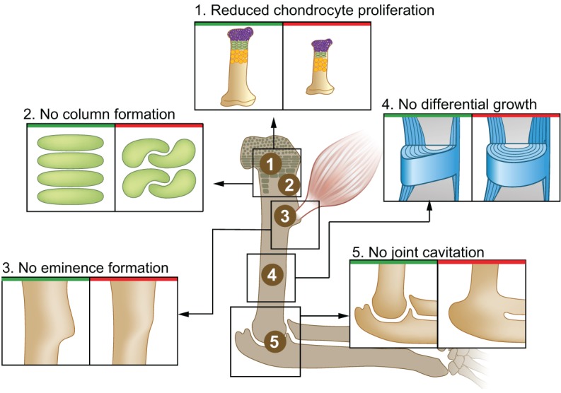
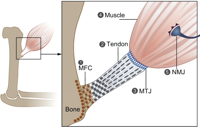
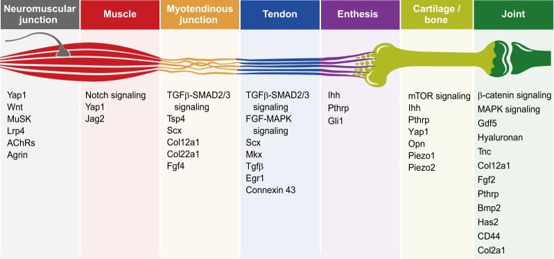
During embryogenesis, the musculoskeletal system develops while containing within itself a force generator in the form of the musculature. This generator becomes functional relatively early in development, exerting an increasing mechanical load on neighboring tissues as development proceeds. A growing body of evidence indicates that such mechanical forces can be translated into signals that combine with the genetic program of organogenesis. This unique situation presents both a major challenge and an opportunity to the other tissues of the musculoskeletal system, namely bones, joints, tendons, ligaments and the tissues connecting them. Here, we summarize the involvement of muscle-induced mechanical forces in the development of various vertebrate musculoskeletal components and their integration into one functional unit.

Keywords: Mechanoregulation; Mechanotransduction; Musculoskeletal development.


Conflict of interest statement

Competing interestsThe authors declare no competing or financial interests.

Figures

Fig. 1.
Fig. 1.
Mechanical forces involved in endochondral bone formation. Bone morphology is regulated by mechanical forces at different levels, as demonstrated by the various developmental and functional aberrations that arise in the absence of muscle contraction. (1) Bone elongation is impaired due to reduced chondrocyte proliferation in the growth plate. (2) Additionally, the organization of resting chondrocytes into columns is impaired, which can also affect skeletal elongation. (3) Bone eminence growth is arrested, resulting in smaller or absent eminences. (4) Differential appositional growth is lost, resulting in a circular circumferential shape. (5) Joint formation is impaired during embryonic development, leading to joint fusion.
Fig. 2.
Fig. 2.
Forces acting during the formation of tendon, muscle and their attachments. The proper formation of tendon, muscle and the attachment between them requires mechanical load. (1) In its absence, mineralized fibrocartilage in the enthesis is lost, and there is increased osteoclast activity and bone resorption in the attachment site. (2) Tendon development is arrested in the absence of muscle, and zeugopod tendons are lost. (3) Proper maturation and the ECM composition of the MTJ, which are regulated by Scx expression, are dependent on muscle contraction. (4) Without muscle contraction, there is a reduction in myotube number, and muscles are smaller than normal and display a delay in splitting. Muscle contraction is also needed to maintain a pool of muscle progenitor cells. (5) In the NMJ, muscle contraction is needed to promote neuronal elimination during development and to prevent NMJ degeneration. MFC, mineralized fibrocartilage; MTJ, myotendinous junction; NMJ, neuromuscular junction.
Fig. 3.
Fig. 3.
Mechanically regulated signaling pathways, factors and genes involved in musculoskeletal system development. The key signaling pathways, factors and genes, as mentioned in this Review, which have been shown to be regulated by mechanical forces during musculoskeletal development.

References

    1. Barik A., Zhang B., Sohal G. S., Xiong W.-C. and Mei L. (2014). Crosstalk between Agrin and Wnt signaling pathways in development of vertebrate neuromuscular junction. Dev. Neurobiol. 74, 828-838. 10.1002/dneu.22190 - DOI - PubMed
    1. Bastow E. R., Lamb K. J., Lewthwaite J. C., Osborne A. C., Kavanagh E., Wheeler-Jones C. P. D. and Pitsillides A. A. (2005). Selective activation of the MEK-ERK pathway is regulated by mechanical stimuli in forming joints and promotes pericellular matrix formation. J. Biol. Chem. 280, 11749-11758. 10.1074/jbc.M414495200 - DOI - PubMed
    1. Bekoff A. (1981). Embryonic development of chick motor behaviour. Trends Neurosci. 4, 181-184. 10.1016/0166-2236(81)90059-X - DOI
    1. Benjamin M., Toumi H., Ralphs J. R., Bydder G., Best T. M. and Milz S. (2006). Where tendons and ligaments meet bone: attachment sites (‘entheses’) in relation to exercise and/or mechanical load. J. Anat. 208, 471-490. 10.1111/j.1469-7580.2006.00540.x - DOI - PMC - PubMed
    1. Bennett M. R., Davey D. F. and Marshall J. J. (1983). The growth of nerves in relation to the formation of premuscle cell masses in the developing chick forelimb. J. Comp. Neurol. 215, 217-227. 10.1002/cne.902150209 - DOI - PubMed

Publication types

LinkOut - more resources