. 2017 Dec 12;114(50):13278-13283.
doi: 10.1073/pnas.1707050114. Epub 2017 Nov 28.

On the role of the corpus callosum in interhemispheric functional connectivity in humans

Affiliations

On the role of the corpus callosum in interhemispheric functional connectivity in humans

Jarod L Roland et al. Proc Natl Acad Sci U S A. .

Abstract

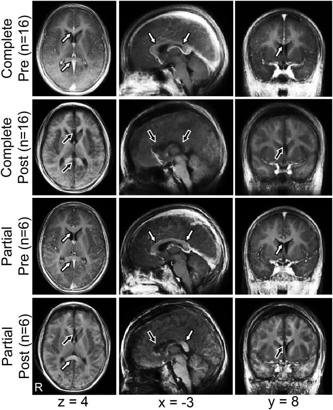
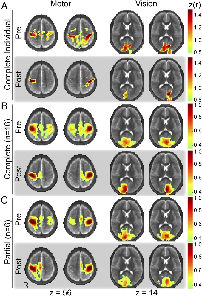
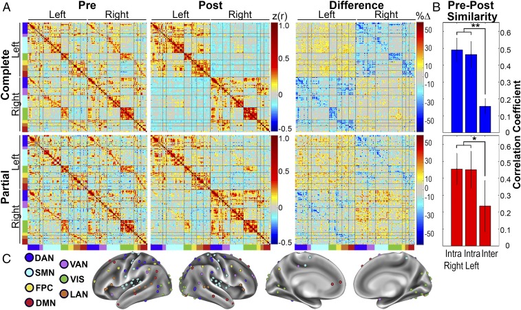
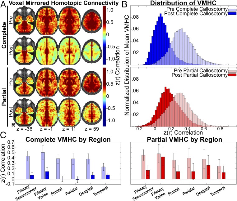
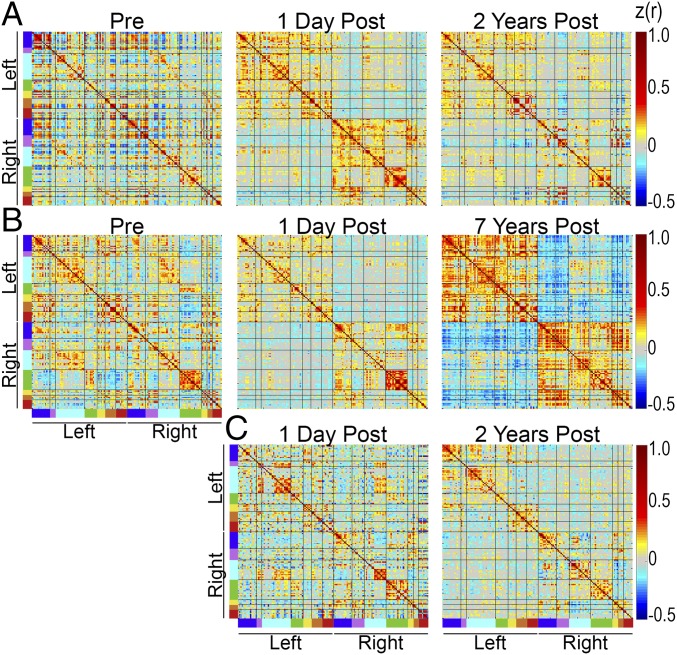
Resting state functional connectivity is defined in terms of temporal correlations between physiologic signals, most commonly studied using functional magnetic resonance imaging. Major features of functional connectivity correspond to structural (axonal) connectivity. However, this relation is not one-to-one. Interhemispheric functional connectivity in relation to the corpus callosum presents a case in point. Specifically, several reports have documented nearly intact interhemispheric functional connectivity in individuals in whom the corpus callosum (the major commissure between the hemispheres) never develops. To investigate this question, we assessed functional connectivity before and after surgical section of the corpus callosum in 22 patients with medically refractory epilepsy. Section of the corpus callosum markedly reduced interhemispheric functional connectivity. This effect was more profound in multimodal associative areas in the frontal and parietal lobe than primary regions of sensorimotor and visual function. Moreover, no evidence of recovery was observed in a limited sample in which multiyear, longitudinal follow-up was obtained. Comparison of partial vs. complete callosotomy revealed several effects implying the existence of polysynaptic functional connectivity between remote brain regions. Thus, our results demonstrate that callosal as well as extracallosal anatomical connections play a role in the maintenance of interhemispheric functional connectivity.

Keywords: callosotomy; corpus callosum; functional connectivity; resting state; structural connectivity.


Conflict of interest statement

Conflict of interest statement: E.C.L. discloses financial relationships with the following companies: Intellectual Ventures, Monteris Medical, Acera Medical, Pear Therapeutics, General Sensing, Immunovalent, Face to Face Biometrics, Neurolutions, and Osteovantage.

Figures

Fig. 1.
Fig. 1.
Anatomic imaging precallosotomy and postcallosotomy. Mean T1-weighted images before (precallosotomy) and after (postcallosotomy) complete and partial callosotomy, represented in atlas space (right hemisphere on Left). MNI152 coordinates of axial, sagittal, and coronal planes are listed. White arrows indicate intact CC, and black arrows indicate areas of divided CC. Note residual splenium after partial callosotomy.
Fig. 2.
Fig. 2.
FC maps corresponding to seeds in primary motor (first and second columns) and primary visual (third and fourth columns) areas of the right (first and third columns) and left (second and fourth columns) hemispheres. (A) Results obtained in an exemplar individual. Note loss of interhemispheric FC after complete callosotomy with preserved intrahemispheric FC in both motor and visual networks. (B) Mean (n = 16) results before and after complete callosotomy. (C) Mean (n = 6) results before and after partial callosotomy. Note maintained visual but not motor interhemispheric FC after partial, but not complete, callosotomy. The FC maps are thresholded at z(r) > 0.80 in the exemplar individual and z(r) > 0.35 in the group results.
Fig. 3.
Fig. 3.
Contrast between interhemispheric vs. intrahemispheric FC. (A) FC matrices representing seven RSNs are organized according to hemisphere of seed. Diagonal and off-diagonal blocks represent intrahemispheric and interhemispheric FC, respectively. RSN color codes are defined in C. (B) Bar graphs representing similarity between precallosotomy and postcallosotomy for intrahemispheric and interhemispheric FC. The error bars represent 95% confidence intervals. **P < 0.001, *P < 0.05. (C) Seeds plotted on an inflated mean brain surface.
Fig. 4.
Fig. 4.
Topography of CC-mediated FC and distribution of VMHC. (A) VMHC computed as the Fisher z-transformed Pearson correlation between voxels mirrored about the midline. By definition, these displays are bilaterally symmetric. Spatial blurring during preprocessing generates artifactually high homotopic FC along the midline. The underlay is the T2-weighted atlas representative image. (B) Distributions of mean VMHC across all voxels after vs. before callosotomy. Note larger shift toward zero after complete relative to partial callosotomy. (C) Bar graphs (mean ± 95% confidence interval) representing homotopic FC organized according to anatomical region. Note partial preservation of FC in primary sensorimotor and visual cortices after complete callosotomy but nearly complete loss of VMHC in multimodal associative areas. Note also more retained VMHC after partial callosotomy.
Fig. 5.
Fig. 5.
FC matrices obtained in three individuals including longitudinal imaging at follow-up intervals of 2–7 y. (A) Partial callosotomy at age 2 y; follow-up (sedated) at age 4 y. (B) Complete callosotomy at age 13 y; follow-up (nonsedated) at age 20 y. (C) Complete callosotomy at age 15 y; follow-up (sedated) at age 17 y. The precallosotomy study in this case was excluded as this patient initially presented with epileptic encephalopathy. Follow-up imaging 2 y after complete callosotomy was obtained under sedation for clinical indications. Note no sign of recovery of interhemispheric FC at follow-up in any of these individuals.

Similar articles

Cited by

References

    1. Shen HH. Core concept: Resting-state connectivity. Proc Natl Acad Sci USA. 2015;112:14115–14116. - PMC - PubMed
    1. Snyder AZ. Intrinsic brain activity and resting state networks. In: Pfaff DW, Volkow ND, editors. Neuroscience in the 21st Century. Springer; New York: 2016. pp. 1–52.
    1. Hacker CD, et al. Resting state network estimation in individual subjects. Neuroimage. 2013;82:616–633. - PMC - PubMed
    1. Leuthardt EC, et al. Resting-state blood oxygen level-dependent functional MRI: A paradigm shift in preoperative brain mapping. Stereotact Funct Neurosurg. 2015;93:427–439. - PubMed
    1. Brier MR, Day GS, Snyder AZ, Tanenbaum AB, Ances BM. N-methyl-D-aspartate receptor encephalitis mediates loss of intrinsic activity measured by functional MRI. J Neurol. 2016;263:1083–1091. - PMC - PubMed

Publication types