. 2018 Jan 12;293(2):557-566.
doi: 10.1074/jbc.M117.817494. Epub 2017 Nov 28.

Structural basis for the recognition of kinesin family member 21A (KIF21A) by the ankyrin domains of KANK1 and KANK2 proteins

Affiliations

Structural basis for the recognition of kinesin family member 21A (KIF21A) by the ankyrin domains of KANK1 and KANK2 proteins

Qiong Guo et al. J Biol Chem. .

Abstract

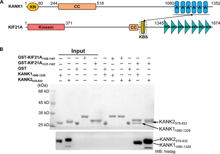
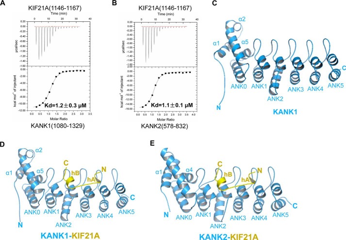
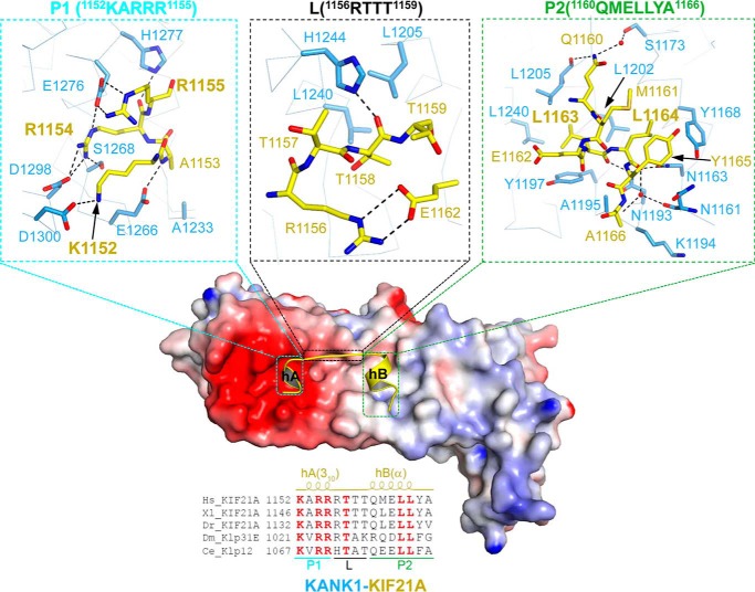
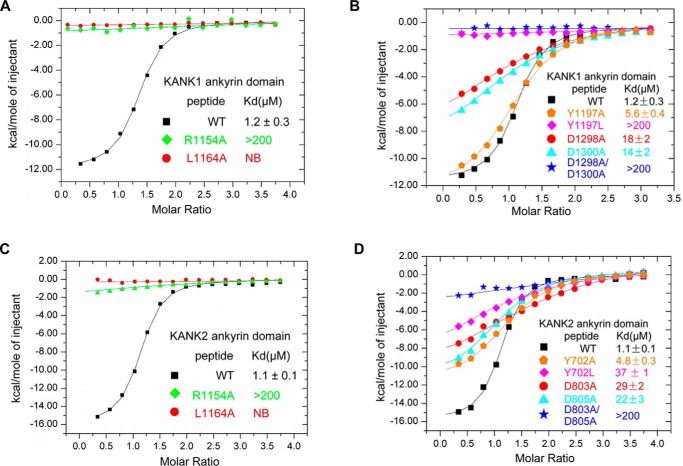
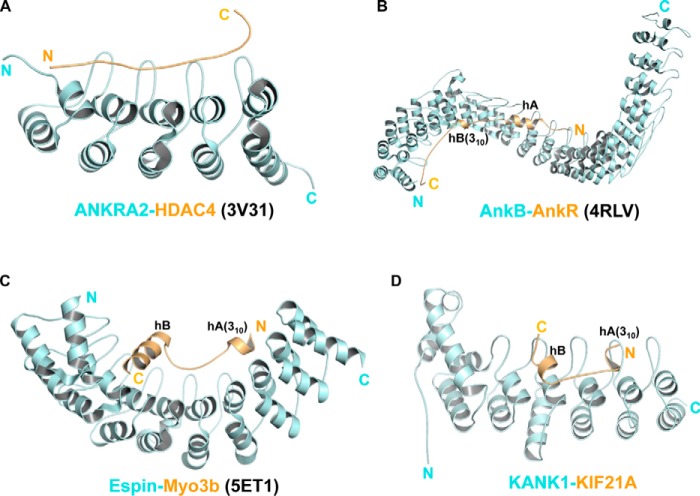
A well-controlled microtubule organization is essential for intracellular transport, cytoskeleton maintenance, and cell development. KN motif and ankyrin repeat domain-containing protein 1 (KANK1), a member of KANK family, recruits kinesin family member 21A (KIF21A) to the cell cortex to control microtubule growth via its C-terminal ankyrin domain. However, how the KANK1 ankyrin domain recognizes KIF21A and whether other KANK proteins can also bind KIF21A remain unknown. Here, using a combination of structural, site-directed mutagenesis, and biochemical studies, we found that a stretch of ∼22 amino acids in KIF21A is sufficient for binding to KANK1 and its close homolog KANK2. We further solved the complex structure of the KIF21A peptide with either the KANK1 ankyrin domain or the KANK2 ankyrin domain. In each complex, KIF21A is recognized by two distinct pockets of the ankyrin domain and adopts helical conformations upon binding to the ankyrin domain. The elucidated KANK structures may advance our understanding of the role of KANK1 as a scaffolding molecule in controlling microtubule growth at the cell periphery.

Keywords: X-ray crystallography; ankyrin domain; cell adhesion; cell signaling; kinesin; structural biology.


Conflict of interest statement

The authors declare that they have no conflicts of interest with the contents of this article

Figures

Figure 1.
Figure 1.
Human KANK1(1080–1329) and KANK2(578–832) ankyrin domains specifically recognize the KIF21A(1137–1167) peptide. A, domain organizations of human KANK1 and KIF21A. KN, KANK N-terminal (KN) motif; CC, coiled-coil domain; AR, ankyrin repeat; Kinesin, kinesin motor domain; KBS, KANK1-binding site; WD, WD40 repeat. The interaction between the KANK1 ankyrin domain and the KANK1-binding site is denoted by the solid black arrow. B, pull-down binding assay between GST fusion peptides (KIF21A(1158–1187) and KIF21A(1137–1167)) and His-tagged ankryin domains of KANK1 and KANK2. GST protein is used as the control. His-tagged recombinant KANK1 and KANK2 were further detected by Western blotting.
Figure 2.
Figure 2.
KIF21A(1146–1167) is sufficient for binding to KANK1/2 and overall structures of the KANK1/2 ankyrin domains with the KIF21A peptide. A, ITC binding between the ankyrin domain of KANK1 and the KIF21A(1146–1167) peptide. B, ITC binding between the ankyrin domain of KANK2 and the KIF21A(1146–1167) peptide. C, structure of the KANK1 ankyrin domain alone, shown in blue, with ANK0–5 and three extra N-terminal helices labeled. D, overall structure of the KANK1-KIF21A complex, with protein and peptide shown in blue and yellow, respectively. ANK0–5 and three extra N-terminal helices of KANK1, as well as hA and hB of KIF21A, are also labeled. E, overall structure of the KANK2-KIF21A complex, with protein and peptide shown in the same way as in Fig. 2D.
Figure 3.
Figure 3.
Detailed interactions between the KANK1 ankyrin domain (residues 1080–1329) and the KIF21A peptide (residues 1146–1167). Center, the electrostatic surface of the KANK1 ankyrin domain bound with the KIF21A peptide (yellow schematic); top, 1152KARR1155 of KIF21A bound to the acidic patch (P1 pocket) of KANK1; top center, 1156RTTT1159 OF KIF21A makes few contacts with the periphery of the acidic patch (L region) of KANK1; top right, 1160QMELLYA1166 bound to the hydrophobic pocket of KANK1 (P2 pocket). The residues involved in the protein-peptide interactions are labeled and shown in stick representation. Bottom, KIF21A orthologs aligned on 1152–1166 of human KIF21A, with the absolutely conserved residues colored in red. The 310- and α-helices in the KIF21A peptide are labeled at the top of the sequences as hA(310) and hB(α), respectively.
Figure 4.
Figure 4.
Comparison of the binding affinity measured by ITC between KANK1/2 and the KIF21A peptide with either that of KANK1/2 and mutant peptide or that of mutant protein and wild type peptide. A, comparison of the KANK1-binding affinities of wild-type and mutant KIF21A peptides. B, comparison of the KIF21A-binding affinities of KANK1 and its mutants. C, comparison of the KANK2-binding affinities of wild-type and mutant KIF21A peptides. D, comparison of the KIF21A-binding affinities of KANK2 and its mutants. NB, no detectable binding.
Figure 5.
Figure 5.
Comparison of different ankyrin-peptide complex structures. A, the structure of the ANKRA2 ankyrin repeats (cyan) in complex with the HDAC4 peptide (yellow) (PDB code 3V31). B, the complex structure of the AnkB ankyrin domain (cyan) and the AnkR peptide (yellow) (PDB code 4RLV). C, the structure of the Espin ankyrin domain (cyan) with the Myo3b peptide (yellow) (PDB code 5ET1). D, the structure of the KANK1 ankyrin domain (cyan) with the KIF21 peptide (yellow). All ankyrin repeats are arranged from N to C (left to right). The 310-helices of AnkR, Myo3b, and KIF21A are also indicated.

Similar articles

Cited by

References

    1. Vale R. D. (1987) Intracellular transport using microtubule-based motors. Annu. Rev. Cell Biol. 3, 347–378 10.1146/annurev.cb.03.110187.002023 - DOI - PubMed
    1. Kapitein L. C., and Hoogenraad C. C. (2015) Building the neuronal microtubule cytoskeleton. Neuron 87, 492–506 10.1016/j.neuron.2015.05.046 - DOI - PubMed
    1. Akhmanova A., Stehbens S. J., and Yap A. S. (2009) Touch, grasp, deliver and control: functional cross-talk between microtubules and cell adhesions. Traffic 10, 268–274 10.1111/j.1600-0854.2008.00869.x - DOI - PubMed
    1. Rodriguez O. C., Schaefer A. W., Mandato C. A., Forscher P., Bement W. M., and Waterman-Storer C. M. (2003) Conserved microtubule-actin interactions in cell movement and morphogenesis. Nat. Cell Biol. 5, 599–609 10.1038/ncb0703-599 - DOI - PubMed
    1. Stehbens S. J., Paszek M., Pemble H., Ettinger A., Gierke S., and Wittmann T. (2014) CLASPs link focal-adhesion-associated microtubule capture to localized exocytosis and adhesion site turnover. Nat. Cell Biol. 16, 561–573 10.1038/ncb2975 - DOI - PMC - PubMed

Publication types