. 2018 Jan 26;293(4):1178-1191.
doi: 10.1074/jbc.RA117.000392. Epub 2017 Nov 28.

Deubiquitylase USP9X suppresses tumorigenesis by stabilizing large tumor suppressor kinase 2 (LATS2) in the Hippo pathway

Affiliations

Deubiquitylase USP9X suppresses tumorigenesis by stabilizing large tumor suppressor kinase 2 (LATS2) in the Hippo pathway

Chu Zhu et al. J Biol Chem. .

Abstract

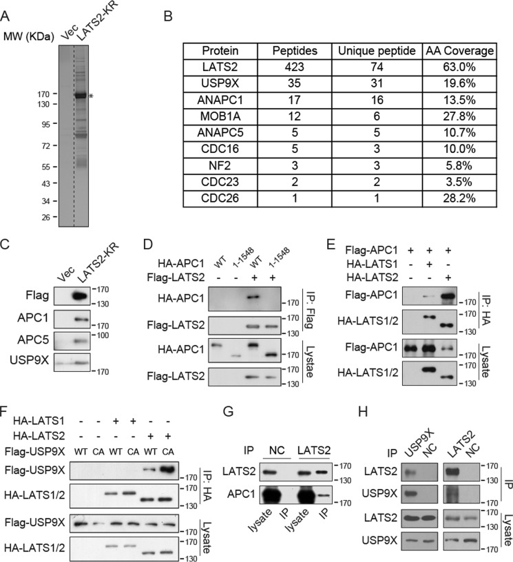
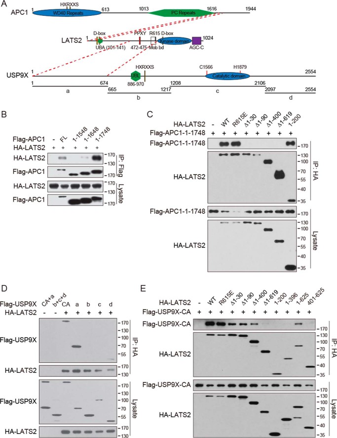
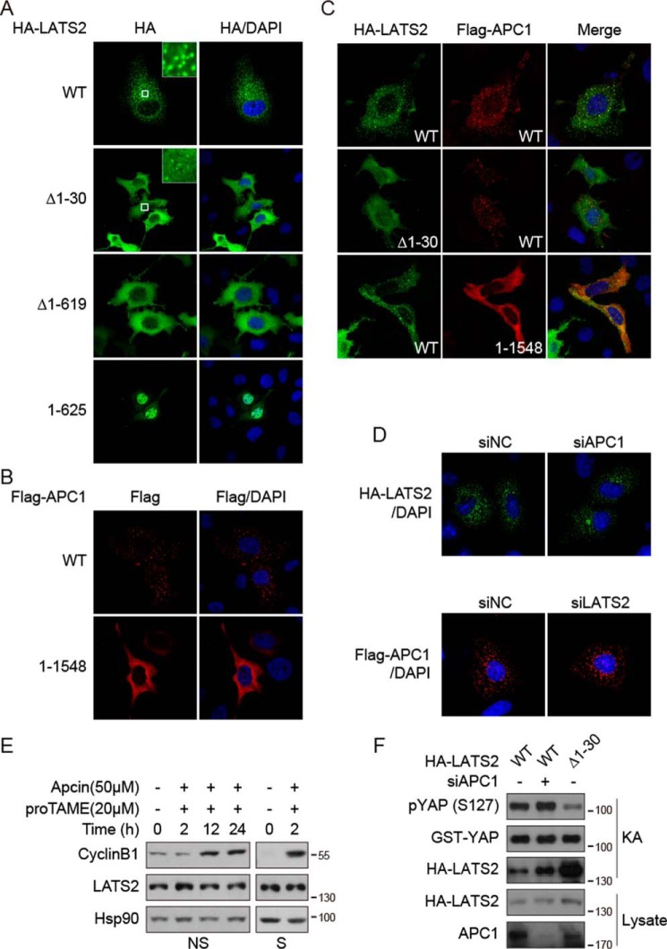
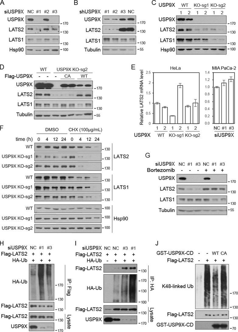
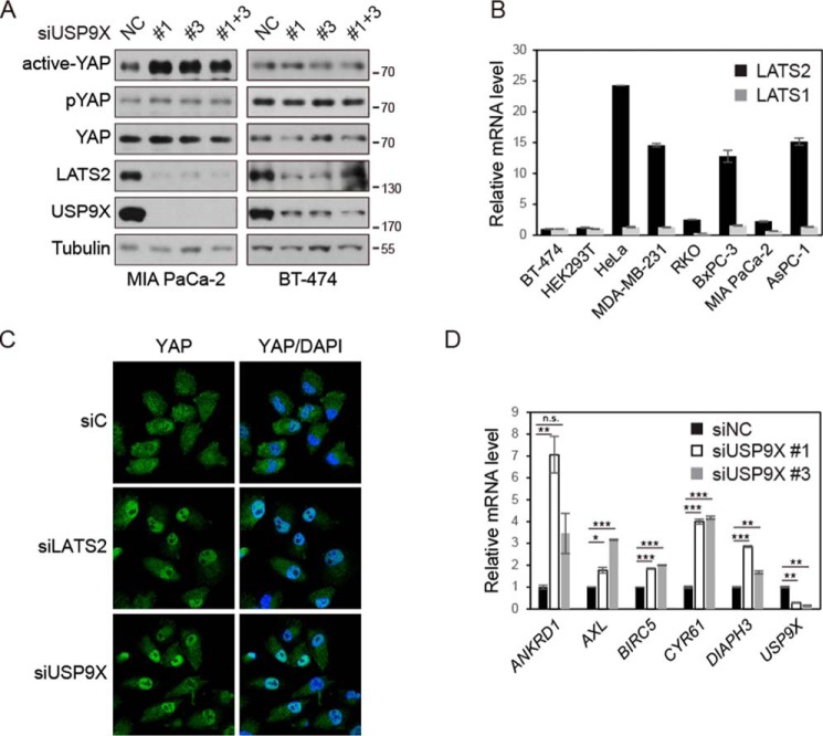
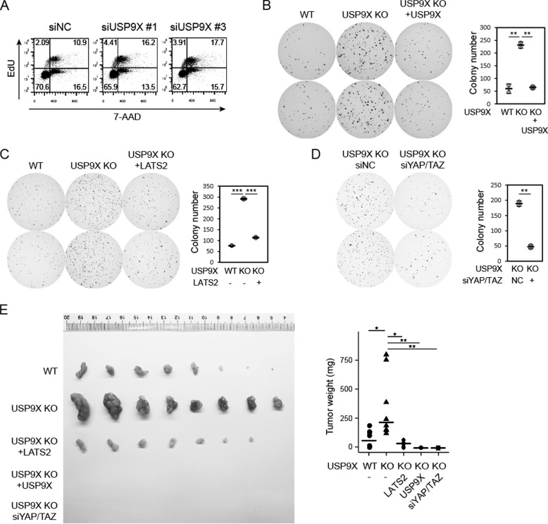
The Hippo pathway plays important roles in controlling organ size and in suppressing tumorigenesis through large tumor suppressor kinase 1/2 (LATS1/2)-mediated phosphorylation of YAP/TAZ transcription co-activators. The kinase activity of LATS1/2 is regulated by phosphorylation in response to extracellular signals. Moreover, LATS2 protein levels are repressed by the ubiquitin-proteasome system in conditions such as hypoxia. However, the mechanism that removes the ubiquitin modification from LATS2 and thereby stabilizes the protein is not well understood. Here, using tandem affinity purification (TAP), we found that anaphase-promoting complex/cyclosome (APC/C), a ubiquitin ligase complex, and USP9X, a deubiquitylase, specifically interact with LATS2. We also found that although APC1 co-localizes with LATS2 to intracellular vesicle structures, it does not regulate LATS2 protein levels and activity. In contrast, USP9X ablation drastically diminished LATS2 protein levels. We further demonstrated that USP9X deubiquitinates LATS2 and thus prevents LATS2 degradation by the proteasome. Furthermore, in pancreatic cancer cells, USP9X loss activated YAP and enhanced the oncogenic potential of the cells. In addition, the tumorigenesis induced by the USP9X ablation depended not only on LATS2 repression, but also on YAP/TAZ activity. We conclude that USP9X is a deubiquitylase of the Hippo pathway kinase LATS2 and that the Hippo pathway functions as a downstream signaling cascade that mediates USP9X's tumor-suppressive activity.

Keywords: Hippo pathway; LATS2; USP9X; anaphase-promoting complex; deubiquitination; protein stability; signal transduction; tumor suppressor gene; ubiquitination.


Conflict of interest statement

The authors declare that they have no conflicts of interest with the contents of this article

Figures

Figure 1.
Figure 1.
LATS2 interacts with APC/C complex and USP9X. A, TAP purification of LATS2-KR. MCF10A cells stably expressing FLAG-SBP–tagged LATS2-KR were established. Cell lysates were purified first with anti-FLAG resin and then with streptavidin resin. The final eluates were resolved on SDS-polyacrylamide gels and silver-stained. *, band of LATS2. MW, molecular weight. B, mass spectrometry analysis identified APC/C complex proteins and USP9X as LATS2-binding proteins. LATS2-KR TAP products were analyzed by MS-MS. The number of peptide hits for APC/C complex proteins, USP9X, and several known LATS2-binding proteins are shown. AA, amino acid. C, Western blotting of TAP samples confirmed that APC1, APC5, and USP9X interact with FLAG-LATS2-KR. TAP samples were examined by Western blotting using the indicated antibodies. D, APC1 co-immunoprecipitates with LATS2. FLAG-LATS2 and HA-APC1 were co-expressed in HEK293T cells, and immunoprecipitation (IP) was performed with anti-FLAG antibody. Samples were analyzed by Western blotting. E, APC1 preferentially binds to LATS2 rather than LATS1. Experiments were similar to those in D. F, USP9X specifically binds to LATS2. Experiments were similar to those in D. CA, C1566A inactive mutant of USP9X. G, LATS2 interacts with APC1 on the endogenous level. Endogenous LATS2 was immunoprecipitated from MCF10A cells with anti-LATS2 antibody, and samples were examined by Western blotting. H, LATS2 interacts with USP9X on the endogenous level. Experiments were similar to those in G. Data are representative of at least duplicate experiments.
Figure 2.
Figure 2.
LATS2 interacts with APC1 and USP9X through distinct regions. A, LATS2, APC1, and USP9X protein domain organization. Domains and key residues of mouse LATS2, human APC1, and human USP9X are illustrated to scale. D box, binding motif recognized by APC/C; HXRXXS, LATS2 phosphorylation target consensus motif; PPXY, motif bound by WW domains; UBA, ubiquitin-associated domain; UBL, ubiquitin-like domain. Protein regions interacting with each other are indicated by dashed red lines. B, interaction of APC1 mutants with LATS2. The indicated plasmids were co-transfected into HEK293T cells. Cell lysates were immunoprecipitated (IP) with anti-FLAG antibody. Samples were examined by Western blotting. C, interaction of LATS2 mutants with APC1. Experiments were similar to those in B. D, interaction of USP9X mutants with LATS2. USP9X was truncated into four fragments as indicated in A. Experiments were similar to those in B. E, interaction of LATS2 mutants with USP9X. Experiments were similar to those in B. Data are representative of triplicate experiments.
Figure 3.
Figure 3.
APC1 does not regulate the localization, protein level, or activity of LATS2. A, subcellular localization of LATS2. The indicated proteins were expressed in HeLa cells and visualized by immunofluorescence staining. B, subcellular localization of APC1. Experiments were similar to those in A. C, co-localization of LATS2 and APC1. Experiments were similar to those in A. D, knockdown of APC1 or LATS2 does not affect the localization of the other. HeLa cells were co-transfected with indicated plasmids and siRNAs. HA-LATS2 and FLAG-APC1 were visualized by immunofluorescence staining. E, inhibition of APC/C does not affect the protein level of LATS2. Non-synchronized (NS) or thymidine-nocodazole–synchronized (S) HeLa cells were treated with APC/C inhibitors as indicated. Samples were examined by Western blotting. F, knockdown of APC1 does not affect the kinase activity of LATS2. LATS2 was immunoprecipitated from transfected HEK293T cells with anti-HA antibody and then subjected to in vitro kinase assay (KA) using recombinant GST-YAP as substrate. Data are representative of triplicate experiments.
Figure 4.
Figure 4.
USP9X deubiquitinates and stabilizes LATS2. A and B, knockdown of USP9X diminishes LATS2 protein level. USP9X was knocked down by siRNAs in MIA PaCa-2 cells (A) or by shRNAs in BxPC-3 cells (B). Cell lysates were examined by Western blotting. C, knockout of USP9X diminishes LATS2 protein level. USP9X was knocked out in HeLa cells by CRISPR-Cas9 technology using two independent sgRNAs. Two clones were examined for each genotype. D, enzymatically inactive USP9X fails to rescue LATS2 protein level in USP9X-knockout cells. FLAG-USP9X-WT and CA mutant were expressed in USP9X-knockout MIA PaCa-2 cells. Cell lysates were examined by Western blotting. E, loss of USP9X does not inhibit LATS2 mRNA expression. The mRNA levels of LATS2 in USP9X-knockout HeLa cells or USP9X knockdown MIA PaCa-2 cells were determined by quantitative RT-PCR. Values represent means ± S.D. (error bars) from three technical repeats. F, LATS2 is destabilized in USP9X-knockout cells. Wild-type and USP9X-knockout HeLa cells were treated with cycloheximide (CHX) as indicated before collection. Cell lysates were examined by Western blotting. G, proteasome inhibitor rescues LATS2 protein level in USP9X knockdown cells. Wild-type and USP9X knockdown MIA PaCa-2 cells were treated with 100 nm bortezomib for 24 h before collection. H and I, knockdown of USP9X promotes LATS2 ubiquitination. LATS2 and siRNAs were transfected into HeLa cells stably expressing HA-Ub or control HeLa cells, as indicated. FLAG-LATS2 (H) or HA-Ub (I) was immunoprecipitated (IP) with anti-FLAG or anti-HA antibodies, respectively. Samples were examined by Western blotting. J, USP9X deubiquitinates LATS2 in vitro. Wild-type or inactive catalytic domain of USP9X was purified from E. coli. FLAG-LATS2 was immunoprecipitated from bortezomib-treated transfected HeLa-Ub stable cells. LATS2 and USP9X catalytic domain were then incubated in deubiquitination assay buffer. Samples were analyzed by Western blotting. Data are representative of triplicate experiments.
Figure 5.
Figure 5.
Knockdown of USP9X activates YAP in pancreatic cancer cells. A, knockdown of USP9X increases dephosphorylated active YAP in MIA PaCa-2 but not in BT-474 cells. Lysates of siRNA-transfected cells were examined by Western blotting. B, LATS1/2 mRNA levels in various cancer cell lines. Relative LATS1 and LATS2 mRNA levels in cancer cell lines were determined by quantitative RT-PCR. The levels of LATS1 and LATS2 in BT-474 cells were set to 1 arbitrary unit. C, knockdown of USP9X increases YAP in cell nuclei. siRNA-transfected MIA PaCa-2 cells were fixed and stained for YAP protein. D, knockdown of USP9X increases the expression of YAP target genes. mRNA levels of the indicated genes in siRNA-transfected MIA PaCa-2 cells were determined by quantitative RT-PCR. Values represent means ± S.D. (error bars) from three technical repeats. p value was calculated by Student's t test. n.s., not significant; *, p < 0.05; **, p < 0.01; ***, p < 0.001. Data are representative of triplicate experiments.
Figure 6.
Figure 6.
USP9X suppresses tumorigenesis via the Hippo pathway. A, USP9X knockdown increases the proportion of proliferating cells. S phase cells were labeled by EdU, and total DNA was labeled by 7-AAD. Samples were analyzed by flow cytometry. B, USP9X knockout promotes anchorage-independent growth. Wild-type, USP9X-knockout, and USP9X-restored MIA PaCa-2 cells were subjected to a soft agar colony formation assay. Colonies were stained by crystal violet and quantified (right). C, re-expression of LATS2 repressed colony formation induced by USP9X knockout. Experiments were similar to those in B. D, YAP/TAZ are required for colony formation induced by USP9X knockout. USP9X-knockout cells were transfected with siRNAs. Experiments were similar to those in B. E, tumorigenesis induced by USP9X knockout is dependent on LATS2 repression and YAP/TAZ activity. Nude mice were injected subcutaneously on the two flanks with cells the same as those in B–D. Tumors were dissected after 6 weeks. Values represent means ± S.D. (error bars). Individual data points are also shown. p value was calculated by Student's t test. n.s., not significant; *, p < 0.05; **, p < 0.01; ***, p < 0.001. Data are representative of at least duplicate experiments.

Similar articles

Cited by

References

    1. Pan D. (2010) The Hippo signaling pathway in development and cancer. Dev. Cell 19, 491–505 10.1016/j.devcel.2010.09.011 - DOI - PMC - PubMed
    1. Yu F. X., Zhao B., and Guan K. L. (2015) Hippo pathway in organ size control, tissue homeostasis, and cancer. Cell 163, 811–828 10.1016/j.cell.2015.10.044 - DOI - PMC - PubMed
    1. Wu S., Huang J., Dong J., and Pan D. (2003) hippo encodes a Ste-20 family protein kinase that restricts cell proliferation and promotes apoptosis in conjunction with salvador and warts. Cell 114, 445–456 10.1016/S0092-8674(03)00549-X - DOI - PubMed
    1. Song H., Mak K. K., Topol L., Yun K., Hu J., Garrett L., Chen Y., Park O., Chang J., Simpson R. M., Wang C. Y., Gao B., Jiang J., and Yang Y. (2010) Mammalian Mst1 and Mst2 kinases play essential roles in organ size control and tumor suppression. Proc. Natl. Acad. Sci. U.S.A. 107, 1431–1436 10.1073/pnas.0911409107 - DOI - PMC - PubMed
    1. Udan R. S., Kango-Singh M., Nolo R., Tao C., and Halder G. (2003) Hippo promotes proliferation arrest and apoptosis in the Salvador/Warts pathway. Nat. Cell Biol. 5, 914–920 10.1038/ncb1050 - DOI - PubMed

Publication types

MeSH terms