. 2017 Nov 28;8(6):e01872-17.
doi: 10.1128/mBio.01872-17.

Vaccination with Recombinant Cryptococcus Proteins in Glucan Particles Protects Mice against Cryptococcosis in a Manner Dependent upon Mouse Strain and Cryptococcal Species

Affiliations

Vaccination with Recombinant Cryptococcus Proteins in Glucan Particles Protects Mice against Cryptococcosis in a Manner Dependent upon Mouse Strain and Cryptococcal Species

Charles A Specht et al. mBio. .

Abstract

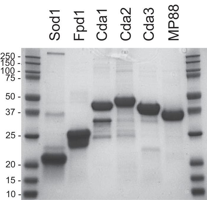
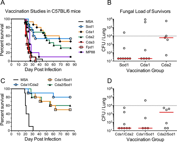
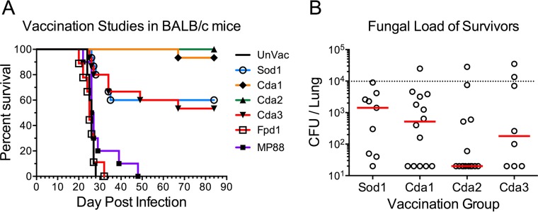
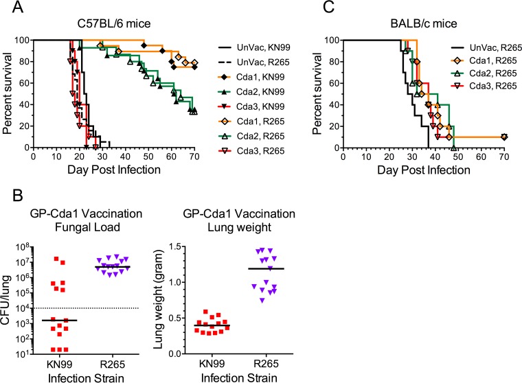
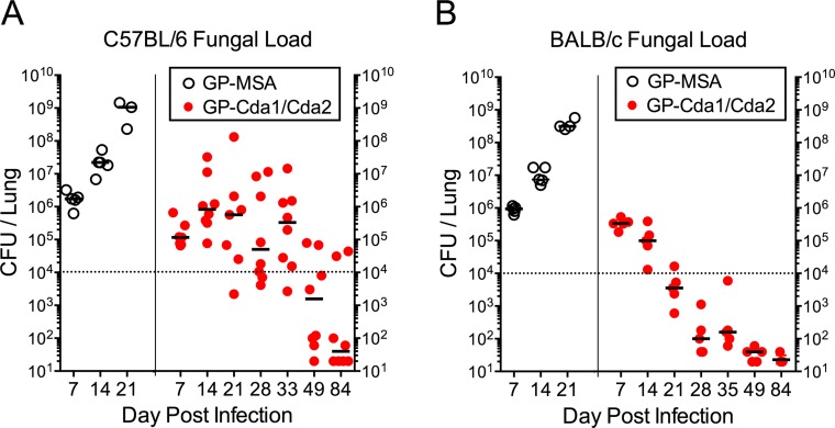
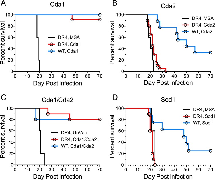
Development of a vaccine to protect against cryptococcosis is a priority given the enormous global burden of disease in at-risk individuals. Using glucan particles (GPs) as a delivery system, we previously demonstrated that mice vaccinated with crude Cryptococcus-derived alkaline extracts were protected against lethal challenge with Cryptococcus neoformans and Cryptococcus gattii The goal of the present study was to identify protective protein antigens that could be used in a subunit vaccine. Using biased and unbiased approaches, six candidate antigens (Cda1, Cda2, Cda3, Fpd1, MP88, and Sod1) were selected, recombinantly expressed in Escherichia coli, purified, and loaded into GPs. Three mouse strains (C57BL/6, BALB/c, and DR4) were then vaccinated with the antigen-laden GPs, following which they received a pulmonary challenge with virulent C. neoformans and C. gattii strains. Four candidate vaccines (GP-Cda1, GP-Cda2, GP-Cda3, and GP-Sod1) afforded a significant survival advantage in at least one mouse model; some vaccine combinations provided added protection over that seen with either antigen alone. Vaccine-mediated protection against C. neoformans did not necessarily predict protection against C. gattii Vaccinated mice developed pulmonary inflammatory responses that effectively contained the infection; many surviving mice developed sterilizing immunity. Predicted T helper cell epitopes differed between mouse strains and in the degree to which they matched epitopes predicted in humans. Thus, we have discovered cryptococcal proteins that make promising candidate vaccine antigens. Protection varied depending on the mouse strain and cryptococcal species, suggesting that a successful human subunit vaccine will need to contain multiple antigens, including ones that are species specific.IMPORTANCE The encapsulated fungi Cryptococcus neoformans and Cryptococcus gattii are responsible for nearly 200,000 deaths annually, mostly in immunocompromised individuals. An effective vaccine could substantially reduce the burden of cryptococcosis. However, a major gap in cryptococcal vaccine development has been the discovery of protective antigens to use in vaccines. Here, six cryptococcal proteins with potential as vaccine antigens were expressed recombinantly and purified. Mice were then vaccinated with glucan particle preparations containing each antigen. Of the six candidate vaccines, four protected mice from a lethal cryptococcal challenge. However, the degree of protection varied as a function of mouse strain and cryptococcal species. These preclinical studies identify cryptococcal proteins that could serve as candidate vaccine antigens and provide a proof of principle regarding the feasibility of protein antigen-based vaccines to protect against cryptococcosis.

Keywords: Cryptococcus; bioinformatics; glucans; immunization; major histocompatibility complex.


Figures

FIG 1
FIG 1
SDS-PAGE of purified recombinant proteins used to make GP vaccines. Each lane contains 10 μg of total protein. Proteins were stained with Coomassie. Molecular masses (in kilodaltons) of protein standards are indicated. The expected molecular masses of the proteins are listed in Table 1.
FIG 2
FIG 2
Vaccination studies with GP-recombinant protein vaccines in C57BL/6 mice. Mice were vaccinated 3 times with 10 µg/vaccine dose of the indicated recombinant protein in GPs and then challenged with 104 CFU of strain KN99. Survival curves and fungal burden in the lungs of mice that survived 84 days p.i. are shown for mice vaccinated with one recombinant protein (A and B) and with two recombinant proteins (C and D). Data were combined from two to three independent vaccination studies with five to six mice per vaccination group in each study. (A) P < 0.001 comparing MSA with Sod1, Cda1, or Cda2. (C) P < 0.001 comparing MSA with any other group. In panels B and D, the dotted line indicates the CFU of KN99 challenge; median CFU per lung for each group are indicated by a solid line.
FIG 3
FIG 3
Vaccination studies with GP-recombinant protein vaccines in BALB/c mice challenged with KN99. Studies were conducted as in Fig. 2A and B, except BALB/c mice were used, and the comparator was unvaccinated (UnVac) mice. Survival curves and fungal burden in the lungs of mice that survived 84 days p.i. are shown. Data were combined from two to three independent vaccination studies with four to five mice per vaccination group in each study. P < 0.001 comparing UnVac with Sod1, Cda1, Cda2, or Cda3. For panel B, the dotted line identifies CFU of KN99 challenge; median CFU per lung for each group are indicated by a solid line.
FIG 4
FIG 4
Survival of vaccinated C57BL/6 and BALB/c mice following infection with C. gattii strain R265. C57BL/6 (A) and BALB/c (C) mice were vaccinated 3 times with 10 µg/vaccine dose of recombinant Cda1, Cda2, or Cda3 in GPs. Control mice were left unvaccinated (UnVac). Mice were then challenged with 104 CFU of KN99 (C. neoformans) or R265 (C. gattii). Panel B shows fungal burden in the lungs (left panel) and lung weights (right panel) of C57BL/6 mice vaccinated with GP-Cda1 that survived 84 days. Data were combined from two to four independent experiments, each with four to five mice per group. (A) P < 0.001 comparing UnVac with Cda1 or Cda2 for both KN99 and R265. (B) P < 0.01 and P < 0.001 comparing fungal load and lung weight, respectively, in KN99 with R265. (C) P < 0.05 comparing UnVac with any other group. In panel B, the dotted line indicates CFU of KN99 challenge; the median CFU per lung for each group are indicated by a solid line.
FIG 5
FIG 5
Lung CFU of C57BL/6 and BALB/c mice vaccinated with GP-Cda1/Cda2 and challenged with C. neoformans strain KN99. C57BL/6 (A) and BALB/c (B) mice were vaccinated three times with 10 μg/vaccine dose of GP-Cda1 and GP-Cda2 (GP-Cda1/Cda2) and then challenged with 104 CFU of C. neoformans strain KN99. Control mice received GP-MSA. At the indicated time points, mice were euthanized and lung CFU determined. For panel A, GP-Cda1/Cda2 data were collected from three independent studies (does not include day 84 data from Fig. 2D), and for panel B, data were from two independent studies. Each datum point represents the CFU from one mouse.
FIG 6
FIG 6
Histology of mouse lungs. Shown are representative H&E- and mucicarmine-stained sections of lungs from vaccinated mice. (A) Mice were vaccinated three times with 10 μg/vaccine dose of GP-Cda1 and GP-Cda2 (GP-Cda1/Cda2) and then challenged with 104 CFU of C. neoformans strain KN99. Control mice received GP-MSA. After 7 (A) and 84 (B) days, mice were euthanized and lung sections prepared. Scale bars on the photomicrographs are as follows: 20× = 1 mm, 100× = 100 µm, 200× = 50 µm, and 400× = 20 µm.
FIG 7
FIG 7
Protection of vaccinated DR4 mice from infection with C. neoformans strain KN99. DR4 and wild-type (WT) C57BL/6 mice were vaccinated three times with 10 μg/vaccine dose of GPs containing (A) Cda1, (B) Cda2, (C) Cda1 and Cda2 (Cda1/Cda2), and (D) Sod1. Control mice received GPs containing MSA or were left unvaccinated (UnVac). Mice were then challenged with 104 CFU of C. neoformans strain KN99 and monitored for survival. Data from vaccinated DR4 mice were combined from at least two independent experiments with a total of 10 to 20 mice per vaccination group. The number of control mice per experimental group ranged from 5 to 10. P < 0.001 comparing DR4 MSA with DR4 Cda1, P < 0.05 comparing DR4 MSA with DR4 Cda2, and P < 0.001 comparing DR4 UnVac with DR4 Cda1/2.

Similar articles

Cited by

References

    1. Heitman J, Kozel TR, Kwon-Chung KJ, Perfect JR, Casadevall A (ed). 2011, Cryptococcus: from human pathogen to model yeast. ASM Press, Washington, DC.
    1. Brown GD, Denning DW, Gow NA, Levitz SM, Netea MG, White TC. 2012. Hidden killers: human fungal infections. Sci Transl Med 4:165rv13. doi:10.1126/scitranslmed.3004404. - DOI - PubMed
    1. Rajasingham R, Smith RM, Park BJ, Jarvis JN, Govender NP, Chiller TM, Denning DW, Loyse A, Boulware DR. 2017. Global burden of disease of HIV-associated cryptococcal meningitis: an updated analysis. Lancet Infect Dis 17:873–881. doi:10.1016/S1473-3099(17)30243-8. - DOI - PMC - PubMed
    1. Singh N, Dromer F, Perfect JR, Lortholary O. 2008. Cryptococcosis in solid organ transplant recipients: current state of the science. Clin Infect Dis 47:1321–1327. doi:10.1086/592690. - DOI - PMC - PubMed
    1. Byrnes EJ III, Li W, Lewit Y, Ma H, Voelz K, Ren P, Carter DA, Chaturvedi V, Bildfell RJ, May RC, Heitman J. 2010. Emergence and pathogenicity of highly virulent Cryptococcus gattii genotypes in the northwest United States. PLoS Pathog 6:e1000850. doi:10.1371/journal.ppat.1000850. - DOI - PMC - PubMed

MeSH terms