. 2017 Nov 28;7(1):16533.
doi: 10.1038/s41598-017-15813-8.

Phosphorylated exogenous alpha-synuclein fibrils exacerbate pathology and induce neuronal dysfunction in mice

Affiliations

Phosphorylated exogenous alpha-synuclein fibrils exacerbate pathology and induce neuronal dysfunction in mice

Mantia Karampetsou et al. Sci Rep. .

Abstract

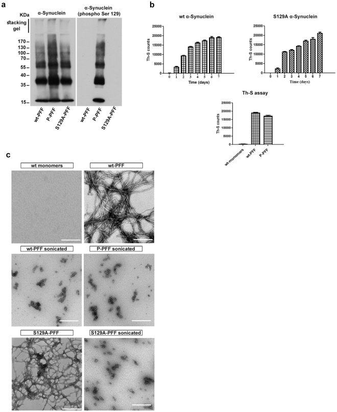
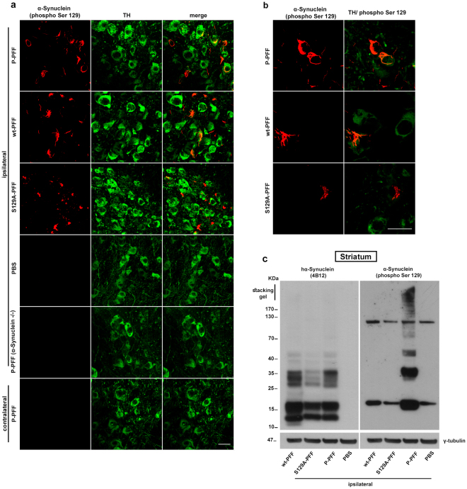
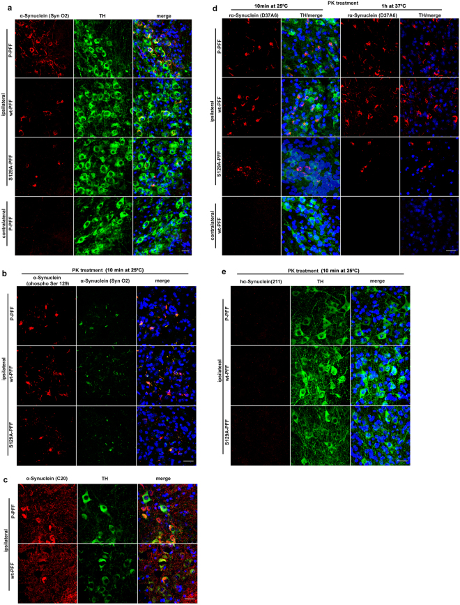
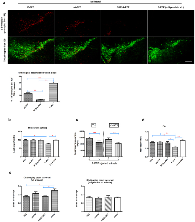
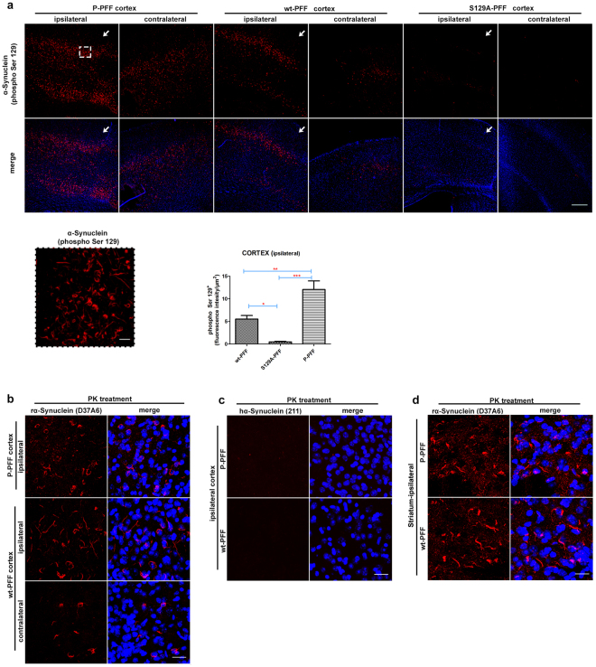
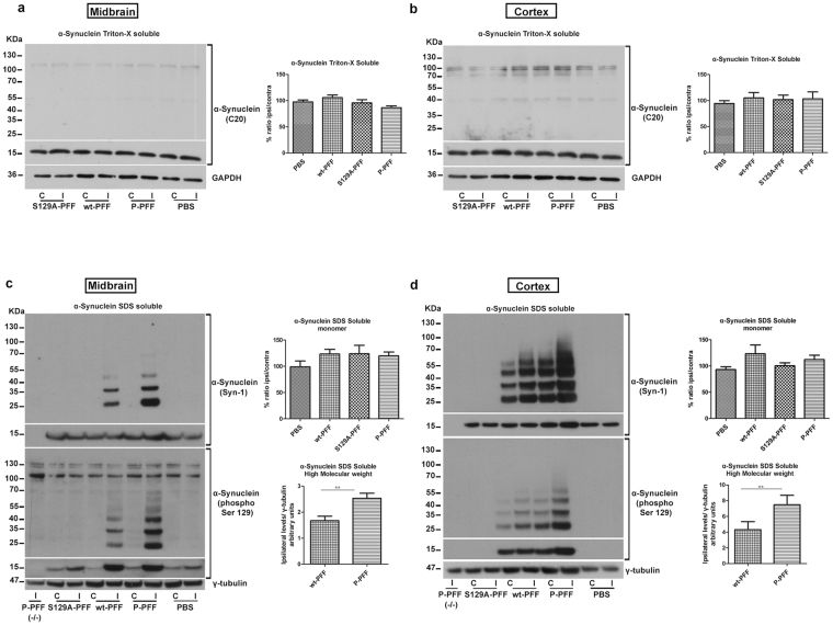
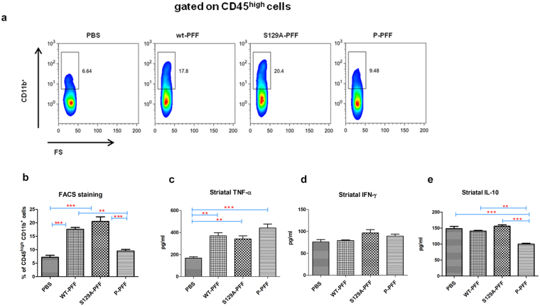
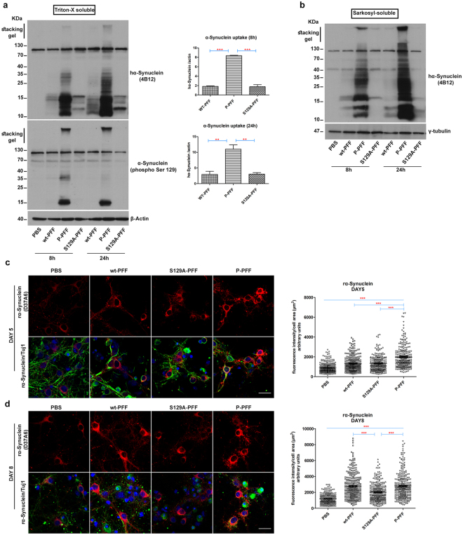
Approximately 90% of alpha-synuclein (α-Synuclein) deposited in Lewy bodies is phosphorylated at serine 129 suggesting that the accumulation of phosphorylated α-Synuclein is critical in the pathogenesis of Parkinson's disease. However, in vivo experiments addressing the role of phosphorylated α-Synuclein in the progression of Parkinson's disease have produced equivocal data. To clarify a role of Ser129 phosphorylation of α-Synuclein in pathology progression we performed stereotaxic injections targeting the mouse striatum with three fibrilar α-Synuclein types: wt-fibrils, phosphorylated S129 fibrils and, phosphorylation incompetent, S129A fibrils. Brain inoculation of all three fibrilar types caused seeding of the endogenous α-Synuclein. However, phosphorylated fibrils triggered the formation of more α-Synuclein inclusions in the Substantia Nigra pars compacta (SNpc), exacerbated pathology in the cortex and caused dopaminergic neuronal loss and fine motor impairment as early as 60 days post injection. Phosphorylated fibril injections also induced early changes in the innate immune response including alterations in macrophage recruitment and IL-10 release. Our study further shows that S129 phosphorylation facilitated α-Synuclein fibril uptake by neurons. Our results highlight the role of phosphorylated fibrilar α-Synuclein in pathology progression in vivo and suggest that targeting phosphorylated α-Synuclein assemblies might be important for delaying inclusion formation.


Conflict of interest statement

The authors declare that they have no competing interests.

Figures

Figure 1
Figure 1
Characterization of wt-, P- and S129A- PFF. (a) Western blotting analysis for wt-, P- and S129A-PFF. Equal amounts of fibrils were analyzed in a 10% SDS-PAGE gel using the C20 antibody. P-PFF were detected with the α-Synuclein (phospho Ser) antibody whereas no signal was observed for the S129A- and wt-PFF. (b) Fibril formation monitoring by Th-S assay. Graphs show: fibril formation monitoring of the wt- and S129A-monomers incubated for 7 days (top). Comparison of the fibril content of wt-PFF and P-PFF (bottom). Monomeric α-Synuclein was used as control. The assays were performed in triplicate. (c) Electron microscopy images of negatively stained samples of the different types of α-Synuclein to confirm the presence of fibrils compared to the monomeric non-fibrilar α-Synuclein. Scale bar, 500 nm.
Figure 2
Figure 2
Pathological α-Synuclein accumulation in the SNpc dopaminergic neurons of wt mice following stereotaxic unilateral striatal injection of three different human-PFF types (P-PFF, wt-PFF, and S129A-PFF). Animals were analyzed 60 dpi. (a) Confocal images showing double immunostaining for P-α-Synuclein and TH in nigral sections of PFF-injected animals. Accumulation of hyper-phosphorylated α-Synuclein (α-Synuclein phospho Ser 129) is evident in dopaminergic neurons (TH) of the ipsilateral SNpc. Pathology is absent in the ipsilateral side of PBS injected animals. The contralateral side of P-PFF injected animals shows no signs of pathologic accumulations. α-Synuclein (phospho Ser129) immunoreactivity is not detected in the ipsilateral nigra of α-Synuclein null (−/−) animals injected with P-PFF (n = 4). (b) Images in higher magnification are showing P-α-Synuclein accumulations induced by the different types of PFF. (c) Striatal tissue of injected animals (3 dpi) was extracted and immunoblotted with the 4B12 and phospho Ser 129 antibodies. Human α-Synuclein was readily detected in the striatal extracts. S129A- and wt-PFF could not be detected with the phospho Ser 129 antibody. γ-tubulin was used as a loading control (cropped gel/blot is shown). Scale bars represent 25 μm.
Figure 3
Figure 3
Characterization of SNpc intraneuronal α-Synuclein accumulations. (a) Double labeling with the conformational specific α-Synuclein antibody SynO2 and TH is showing the fibrilar nature of the α-Synuclein cytoplasmic accumulations restricted to TH neurons of the ipsilateral SNpc following injections with P-, wt- and mutant S129A-PFF at 60 dpi. Contralateral side shows only background staining with the SynO2 antibody. (b) Representative sections of SNpc from all PFF injected animals showing the co-staining of α-Synuclein accumulations with the α-Synuclein (phospho Ser 129) and SynO2 antibodies following PK treatment for 10 min at 25 °C to expose antigenic sites. (c) α-Synuclein accumulations also stained positive with the C20 antibody. (d) Host α-Synuclein expression is essential for the formation of pathological α-Synuclein accumulations. TH-stained nigral sections (PK-treated, 10 min at 25 °C) are exhibiting α-Synuclein accumulations that are positive for the endogenous rodent α-Synuclein (D37A6) antibody. PK resistant D37A6-positive accumulations were also evident following prolonged PK treatment (1 h at 37 °C) in nigral sections until the TH signal is not detectable (e) α-Synuclein accumulations do not stain with the human specific anti-α-Synuclein (211) antibody. TO-PRO-3 (blue) was used as a cell nuclear marker (n = 4). Scale bars represent 25 μm.
Figure 4
Figure 4
P-PFF exacerbate the pathology within the SNpc and significantly impair the integrity of the dopaminergic neurons. (a) Coronal nigral sections were immunostained for α-Synuclein (phospho Ser S129) and TH. The absolute numbers of P-accumulations that were formed within the TH positive neurons in the different fibril-injected brains were counted. As shown in representative tiled images for each of the P-, wt-, and S129A-PFF injected animals, P-PFF induced a more widespread pathology compared to the other two fibrilar types. P-PFF injected α-Synuclein null mice did not show any sign of pathology. Graph depicts the percentage of TH neurons containing P-α-Synuclein positive accumulations for each treatment group (n = 4 animals per group, 3 sections per animal). (b) Stereological analysis of TH-positive neurons is showing a significant loss of dopaminergic neurons in P-PFF injected animals compared to the PBS, wt-, S129A-PFF injected animals and to P-PFF α-Synuclein null (−/−) injected animals. The data are presented as a percentage of ipsilateral to contralateral side (n = 5–6 animals per group). (c) Decreased nigral TH positive neuron number was confirmed with VMAT2 stereological analysis following P-PFF injections (4–6 animals per group, paired Student’s t-test analysis). (d) Significant decrease in striatal DA levels in wt animals injected with P-PFF. The data are presented as a ratio of ipsilateral to contralateral side (n = 5–7 animals per group). (e) Fine motor impairment as increased errors/step in the challenging beam traversal test in P-PFF- vs. S129A- and control PBS- injected animals (n = 7–8 animals/group). Similar injections did not cause any motor impairment in null α-Synuclein (−/−) mice (n = 5 animals/group). Data represent mean values ± SEM. Differences were estimated using one-way ANOVA followed by Tukey’s post-hoc test. (a) p < 0,0001 (b) p = 0,0052 (c) For TH p = 0,0002, and VMAT p = 0,0062 two-tailed paired t-test (d) p = 0,0009 (e) p = 0,0163. Scale bar in (a) represents 250 μm.
Figure 5
Figure 5
Robust endogenous α-Synuclein accumulation is also evident in the cortex of injected animals. Coronal sections of P-, wt- and S129A- PFF injected animals were stained for α-Synuclein (phospho Ser129). (a) P-α-Synuclein positive inclusions (highlighted in magnification within the dashed line frame) were evident in the ipsilateral cortex of P- and wt- PFF-injected animals. Progression of pathology but in a more dispersed pattern is also evident in the contralateral cortex. S129A- PFF are not as efficient in inducing α-Synuclein accumulation on either sides. Arrows indicate the needle entry point. Graph shows phospho Ser129 mean fluorescence intensity of the ipsilateral cortex, normalized to the measured area (intensity/μm2) (n = 4 animals/group, p = 0,0002). (b) The rodent specific anti-α-Synuclein (D37A6) antibody and (c) the human specific anti-α-Synuclein (211) were used for immunostaining of PK-treated sections (10 min at 25 °C to expose the antigenic sites). Pathological accumulations in the area of the cortex of both hemispheres were positively stained with the rodent specific antibody. α-Synuclein accumulations did not stain with the human specific anti-α-Synuclein (211) antibody in the ipsilateral cortex of P- and wt-PFF injected animals (n = 4). (d) Coronal striatal sections of P- and wt-PFF injected animals were also positively stained with the rodent specific α-Synuclein (D37A6) antibody. TO-PRO-3 (blue) was used as a cell nuclear marker. Scale bars represent 250 μm in (a) and 25 μm in (b,c,d).
Figure 6
Figure 6
Biochemical profile of α-Synuclein in PFF-injected animals. (a) Midbrain Triton-X soluble samples of injected animals showed no difference in α-Synuclein levels between the ipsi-and contralateral side in all fibrilar types and the control PBS- injected animals (α-Synuclein monomer is shown in cropped gel/blot). GAPDH was used as a loading control (cropped gel/blot is shown) (n = 5–7 brains/group). Similar in (b) no differences were found for the Triton-X soluble fraction in the area of the cortex (α-Synuclein monomer is shown in cropped gel/blot) (n = 4 brains/group). (c) P- and wt-PFF injections resulted in a shift of the SDS-soluble α-Synuclein in higher molecular weight species ipsilaterally in the midbrain. These SDS-soluble α-Synuclein fraction was significantly enriched in the P-PFF injected side compared to the wt-PFF treatment (n = 5 brains/group). High molecular weight species in both treatments were also positive for the phospho Ser 129 α-Synuclein antibody. No difference was observed in the SDS-soluble α-Synuclein monomer levels between the ipsi- and the contralateral side for the P-, wt-, S129A-PFF and the PBS-injected animals (α-Synuclein monomer is shown in cropped gel/blot) (n-6–7 animals/group). γ-tubulin was used as a loading control (cropped gel/blot is shown). (d) Immunoblot for the SDS-soluble fraction extracted from the cortex of injected animals showed that α-Synuclein SDS-soluble high molecular weight species are formed readily in P-PFF-injected animals in both sides compared to the wt treatment. Densitomentry of the ipsilateral α-Synuclein levels confirmed the significant difference between the treatments (n = 5 animals/group). α-Synuclein null mice (−/−) injected with P-PFF did not show any positive signal with the Syn-1 antibody. The observed α-Synuclein species were phosphorylated in nature as seen following immunoblotting with the phospho Ser 129 antibody. No difference was observed in the SDS-soluble α-Synuclein monomer levels between the ipsi- and the contralateral side for all types of injected animals (α-Synuclein monomer is shown in cropped gel/blot) (n = 5–6 animals/group). γ-tubulin was used as a loading control (cropped gel/blot is shown). Data represent mean values ± SEM. Differences were estimated using one-way ANOVA followed by Tukey’s post-hoc test and paired two-tailed Student’s t-test. (c) p = 0,0037 (d) p = 0,0053.
Figure 7
Figure 7
Reduced innate immune response following P-PFF treatments in vivo. (a) Cells obtained from ipsilateral striatal tissue of 3 dpi injected mice were stained and analyzed by flow-cytometry. Each animal was analyzed individually. Representative FACS plots showing the percentages of CD11b+ cells gated on CD45high leukocytes. (b) Cumulative data showing the percentages of CD45highCD11b+ macrophages in injected animals. Data are pooled from three independent experiments. (c) TNF-α (d) INF-γ and (e) IL-10 levels were measured in ipsilateral striatum homogenates by ELISA. Data are expressed as mean ± SEM of triplicate wells. Data shown for cytokine release are pooled from three independent experiments. Statistical significance was obtained by ANOVA followed by Tukey’s post-hoc test. (b) p < 0,0001 (c) p = 0,0007 (e) p = 0,0001.
Figure 8
Figure 8
Increased uptake and faster seeding of the endogenous α-Synuclein in primary neurons following P-PFF treatment (a) Mouse primary cultures (6div) were treated with PFF for 8 and 24 hours. Following Triton-X extraction, internalized fibrils were visualized with the human specific α-Synuclein antibody 4B12. Increased uptake of P-PFF was observed as early as 8 h. The uptake was quantified by densitometry and found to be significantly increased for P-PFF in both time points compared to wt- or S129A- PFF. P-PFF uptake was further verified using the phospho Ser 129 antibody. β-Actin was used as a loading control (cropped gel/blot is shown). (b) Sarcosyl-soluble α-Synuclein species were also increased following P-PFF treatment of primary cortical neurons. γ-tubulin was used as a loading control (cropped gel/blot is shown). (c) Immunocytochemistry with the rodent specific α-Synuclein antibody (D37A6) in primary mouse cortical cultures treated with the three fibrilar types. P-PFF seed the endogenous α-Synuclein more effectively compared to the other fibrilar types and PBS control treated neurons for 5 days. However (d) 8-day-treated neurons exhibit similar levels of endogenous α-Synuclein signal for both the wt- and P-PFF treatments, in contrast to the significant lower levels of S129A-treated neurons. Scatter plots present the mean fluorescence intensity/cell of three independent experiments (n≈90 single cells/condition per replicate). β Tubulin III (Tuj 1) was used as a neuronal marker. TO-PRO-3 (blue) was used as a cell nuclear marker. All data represent mean values ± SEM from three independent experiments. Statistical significance was obtained by ANOVA followed by Tukey’s post-hoc test for (a) and non parametric Kruskal-Wallis test followed by Dunn’s post-hoc test for (c and d). (a) For 8 h p < 0,0001 and for 24 h p = 0,002, (c) p < 0,0001 (d) p < 0,0001.Scale bar in (c and d) represent 25 μm.

Similar articles

Cited by

References

    1. Polymeropoulos MH, et al. Mutation in the alpha-synuclein gene identified in families with Parkinson’s disease. Science. 1997;276:2045–2047. doi: 10.1126/science.276.5321.2045. - DOI - PubMed
    1. Singleton AB, et al. alpha-Synuclein locus triplication causes Parkinson’s disease. Science. 2003;302:841. doi: 10.1126/science.1090278. - DOI - PubMed
    1. Fujiwara H, et al. alpha-Synuclein is phosphorylated in synucleinopathy lesions. Nature cell biology. 2002;4:160–164. doi: 10.1038/ncb841. - DOI - PubMed
    1. Kahle PJ, et al. Hyperphosphorylation and insolubility of alpha-synuclein in transgenic mouse oligodendrocytes. EMBO reports. 2002;3:583–588. doi: 10.1093/embo-reports/kvf109. - DOI - PMC - PubMed
    1. Anderson JP, et al. Phosphorylation of Ser-129 is the dominant pathological modification of alpha-synuclein in familial and sporadic Lewy body disease. The Journal of biological chemistry. 2006;281:29739–29752. doi: 10.1074/jbc.M600933200. - DOI - PubMed

Publication types

MeSH terms