. 2017 Nov 28;7(1):16494.
doi: 10.1038/s41598-017-16392-4.

Deciphering how Cpl-7 cell wall-binding repeats recognize the bacterial peptidoglycan

Affiliations

Deciphering how Cpl-7 cell wall-binding repeats recognize the bacterial peptidoglycan

Noemí Bustamante et al. Sci Rep. .

Abstract

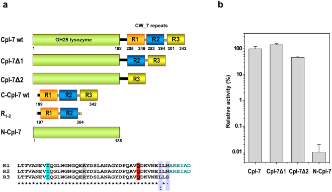
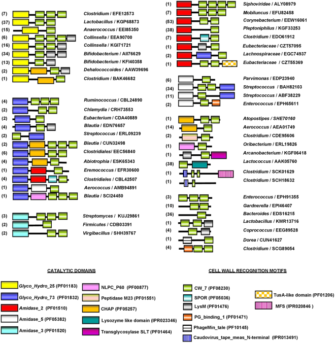
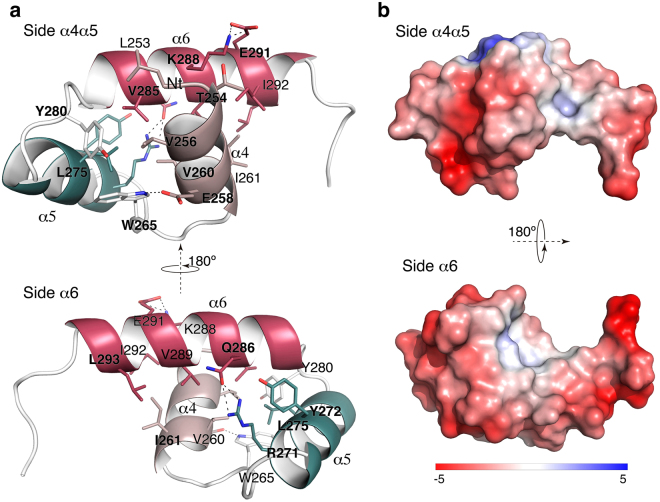
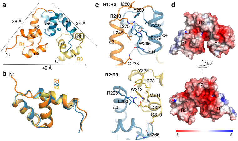
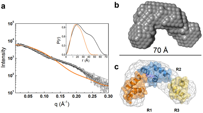
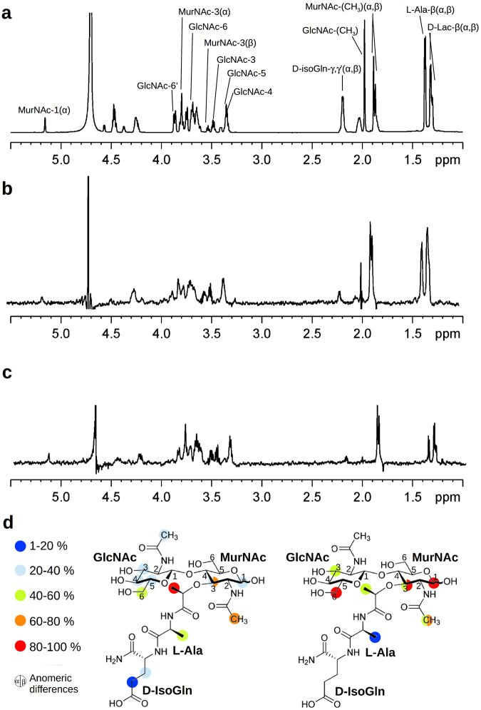
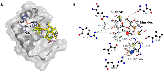
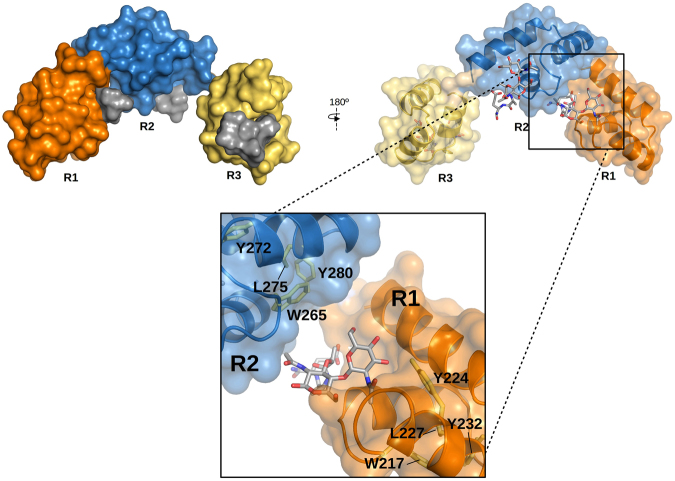
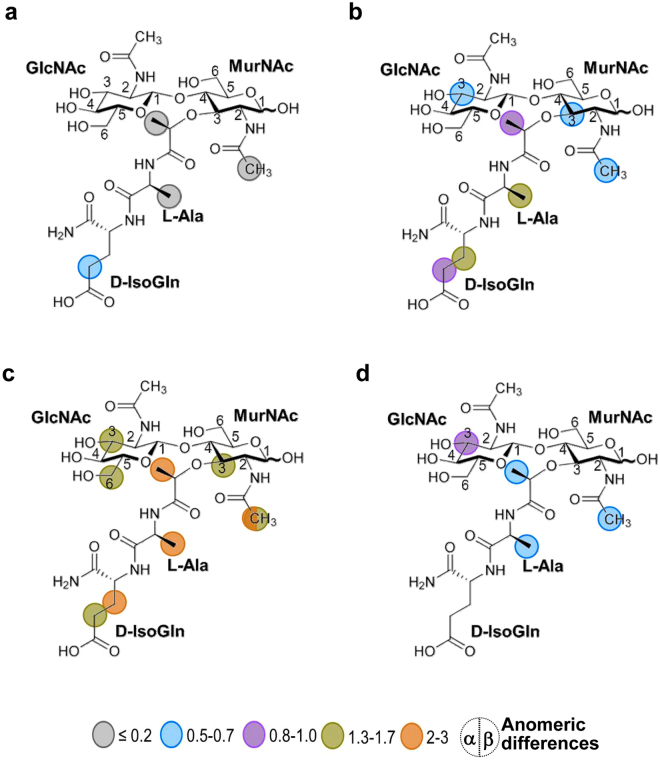
Endolysins, the cell wall lytic enzymes encoded by bacteriophages to release the phage progeny, are among the top alternatives to fight against multiresistant pathogenic bacteria; one of the current biggest challenges to global health. Their narrow range of susceptible bacteria relies, primarily, on targeting specific cell-wall receptors through specialized modules. The cell wall-binding domain of Cpl-7 endolysin, made of three CW_7 repeats, accounts for its extended-range of substrates. Using as model system the cell wall-binding domain of Cpl-7, here we describe the molecular basis for the bacterial cell wall recognition by the CW_7 motif, which is widely represented in sequences of cell wall hydrolases. We report the crystal and solution structure of the full-length domain, identify N-acetyl-D-glucosaminyl-(β1,4)-N-acetylmuramyl-L-alanyl-D-isoglutamine (GMDP) as the peptidoglycan (PG) target recognized by the CW_7 motifs, and characterize feasible GMDP-CW_7 contacts. Our data suggest that Cpl-7 cell wall-binding domain might simultaneously bind to three PG chains, and also highlight the potential use of CW_7-containing lysins as novel anti-infectives.


Conflict of interest statement

The authors declare that they have no competing interests.

Figures

Figure 1
Figure 1
Module composition of Cpl-7 endolysin, deletion constructs and contribution of CW_7 repeats to the murolytic activity. (a) Cartoon representation of Cpl-7 variants used in this study. Numbers indicate residues comprised in each structural element. The catalytic domain, in green, belongs to the GH-25 family. The cell wall-binding domain comprises three almost identical CW_7 repeats of 42 residues (R1, R2 and R3; sequence alignment is shown at the bottom) connected by six-residue-long inter-repeat linkers (AREIAD). The residues substituted by alanine in C-Cpl-7R, C-Cpl-7Q and C-Cpl-7I mutants are highlighted in grey, red and cyan, respectively. (b) Variation of the percentage of specific activity on pneumococcal cell walls with serial deletion of the CW_7 repeats from Cpl-7 sequence. Full-length Cpl-7 specific activity was taken as 100%. Data are the average of three independent assays of four-to-six replicas each; bars indicated standard deviations.
Figure 2
Figure 2
Schematic representation of protein architectures containing the CW_7 motif. The accession number of representative proteins and the respective coding bacterium are indicated. Figures in parenthesis are the number of sequences within each architecture. Comprised modules are colour coded and Pfam or INTERPRO accession codes are shown at the bottom.
Figure 3
Figure 3
Crystal structure of the CW_7 repeat of Cpl-7. (a) Ribbon representation of the R2 repeat showing views of the side α4α5 (top) and the side α6 (bottom). Relevant residues are depicted as capped-sticks. A hydrophobic patch is observed at the core of the three-helix bundle. Hydrogen bonds and salt-bridge interactions are displayed with dot lines. (b) Poisson-Boltzmann electrostatic-potential surface (color bar range ± 5 kT/e) generated by PyMOL APBS tool for side α4α5 (top) and side α6 (bottom) of the R2 repeat.
Figure 4
Figure 4
Structure of full-length C-Cpl-7R domain. (a) Crystal structure of C-Cpl-7R with repeats coloured in orange (R1), blue (R2) and yellow (R3). Comprised helices are labelled from α1 to α9. Bars indicate the side dimensions of the C-Cpl-7R triangular structure in angstroms. (b) Structural superimposition of the three repeats of C-Cpl-7R. (c) Interactions between repeats R1 and R2 repeats (top panel) and R2 and R3 repeats (bottom panel) coloured as in (a). Residues involved in inter-repeat interactions are depicted as sticks, and polar interactions as dotted lines. (d) Poisson-Boltzmann electrostatic-potential surface (color bar range ± 5 kT/e) generated by PyMOL APBS tool for the full-length C-Cpl-7R domain.
Figure 5
Figure 5
Determination of C-Cpl-7 configuration in solution by SAXS. (a) Overlay of the scattering curve of C-Cpl-7 wt at infinite dilution (black dots) with the scattering profile calculated from the full-length domain crystal structure using CRYSOL and the fit obtained by rigid-body modelling (BUNCH) (orange and grey traces, respectively). The inset shows the real-space distance-distribution functions P(r) for the configuration in solution (black trace) and the crystal structure (orange trace). (b) Ab initio bead model of C-Cpl-7 reconstructed from SAXS data using DAMMIF. (c) Superposition of the DAMMIF model in mesh representation with the best rigid model (cartoon) produced by BUNCH.
Figure 6
Figure 6
Characterization of GMDP binding to C-Cpl-7 by STD-NMR in PB buffer. (a) 1H NMR spectrum of free GMDP as reference. Proton accounting for relevant peaks are marked as in Supplementary Table S2 where chemical shifts of 1H NMR spectra of GMDP α:β anomer mixture (2:1 ratio) in D2O are shown. (b) and (c) STD-NMR spectra of GMDP (2.5 mM) in the presence of C-Cpl-7 wt (33 μM) upon irradiation at the methyl or the aromatic region, respectively. The 2:1 ratio between the α and β anomers of free GMDP changes to 1.3:1 in STD spectra, unveiling the preferential binding of the β anomer. (d) GMDP binding epitopes as deduced from STD data upon irradiation at methyl (left) or aromatic (right) protein regions. Colour labels indicate the STD intensity (I) of each signal relative to the most intense signal observed. Red circles: 100% > I > 80%; orange: 80% > I > 60%; green: 60% > I > 40%; cyan: 40% > I > 20%; blue: 20% > I > 1%.
Figure 7
Figure 7
Structural model of the complex of GMDP with the R2 repeat of C-Cpl-7 wt based on docking analysis and STD-NMR data. (a) Stick representation of GMDP conformers in the best complex models obtained with AutoDock4.2 (green), CRDOCK (grey), AutoDock Vina (yellow), and DOCK6 (magenta). R2 surface is coloured in grey and protein residues irradiated during STD experiments are drawn as sticks (aromatics in blue and Leu275 in orange). (b) Ligplot representation of the hydrogen bonds and hydrophobic contacts in the R2:GMDP complex generated with AutoDock4.2, the one which agreed best with the GMDP epitope map derived from STD measurements. Overlapping circles show the STD epitope mapping using the colour code of Fig. 6.
Figure 8
Figure 8
Model of C-Cpl-7 wt in complex with GMDP based on the BUNCH model generated from SAXS data and the three-fold transfer of the GMDP pose in the best docking model to R2 repeat. GMDP molecules are shown in solid surface (left panel) or in stick representation (right panel). A detail of the interface between R1 and R2 repeats is shown in the close-up. R2 likely contributes to shape the binding pocket of R1, thereby explaining the STD signals of the anomeric proton and the acetamido group of MurNAc, unaccounted for in the model of GMDP in complex with one isolated repeat. Protein residues relevant for the STD measurements observable in this view (Y224, Y280, W265 L227, L275) are shown in stick representation.
Figure 9
Figure 9
Contribution of the residues mutated in C-Cpl-7R, C-Cpl-7Q and C-Cpl-7I to the GMDP binding. Binding epitope of GMDP in the presence of (a) C-Cpl-7R (b) C-Cpl-7Q and (c) C-Cpl-7I upon irradiation at the methyl region. (d) STD-NMR spectrum and binding epitope in the presence of C-Cpl-7I upon irradiation at the aromatic region. The intensity ratio of the STD effect induced by C-Cpl-7R, C-Cpl-7Q and C-Cpl-7I with respect to that of C-Cpl-7 wt is indicated by colours: grey circles 0.2; blue: 0.1–0.5; magenta: 0.8–1.0; green: 1.3–1.7; orange: 2–3. Measurements were carried out in PB-D2O buffer at 2.5 mM GMDP and 50 μM mutant concentration.

Similar articles

Cited by

References

    1. Pastagia M, Schuch R, Fischetti VA, Huang DB. Lysins: the arrival of pathogen-directed anti-infectives. J. Med. Microbiol. 2013;62:1506–1516. doi: 10.1099/jmm.0.061028-0. - DOI - PubMed
    1. Rodríguez-Cerrato V, et al. Pneumococcal LytA autolysin, a potent therapeutic agent in experimental peritonitis-sepsis caused by highly β-lactam-resistant Streptococcus pneumoniae. Antimicrob. Agents Chemother. 2007;51:3371–3373. doi: 10.1128/AAC.00137-07. - DOI - PMC - PubMed
    1. Borysowski, J. & Górski, A. Anti-staphylococcal lytic enzymes. In: Enzybiotics: Antibiotic Enzymes as Drugs and Therapeutics. pp. 149–172 Villa, T. G., Veiga-Crespo, P, editors, John Wiley & Sons, Inc., Hoboken, NJ, USA. (2010).
    1. Fischetti VA. Bacteriophage endolysins: A novel anti-infective to control Gram-positive pathogens. Inter. Med. Microbiol. 2010;300:357–362. doi: 10.1016/j.ijmm.2010.04.002. - DOI - PMC - PubMed
    1. Nelson DC, et al. Endolysins as antimicrobials. Adv. Virus Res. 2012;83:299–365. doi: 10.1016/B978-0-12-394438-2.00007-4. - DOI - PubMed

Publication types