Review
. 2017 Nov;30(5):297-312.
doi: 10.1055/s-0037-1606107. Epub 2017 Nov 27.

How Should Imaging Direct/Orient Management of Rectal Cancer?

Affiliations
Review

How Should Imaging Direct/Orient Management of Rectal Cancer?

Jemma Bhoday et al. Clin Colon Rectal Surg. 2017 Nov.

Abstract

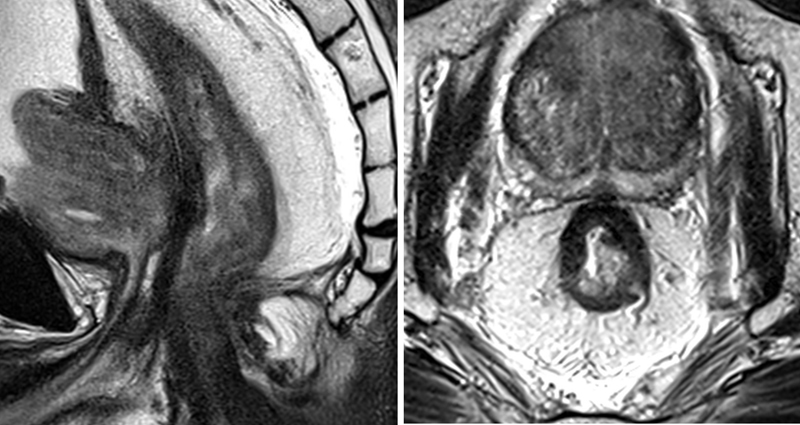
Modern rectal cancer management is dependent on preoperative staging, and radiological assessment is a crucial part of this process. Imaging must provide sufficient information to guide preoperative decision-making that is reliable and reproducible. Different methods have been used for local staging; however, magnetic resonance imaging (MRI) has shown to be the most reliable tool for this purpose. MRI offers prognostic information about the patients and guides the decision between neoadjuvant treatment and total mesorectal excision alone. Also, not only the initial staging but also restaging by MRI can provide significant information regarding tumor response that is essential when considering alternative approaches.

Keywords: local staging; magnetic resonance imaging; rectal cancer.


Figures

Fig. 1
Fig. 1
The yellow line demonstrates the level of the peritoneal reflection in a female patient (left sagittal image) and a male patient (right sagittal image). The green arrows and the green lines delineate the presacral fascia with red dashed lines indicating the mesorectal fascia.
Fig. 2
Fig. 2
Peritoneal reflection: (left) sagittal T2 image and (right) upper T2-axial image; (center) mesorectal fascia. The red dashed line shows mesorectal fascia, and the orange line demarcates the peritoneal reflection.
Fig. 3
Fig. 3
Hyperintense tubular/cystic structures along the seminal vesicles (yellow arrows on sagittal and axial images) show the level of the neurovascular bundles.
Fig. 4
Fig. 4
The purple line indicates Denonvilliers' fascia in a male patient.
Fig. 5
Fig. 5
The coronal image shows the anatomical structures of the anal canal: Levators (orange) continue to the external sphincter (red arrow), and muscularis propria layer of the rectal wall (yellow arrows) continues as the internal sphincter (green arrow). The tiny hyperintense line between the internal and external sphincters indicates an intersphincteric plane (purple line) and the purple arrow indicates the interspnincteric groove which corresponds to the dentate line not visible on magnetic resonance imaging. The blue oval demonstrates the level of puborectalis sling.
Fig. 6
Fig. 6
Axial pretreatment T2 magnetic resonance imaging (MRI) (left) shows a semiannular lesion which infiltrated the rectal wall at 9 to 3'o clock position with evidence of extramural spread at the site of infiltrating border. Tumor infiltrates the mesorectal fascia (distance to circumferential resection margin is < 1 mm) and abuts the prostate. (Right) Posttreatment magnetic resonance image, persistence intermediate signal intensity tumor (yellow arrow) at the site of the primary disease, suggesting mrTRG4. mrTRG, MRI tumor regression grade.
Fig. 7
Fig. 7
Left coronal image shows a polypoidal lesion within the lower/mid rectum. The fibromuscular stalk arises at the left lateral wall, and the maximum degree of invasion would be assessed at this very level (green arrow and line). (Right) The image indicates a low rectal advanced tumor is infiltrating rectal wall at 7 to 10 o'clock position with depression portion at 9 o'clock (green arrow) and rolled ages (dashed green lines). The intramural and extramural contiguous invasion should be assessed at this level and measured in millimeters.
Fig. 8
Fig. 8
A mesorectal lymph node with a heterogeneous signal intensity seen at 9 o'clock position (yellow circle).
Fig. 9
Fig. 9
Extramural vascular invasion, affecting the large diameter superior rectal veins (blue arrows). The yellow circle encases the left internal iliac node with irregular borders.
Fig. 10
Fig. 10
Pelvic floor and low rectum (oblique coronal view). The solid green line indicates intersphincteric plane; solid yellow line indicates plane for extralevator abdominoperineal excision (ELAPE). Early-stage tumor ( A ) has not invaded beyond the muscularis propria; overtreatment with ELAPE, yellow line. Late-stage tumor ( B ) has breached the muscularis propria/internal sphincter and has invaded beyond the intersphincteric space into the levator ani and puborectalis muscle; following the total mesorectal excision plane (green line) would lead to an involved circumferential resection margin.
Fig. 11
Fig. 11
Upper row (pre-CRT): a midrectal tumor infiltrating rectal wall at 5 to 8 o'clock position and discontinuous vascular spread (extramural venous invasion [EMVI]) at 5 o'clock (blue arrow) that abuts the mesorectal fascia. Lower row (post-CRT): a tiny scar at the site of treated tumor (barely visible and a completely fibrotic vascular deposit in the mesorectum at the level of EMVI (blue arrow), suggesting mrTRG1. mrTRG, MRI tumor regression grade.
Fig. 12
Fig. 12
Pretreatment (left) semiannular mass infiltrating rectal wall at 4 to 7 o'clock at which level tumor borders the interspincteric plane. Posttreatment image shows a good response to treatment with dense fibrotic scar (yellow arrow) at the site of the treated disease (mrTRG2). mrTRG, MRI tumor regression grade.
Fig. 13
Fig. 13
Midrectal tumor invading the anterior quadrant of the rectal wall (left). Posttreatment MRI shows predominating dense fibrosis with some minimal intermediate signal (right), suggesting mrTRG3. MRI, magnetic resonance imaging; mrTRG, MRI tumor regression grade.

Similar articles

Cited by

References

    1. Battersby N J, Moran B, Yu S, Tekkis P, Brown G. MR imaging for rectal cancer: the role in staging the primary and response to neoadjuvant therapy. Expert Rev Gastroenterol Hepatol. 2014;8(06):703–719. - PubMed
    1. Balyasnikova S, Brown G. Optimal imaging strategies for rectal cancer staging and ongoing management. Curr Treat Options Oncol. 2016;17(06):32. - PMC - PubMed
    1. Patel U B, Taylor F, Blomqvist L et al.Magnetic resonance imaging-detected tumor response for locally advanced rectal cancer predicts survival outcomes: MERCURY experience. J Clin Oncol. 2011;29(28):3753–3760. - PubMed
    1. Taylor F G, Swift R I, Blomqvist L, Brown G. A systematic approach to the interpretation of preoperative staging MRI for rectal cancer. AJR Am J Roentgenol. 2008;191(06):1827–1835. - PubMed
    1. Glynne-Jones R, Harrison M, Hughes R. Challenges in the neoadjuvant treatment of rectal cancer: balancing the risk of recurrence and quality of life. Cancer Radiother. 2013;17(07):675–685. - PubMed