. 2017 Nov 14:8:805.
doi: 10.3389/fphar.2017.00805. eCollection 2017.

Polygalae Radix Extract Prevents Axonal Degeneration and Memory Deficits in a Transgenic Mouse Model of Alzheimer's Disease

Affiliations

Polygalae Radix Extract Prevents Axonal Degeneration and Memory Deficits in a Transgenic Mouse Model of Alzheimer's Disease

Tomoharu Kuboyama et al. Front Pharmacol. .

Abstract

Memory impairments in Alzheimer's disease (AD) occur due to degenerated axons and disrupted neural networks. Since only limited recovery is possible after the destruction of neural networks, preventing axonal degeneration during the early stages of disease progression is necessary to prevent AD. Polygalae Radix (roots of Polygala tenuifolia; PR) is a traditional herbal medicine used for sedation and amnesia. In this study, we aimed to clarify and analyze the preventive effects of PR against memory deficits in a transgenic AD mouse model, 5XFAD. 5XFAD mice demonstrated memory deficits at the age of 5 months. Thus, the water extract of Polygalae Radix (PR extract) was orally administered to 4-month-old 5XFAD mice that did not show signs of memory impairment. After consecutive administrations for 56 days, the PR extract prevented cognitive deficit and axon degeneration associated with the accumulation of amyloid β (Aβ) plaques in the perirhinal cortex of the 5XFAD mice. PR extract did not influence the formation of Aβ plaques in the brain of the 5XFAD mice. In cultured neurons, the PR extract prevented axonal growth cone collapse and axonal atrophy induced by Aβ. Additionally, it prevented Aβ-induced endocytosis at the growth cone of cultured neurons. Our previous study reported that endocytosis inhibition was enough to prevent Aβ-induced growth cone collapse, axonal degeneration, and memory impairments. Therefore, the PR extract possibly prevented axonal degeneration and memory impairment by inhibiting endocytosis. PR is the first preventive drug candidate for AD that inhibits endocytosis in neurons.

Keywords: 5XFAD mice; Alzheimer’s disease; Polygalae Radix; amyloid β; axon degeneration; endocytosis; growth cone collapse.


Figures

FIGURE 1
FIGURE 1
Memory function in 5XFAD mice before the administration of Polygalae Radix (PR) extract. Novel object recognition tests were performed using 4-month-old wild-type (WT) and 5XFAD mice. Preference indices of objects A2 (training session) and B2 (test session) were quantified. The values of preference indices of each mouse are shown. ∗∗P < 0.01, ∗∗∗P < 0.001, paired t-test, n = 5–14 mice.
FIGURE 2
FIGURE 2
Effects of Polygalae Radix (PR) extract on memory deficits in 5XFAD mice. Vehicle solution (Veh) or PR extract [at doses of 12 or 60 mg kg-1 day-1; PR (12), PR (60), respectively] were orally administered daily for 56 days to 4-month-old WT or 5XFAD mice. A novel object recognition test was performed the day after the last administration. Preference indices of objects C1 (training session) and D1 (test session) were quantified. The values of preference indices of each mouse are shown. P < 0.05, ∗∗P < 0.01; paired t-test, n = 4–5 mice.
FIGURE 3
FIGURE 3
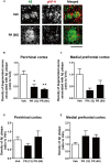
Effects of Polygalae Radix (PR) extract on amyloid β (Aβ) plaques and degenerated axons in 5XFAD mice. Vehicle solution (Veh) or PR extract [at doses of 12 or 60 mg kg-1 day-1, PR (12), PR (60), respectively] were orally administered daily for 56 days to 4-month-old wild-type (WT) or 5XFAD mice. The mice were euthanized 2 days after the last administration, and the brains were sectioned and immunostained for phosphorylated neurofilament-H (pNF-H, an axonal marker, red) and Aβ (green). (A) Representative fluorescence images of the perirhinal cortex are shown. Scale bar, 10 μm. (B,C) The density of degenerated axons associated with the Aβ plaques was quantified in the perirhinal cortex (B) and medial prefrontal cortex (C). (D,E) The density of Aβ plaques was quantified in the perirhinal cortex (D) and medial prefrontal cortex (E). The mean values of the data are presented together with the standard error. P < 0.05, ∗∗P < 0.01 vs. Veh; Bonferroni’s multiple comparison test, n = 4–5 mice.
FIGURE 4
FIGURE 4
Effects of the Polygalae Radix (PR) extract on amyloid β (Aβ)-induced growth cone collapse. Cortical neurons were cultured for 3 days, and treated for 30 min with vehicle solution (Cont, Veh) or PR extract [at doses of 10 or 100 μg ml-1; PR (10), PR (100), respectively]. Next, they were treated with vehicle solution (Cont), Aβ1–42 (1 μM), or Aβ25–35 (10 μM) for 1 h. The neurons were then fixed. Representative bright field images with oblique illumination are shown (upper). Black scale bar, 20 μm. Blue scale bar, 10 μm. The growth cone shapes were quantified as collapse scores. The mean values of the data are presented together with the standard error (lower). The numbers in parentheses indicate the measured numbers of growth cones. ∗∗P < 0.01, ∗∗∗P < 0.001 vs. Veh/Aβ; Dunn’s multiple comparison test.
FIGURE 5
FIGURE 5
Effects of Polygalae Radix (PR) extract on amyloid β (Aβ)-induced axonal atrophy. Cortical neurons were cultured for 3 days and treated simultaneously with vehicle solution (Cont) or Aβ25–35 (10 μM), and vehicle solution (Cont, 0 μg ml-1) or PR extract (at doses of 0.1, 1, 10, or 100 μg ml-1) for 4 days. Thereafter, the neurons were fixed and immunostained for phosphorylated neurofilament-H (pNF-H, an axonal marker, red) and microtubule associated protein 2 (MAP2, a neuronal marker, green). DAPI (blue) was used for counterstaining. Representative fluorescence images are shown (upper). Scale bar, 200 μm. The total axonal length per neuron were quantified. The mean values of the data are presented together with the standard error (lower). ∗∗P < 0.01 vs. 0 μg/mL PR + Aβ25–35; Bonferroni’s multiple comparison test, n = 10 images.
FIGURE 6
FIGURE 6
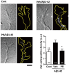
Effects of Polygalae Radix (PR) extract on amyloid β (Aβ)-induced endocytosis. Cortical neurons were cultured for 3 days, and treated with vehicle solution (Cont) or Aβ1–42 (1 μM) for 30 min. Next, they were treated with FM1-43FX (20 μM) for 1 min, and vehicle solution (Cont, Veh) or PR extract (100 μg ml-1) for 20 min, before the neurons were fixed. Representative differential interference contrast and fluorescence images are shown. The yellow dotted lines represent outlines of the growth cone. Scale bar, 5 μm. The density of FM1-43FX-positive area in each growth cone were quantified. The mean values of the data are presented together with the standard error. The numbers in parentheses indicate the measured numbers of growth cones. P < 0.05, ∗∗P < 0.01 vs. Veh/Aβ1–42; Bonferroni’s multiple comparison test.

References

    1. Barker G. R., Warburton E. C. (2011). When is the hippocampus involved in recognition memory? J. Neurosci. 31 10721–10731. 10.1523/jneurosci.6413-10.2011 - DOI - PMC - PubMed
    1. Benes F. M., Farol P. A., Majocha R. E., Marotta C. A., Bird E. D. (1991). Evidence for axonal loss in regions occupied by senile plaques in Alzheimer cortex. Neuroscience 42 651–660. 10.1016/0306-4522(91)90034-L - DOI - PubMed
    1. Chinese Pharmacopoeia Commission (2015). “Polygalae radix,” in Pharmacopoeia of the People’s Republic of China eds Zhao Y., Fan Z., Huang K., Li Z., Gao Y. (Beijing: People’s Medicine Publishing House Press; ).
    1. Cirrito J. R., Kang J. E., Lee J., Stewart F. R., Verges D. K., Silverio L. M., et al. (2008). Endocytosis is required for synaptic activity-dependent release of amyloid-beta in vivo. Neuron 58 42–51. 10.1016/j.neuron.2008.02.003 - DOI - PMC - PubMed
    1. Dickson T. C., Vickers J. C. (2001). The morphological phenotype of beta-amyloid plaques and associated neuritic changes in Alzheimer’s disease. Neuroscience 105 99–107. 10.1016/S0306-4522(01)00169-5 - DOI - PubMed