. 2017 Nov 14:8:2238.
doi: 10.3389/fmicb.2017.02238. eCollection 2017.

Zinc Limitation Induces a Hyper-Adherent Goliath Phenotype in Candida albicans

Affiliations

Zinc Limitation Induces a Hyper-Adherent Goliath Phenotype in Candida albicans

Dhara Malavia et al. Front Microbiol. .

Abstract

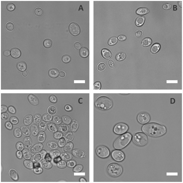
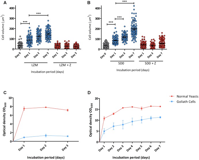
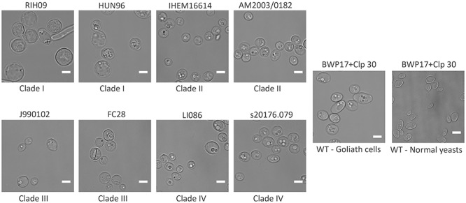
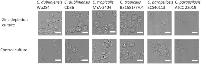
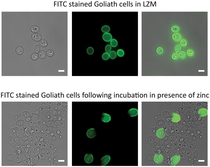
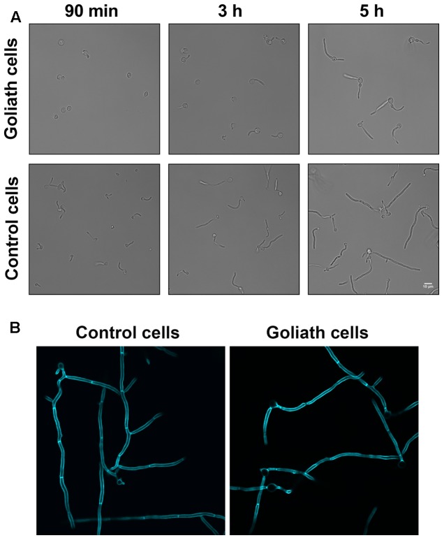
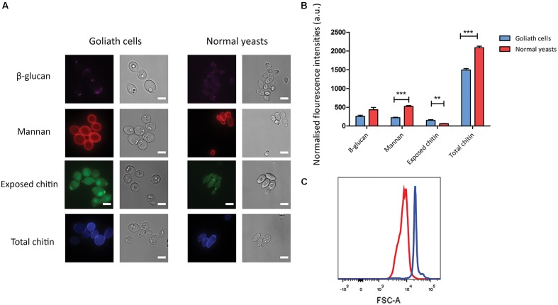
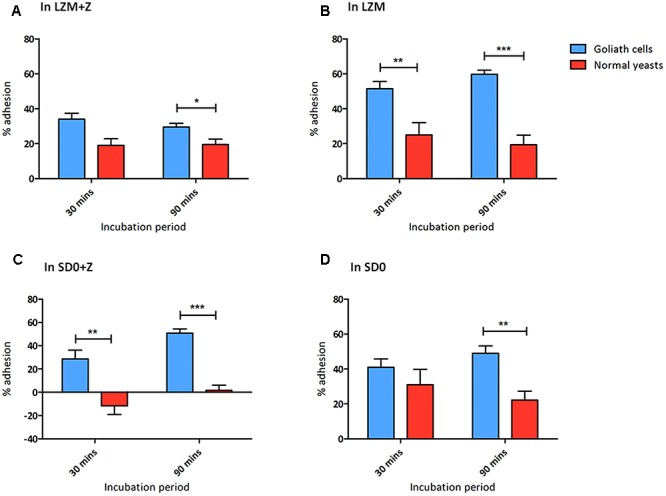
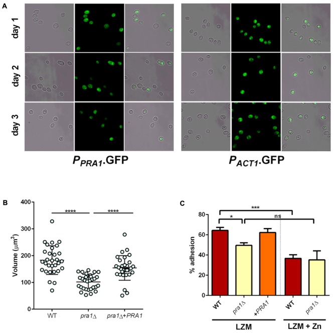
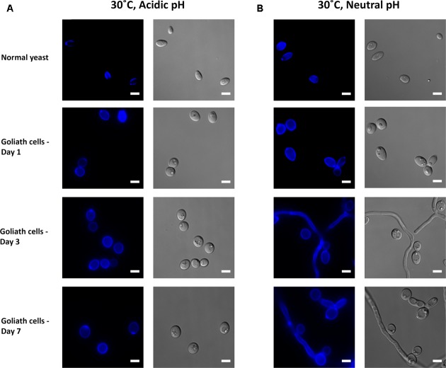
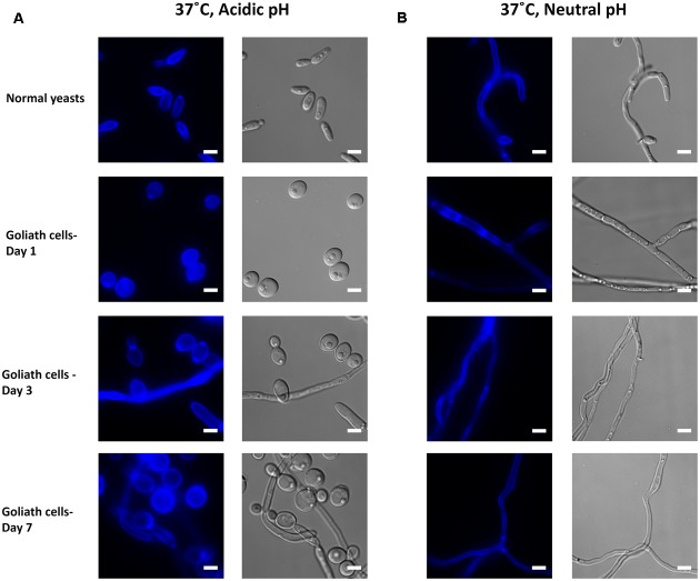
Pathogenic microorganisms often face acute micronutrient limitation during infection due to the action of host-mediated nutritional immunity. The human fungal pathogen Candida albicans is polymorphic and its morphological plasticity is one of its most widely recognized pathogenicity attributes. Here we investigated the effect of zinc, iron, manganese, and copper limitation on C. albicans morphology. Restriction of zinc specifically resulted in the formation of enlarged, spherical yeasts, a phenotype which we term Goliath cells. This cellular response to zinc restriction was conserved in C. albicans, C. dubliniensis and C. tropicalis, but not in C. parapsilosis, C. lusitaniae or Debaryomyces hansenii, suggesting that it may have emerged in the last common ancestor of these related pathogenic species. Cell wall analysis revealed proportionally more chitin exposure on the Goliath cell surface. Importantly, these cells were hyper-adherent, suggesting a possible role in pathogenicity. Interestingly, the zincophore-encoding gene PRA1 was expressed by Goliath cells in zinc limited media and lack of Pra1 inhibited both cellular enlargement and adhesion. Goliath cells represent a further layer of Candida phenotypic plasticity.

Keywords: Candida albicans; adhesion; fungal pathogen; micronutrients; morphogenesis; morphology; nutritional immunity; zinc.


Figures

FIGURE 1
FIGURE 1
Physiological response of C. albicans to metal starvation. C. albicans (BWP17 + Clp30) cells subjected to copper (A), iron (B), manganese (C), and zinc (D) starvation by incubating in limiting medium independently lacking these metals at 30°C, 200 rpm for 3 days. Experiment performed twice. DIC images show that of the metals tested only zinc starvation resulted in cellular enlargement in C. albicans. Scale bar represents 5 μm.
FIGURE 2
FIGURE 2
Developmental kinetics of C. albicans Goliath cell formation under zinc limitation. C. albicans cells pre-grown in SD medium were (A) incubated in LZM or LZM + Z over 3 days or (B) in SD0 or SD0 + Z over 7 days. Cells were imaged at indicated time points and axes diameters measured using ImageJ. Cell volumes were calculated by V = 4/3 π ab2. Each data column shows >100 cells from at least two independent experiments. Both zinc starvation media induced significant cellular enlargement (∗∗∗ < 0.001, Stat. test [ANOVA]). One data point (530 μm3) is omitted from SD0 Day 7 (B). Optical density (OD600) was determined for cultures incubated in (C) LZM (±zinc) every 24 h for 3 days and (D) SDw/oZ (±zinc) every 24 h for 7 days. Data are from three independent experiments. Error bars represent the standard error of the mean.
FIGURE 3
FIGURE 3
Cellular gigantism in multiple C. albicans clinical isolates. Indicated C. albicans clinical isolates pre-grown in SD medium were incubated in LZM for 3 days at 30°C, 200 rpm. DIC images show that all the tested clinical isolates can enlarge to varying degrees upon zinc depletion. Clinical isolates belonging to Clade 1 enlarged to a greater degree than other isolates. All the strains are distinctly bigger than the LZM + Z control. Scale bar represents 5 μm.
FIGURE 4
FIGURE 4
Zinc restriction-induced cellular enlargement is conserved in closely related Candida species. Multiple strains of C. dubliniensis, C. tropicalis, and C. parapsilosis were also tested for their capacity to enlarge by incubating SD-pre-grown cells in LZM (±zinc) for 3 days at 30°C, 200 rpm. All the tested strains of C. dubliniensis and C. tropicalis enlarged in response to zinc limitation, whereas C. parapsilosis strains did not enlarge upon zinc deprivation. Scale bar represents 5 μm.
FIGURE 5
FIGURE 5
Effects of zinc restoration on Goliath cell morphology. C. albicans cells pre-grown in SD were incubated in LZM for 2 days to generate Goliath cells. These Goliath cells were FITC labeled (upper panel) and subsequently inoculated into SD (zinc-replete) medium to an OD600 = 0.5. Lower panel shows that non-labeled cells exhibit regular yeast morphology. Labeled cells remained large. Scale bar represents 5 μm.
FIGURE 6
FIGURE 6
Hyphae induction. Goliath cells and normal yeast cells incubated in the presence of hyphae inducing media (RPMI) for up to 5 h (A) or 6 h at 37°C and stained with Calcofluor White (B). Goliath cells exhibit a delay in germination and hyphae formation as compared to normal yeasts. Scale bar represents 10 μm.
FIGURE 7
FIGURE 7
Cell wall analysis of Goliath cells. (A) Goliath cells stained for exposed β-glucan (Fc-Dectin), mannan (concanavalin A), exposed (WGA) and total (Calcofluor White) chitin and compared with normal yeast cells. Scale bar represents 5 μm. (B) Mean fluorescence intensities of respective cell wall components for Goliath cells and normal yeast cells as determined by flow cytometry represented as a bar graph. Data were obtained from three independent experiments and normalized by cell size (C), error bars represent standard deviation and p-values were calculated by t-test. A significant increase in mean fluorescence intensity of WGA was observed Goliath cells as compared to normal yeast cells. (P ≤ 0.05, ∗∗P ≤ 0.01, ∗∗∗P ≤ 0.0001). (C) Increase in cell size determined using flow cytometry by measuring forward scatter (FSC) intensities of Goliath cells (blue) and normal yeast cells (red) (n > 10000). A shift in FCS peak shows that Goliath cells are significantly larger than normal yeast cells. Mean FCS intensities were compared to determine fold increase in cell size. Based on the FCS intensities, C. albicans Goliath cells exhibit a 3.3-fold increase in size as compared to normal yeasts. Note that relative to cell size, only chitin exposure is relative higher for Goliath cells vs. control yeasts.
FIGURE 8
FIGURE 8
Goliath cells exhibit increased adhesion to plastic surfaces. Adherence of C. albicans Goliath cells formed in LZM (A,B) or in SD0 (C,D) to plastic in the presence (A,C) or absence (B,D) of zinc in the adhesion assay medium. Relevant control yeast cells formed in LZM + Z or SD0 + Z. Following incubation for 30 or 90 min, wells were washed three times with ultrapure water. CFUs in the initial supernatant and in all three wash steps were determined to calculate % adhesion. Error bars represent standard deviation and p-values were determined by t-test. Data are from three independent experiments (A,B); and two independent experiments (C,D). Goliath cells are more adherent than normal yeast cells in all the tested media type at both the time points. The adhesion capacity of Goliath cells is reduced upon incubation in LZM + Z (A vs. B). However, the presence of zinc during adhesion had a less pronounced effect on Goliath cell adhesion in SD0 + Z (C vs. D). Normal yeast cells show increased adhesion when incubated in the absence of zinc (D vs. C) (P ≤ 0.05, ∗∗P ≤ 0.01, ∗∗∗P ≤ 0.0001).
FIGURE 9
FIGURE 9
Pra1 contributes to enlargement and adhesion properties of Goliath cells. (A) PRA1 is expressed in limited zinc medium (LZM). Cells harboring PPRA1-GFP (Citiulo et al., 2012) or PACT1-GFP were analyzed at indicated time points. (B) C. albicans cells were cultured for 2 days in LZM and cell volumes were calculated by V = 4/3 π ab2. Data in each column represent at least 30 independent measurements from three independent experiments. pra1Δ cells were significantly smaller than wild type and pra1Δ+PRA1 cells (P < 0.0001, one-way ANOVA). (C) Adherence of indicated C. albicans cells cultured for 2 days in LZM to induce cellular enlargement or for 1 day in LZM + Z to generate yeast cells. Wild type, but not pra1Δ exhibited enhanced adhesion following incubation in LZM (P < 0.05, ∗∗P < 0.01, ∗∗∗P < 0.001, ∗∗∗∗ P < 0.0001).
FIGURE 10
FIGURE 10
Impact of pH on Goliath morphology. C. albicans cells were incubated in limited zinc media (LZM) at 30°C at acidic (pH 4.6) or neutral-alkaline (pH 7.3) and morphology assessed. (A) In acidic medium zinc limitation induces cellular enlargement. (B) At pH 7.3 zinc limitation induces yeast cell enlargement and triggers filamentation. Experiment was performed twice.
FIGURE 11
FIGURE 11
Impact of temperature on Goliath morphology. C. albicans cells were incubated in limited zinc media (LZM) at 37°C at acidic (pH 4.6) or neutral-alkaline (pH 7.3) and morphology assessed. (A) In acidic medium at 37°C zinc limitation induces a greater degree of cellular enlargement than at 30°C (compare Figure 10A) and pseudohypha development. (B) At pH 7.3 and 37°C filamentation occurs independent of media zinc status. Experiment was performed twice.

References

    1. Alasio T. M., Lento P. A., Bottone E. J. (2003). Giant blastoconidia of Candida albicans. A case report and review of the literature. Arch. Pathol. Lab. Med. 127 868–871. - PubMed
    1. Ameglio F., Di Giorgio C., Terzaroli P., Gandolfo G. M. (1990). Giant cell production by C. albicans cultured in xylitol. Microbiologica 13 343–346. - PubMed
    1. Amich J., Vicentefranqueira R., Mellado E., Ruiz-Carmuega A., Leal F., Calera J. A. (2014). The ZrfC alkaline zinc transporter is required for Aspergillus fumigatus virulence and its growth in the presence of the Zn/Mn-chelating protein calprotectin. Cell Microbiol. 16 548–564. 10.1111/cmi.12238 - DOI - PubMed
    1. Andreini C., Banci L., Bertini I., Rosato A. (2006). Zinc through the three domains of life. J. Proteome Res. 5 3173–3178. 10.1021/pr0603699 - DOI - PubMed
    1. Andreini C., Bertini I., Rosato A. (2009). Metalloproteomes: a bioinformatic approach. Acc. Chem. Res. 42 1471–1479. 10.1021/ar900015x - DOI - PubMed