

Interleukin-4 Supports the Suppressive Immune Responses Elicited by Regulatory T Cells
- PMID: 29184551
- PMCID: PMC5694475
- DOI: 10.3389/fimmu.2017.01508
Interleukin-4 Supports the Suppressive Immune Responses Elicited by Regulatory T Cells
Abstract
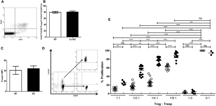
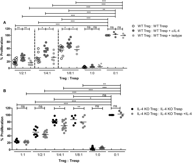
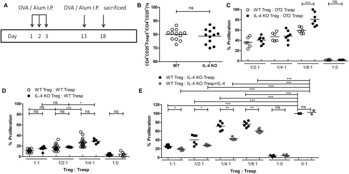
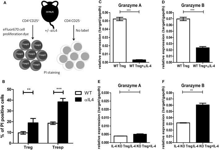
Interleukin-4 (IL-4) has been considered as one of the tolerogenic cytokines in many autoimmune animal models and clinical settings. Despite its role in antagonizing pathogenic Th1 responses, little is known about whether IL-4 possesses functions that affect regulatory T cells (Tregs). Tregs are specialized cells responsible for the maintenance of peripheral tolerance through their immune modulatory capabilities. Interestingly, it has been suggested that IL-4 supplement at a high concentration protects responder T cells (Tresps) from Treg-mediated immune suppression. In addition, such supplement also impedes TGF-β-induced Treg differentiation in vitro. However, these phenomena may contradict the tolerogenic role of IL-4, and the effects of IL-4 on Tregs are therefore needed to be further elucidated. In this study, we utilized IL-4 knockout (KO) mice to validate the role of IL-4 on Treg-mediated immune suppression. Although IL-4 KO and control animals harbor similar frequencies of Tregs, Tregs from IL-4 KO mice weakly suppressed autologous Tresp activation. In addition, IL-4 deprivation impaired the ability of Tregs to modulate immune response, whereas IL-4 supplementation reinforced IL-4 KO Tregs in their function in suppressing Tresps. Finally, the presence of IL-4 was associated with increased cell survival and granzyme expression of Tregs. These results suggest the essential role of IL-4 in supporting Treg-mediated immune suppression, which may benefit the development of therapeutic strategies for autoimmune diseases.
Keywords: cell survival; granzyme; immunosuppression; interleukin-4; regulatory T cell.
Figures
Similar articles
-
The role of age-related T-cell differentiation in patients with renal replacement therapy.Immunol Cell Biol. 2017 Nov;95(10):895-905. doi: 10.1038/icb.2017.57. Epub 2017 Jul 19. Immunol Cell Biol. 2017. PMID: 28722017
-
Feedback loop of immune regulation by CD4+CD25+ Treg.Immunobiology. 2009;214(4):291-302. doi: 10.1016/j.imbio.2008.09.004. Epub 2008 Nov 1. Immunobiology. 2009. PMID: 19327546
-
TGF-β1 and contact mediated suppression by CD4+CD25+CD127- T regulatory cells of patients with self-limiting hepatitis E.Hum Immunol. 2016 Dec;77(12):1254-1263. doi: 10.1016/j.humimm.2016.10.001. Epub 2016 Oct 6. Hum Immunol. 2016. PMID: 27720959
-
Alloantigen specific T regulatory cells in transplant tolerance.Int Immunopharmacol. 2009 May;9(5):570-4. doi: 10.1016/j.intimp.2009.01.016. Epub 2009 Jan 29. Int Immunopharmacol. 2009. PMID: 19539571 Review.
-
Stability of Regulatory T Cells Undermined or Endorsed by Different Type-1 Cytokines.Adv Exp Med Biol. 2015;850:17-30. doi: 10.1007/978-3-319-15774-0_2. Adv Exp Med Biol. 2015. PMID: 26324343 Review.
Cited by
-
Hepatitis C Virus Improves Human Tregs Suppressive Function and Promotes Their Recruitment to the Liver.Cells. 2019 Oct 22;8(10):1296. doi: 10.3390/cells8101296. Cells. 2019. PMID: 31652598 Free PMC article.
-
Vaccination against Atherosclerosis: Is It Real?Int J Mol Sci. 2022 Feb 22;23(5):2417. doi: 10.3390/ijms23052417. Int J Mol Sci. 2022. PMID: 35269559 Free PMC article. Review.
-
Anti-Neuroinflammatory Effects of the Human Milk Oligosaccharide, 2'-Fucosyllactose, Exerted via Modulation of M2 Microglial Activation in a Mouse Model of Ischemia-Reperfusion Injury.Antioxidants (Basel). 2023 Jun 15;12(6):1281. doi: 10.3390/antiox12061281. Antioxidants (Basel). 2023. PMID: 37372011 Free PMC article.
-
Construction of a neutrophil extracellular trap formation-related gene model for predicting the survival of lung adenocarcinoma patients and their response to immunotherapy.Transl Lung Cancer Res. 2024 Dec 31;13(12):3407-3425. doi: 10.21037/tlcr-24-463. Epub 2024 Dec 27. Transl Lung Cancer Res. 2024. PMID: 39830760 Free PMC article.
-
Cytokine and Chemokine Signals of T-Cell Exclusion in Tumors.Front Immunol. 2020 Dec 14;11:594609. doi: 10.3389/fimmu.2020.594609. eCollection 2020. Front Immunol. 2020. PMID: 33381115 Free PMC article. Review.
References
-
- Kidd P. Th1/Th2 balance: the hypothesis, its limitations, and implications for health and disease. Altern Med Rev (2003) 8(3):223–46. - PubMed
LinkOut - more resources
Full Text Sources
Other Literature Sources
Research Materials