Review
. 2017 Oct;6(5):833-838.
doi: 10.21037/tau.2017.04.36.

The role of PET/CT imaging in penile cancer

Affiliations
Review

The role of PET/CT imaging in penile cancer

Sarah R Ottenhof et al. Transl Androl Urol. 2017 Oct.

Abstract

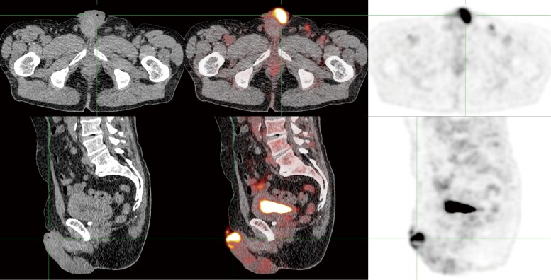
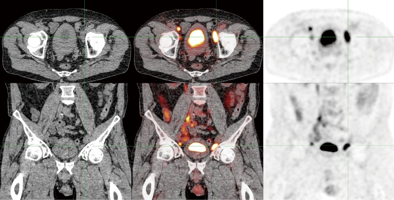
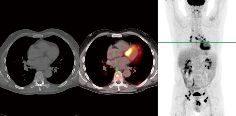
Positron emission tomography (PET) imaging with 18F-fluorodeoxyglucose (FDG) combined with computed tomography (CT) provides functional imaging combined with anatomic information, improving diagnostic accuracy and confidence. Although virtually all primary penile tumors are FDG-avid, PET/CT is not recommended for primary tumor staging as it has limited spatial resolution and is hampered by urinary FDG excretion. The accuracy of PET/CT for lymph node staging seems to improve with the pretest likelihood of metastatic nodes. In groins with normal physical examination, sensitivity is only 57%. In groins with palpably enlarged lymph nodes, sensitivity of PET/CT reaches 96%. For pelvic lymph nodes and distant metastases, PET/CT is more accurate if inguinal metastases are present. However, these results are based on a very limited number of studies. Overall, the role of PET/CT imaging in penile cancer remains ambiguous, especially in inguinal lymph nodes. During staging and follow-up, it may be particularly useful in detecting pelvic lymph node metastases and occult distant metastases prior to systemic chemotherapy and/or extensive surgery, improving selection of patients that are most likely to benefit from such therapies.

Keywords: 18F-fluorodeoxyglucose (FDG); PET/CT; Penile cancer; fluorodeoxyglucose; imaging.


Conflict of interest statement

Conflicts of Interest: The authors have no conflicts of interest to declare.

Figures

Figure 1
Figure 1
FDG-PET/CT of a penile cancer patient. The primary tumor exhibits high FDG uptake (SUVmax 11.7).
Figure 2
Figure 2
FDG-PET/CT of the same penile cancer patient, who was clinically staged as positive for inguinal lymph node metastases (cN+). The PET shows extensive pelvic lymph node metastases. Bilateral inguinal metastases were also FDG-positive.
Figure 3
Figure 3
FDG-PET/CT of the same penile cancer patient, showing a solitary skeletal metastasis in the 8th thoracic vertebra, which was not visible on CT. In addition, extensive mediastinal and hilar lymphadenopathy was visible with small pulmonary and pleural lesions, which were thought to be possible sarcoidosis or metastases. Follow-up CT after 3 months showed gross progression, with multiple metastases in bone, liver, spleen and pelvic lymph nodes. In contrast, the lesions in the lungs and mediastinum were stable, increasing the likelihood of those being caused by a separate process such as sarcoidosis.

References

    1. Kirrander P, Sherif A, Friedrich B, et al. Swedish National Penile Cancer Register: incidence, tumour characteristics, management and survival. BJU Int 2016;117:287-92. 10.1111/bju.12993 - DOI - PubMed
    1. Barnholtz-Sloan JS, Maldonado JL, Pow-Sang J, et al. Incidence trends in primary malignant penile cancer. Urol Oncol 2007;25:361-7. 10.1016/j.urolonc.2006.08.029 - DOI - PubMed
    1. Arya M, Li R, Pegler K, et al. Long-term trends in incidence, survival and mortality of primary penile cancer in England. Cancer Causes Control 2013;24:2169-76. 10.1007/s10552-013-0293-y - DOI - PubMed
    1. Fletcher JW, Djulbegovic B, Soares HP, et al. Recommendations on the use of 18F-FDG PET in oncology. J Nucl Med 2008;49:480-508. 10.2967/jnumed.107.047787 - DOI - PubMed
    1. Lardinois D, Weder W, Hany TF, et al. Staging of non-small-cell lung cancer with integrated positron-emission tomography and computed tomography. N Engl J Med 2003;348:2500-7. 10.1056/NEJMoa022136 - DOI - PubMed

LinkOut - more resources