. 2017 Nov 14:7:469.
doi: 10.3389/fcimb.2017.00469. eCollection 2017.

Internalization of the Active Subunit of the Aggregatibacter actinomycetemcomitans Cytolethal Distending Toxin Is Dependent upon Cellugyrin (Synaptogyrin 2), a Host Cell Non-Neuronal Paralog of the Synaptic Vesicle Protein, Synaptogyrin 1

Affiliations

Internalization of the Active Subunit of the Aggregatibacter actinomycetemcomitans Cytolethal Distending Toxin Is Dependent upon Cellugyrin (Synaptogyrin 2), a Host Cell Non-Neuronal Paralog of the Synaptic Vesicle Protein, Synaptogyrin 1

Kathleen Boesze-Battaglia et al. Front Cell Infect Microbiol. .

Abstract

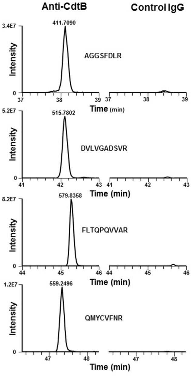
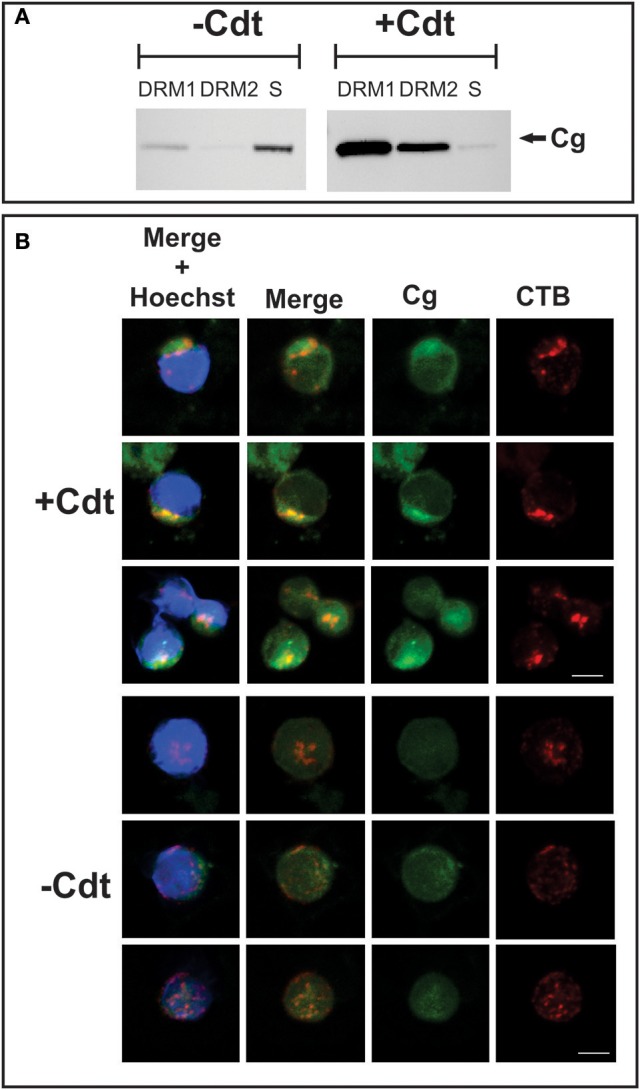
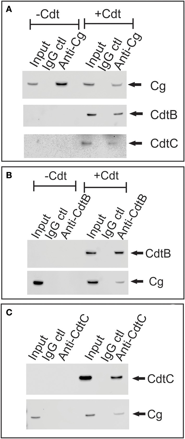
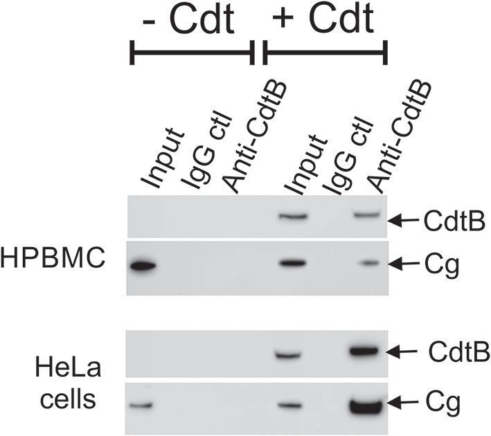
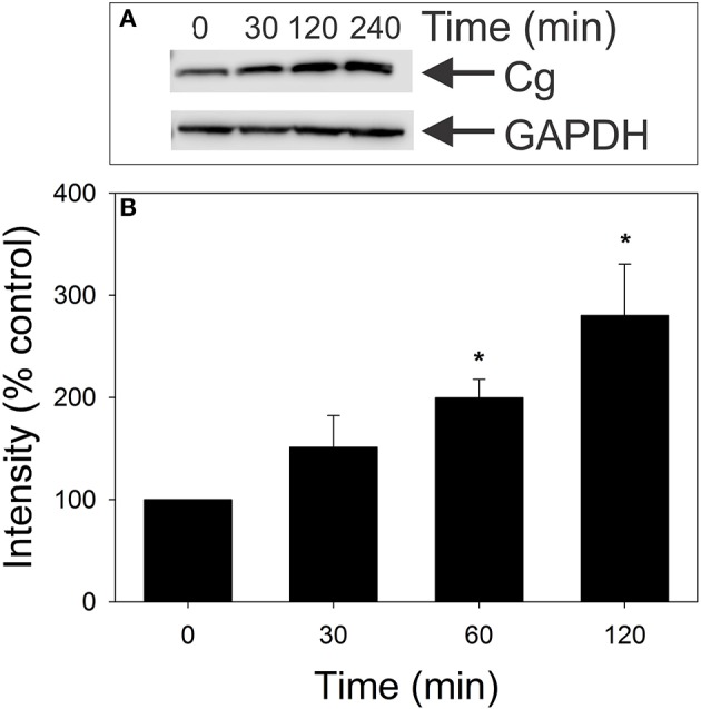
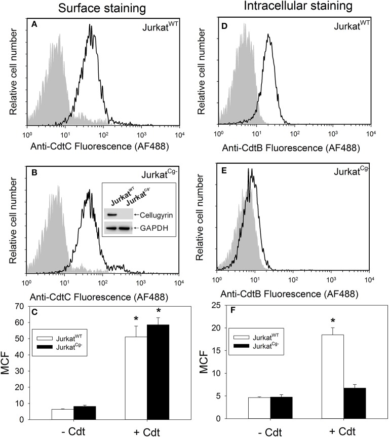
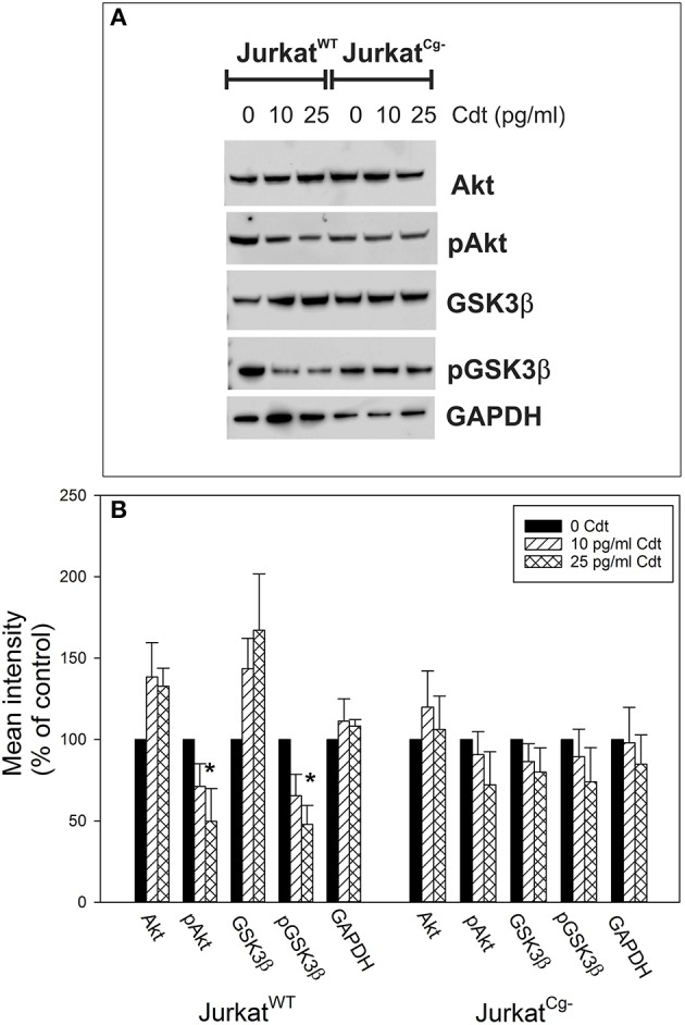
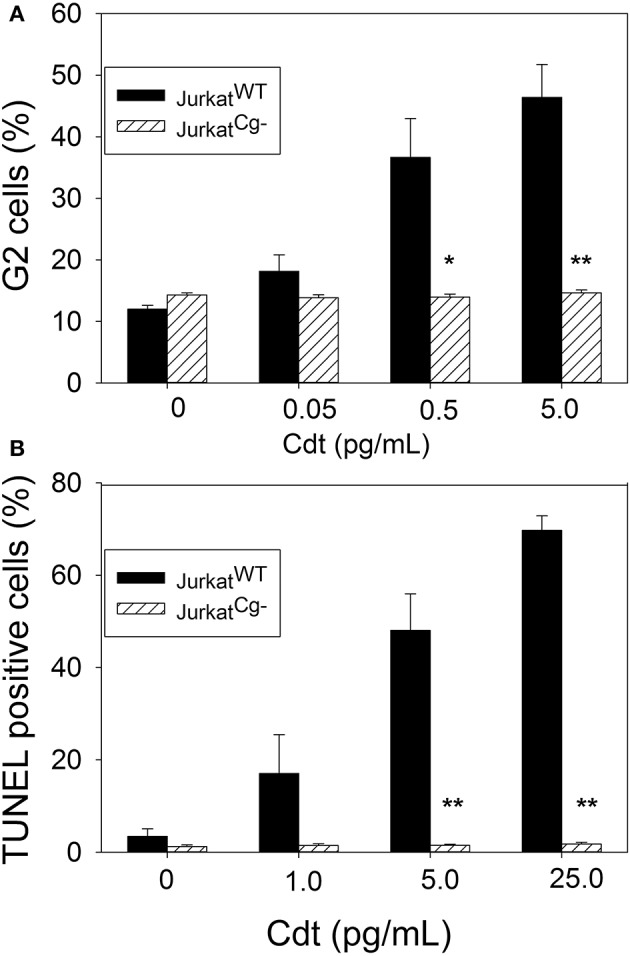
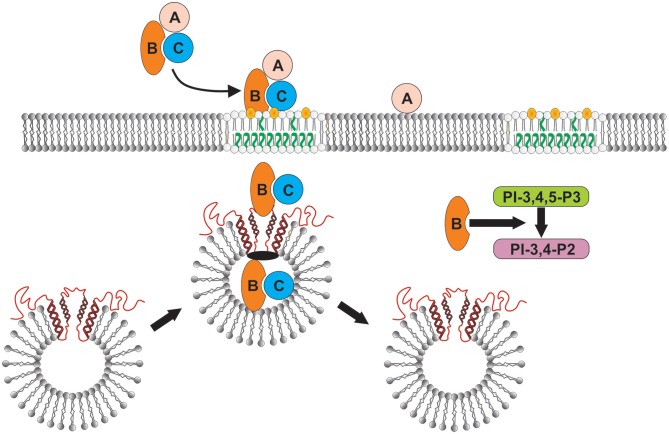
The Aggregatibacter actinomycetemcomitans cytolethal distending toxin (Cdt) is a heterotrimeric AB2 toxin capable of inducing lymphocytes, and other cell types, to undergo cell cycle arrest and apoptosis. Exposure to Cdt results in binding to the cell surface followed by internalization and translocation of the active subunit, CdtB, to intracellular compartments. These events are dependent upon toxin binding to cholesterol in the context of lipid rich membrane microdomains often referred to as lipid rafts. We now demonstrate that, in addition to binding to the plasma membrane of lymphocytes, another early and critical event initiated by Cdt is the translocation of the host cell protein, cellugyrin (synaptogyrin-2) to the same cholesterol-rich microdomains. Furthermore, we demonstrate that cellugyrin is an intracellular binding partner for CdtB as demonstrated by immunoprecipitation. Using CRISPR/cas9 gene editing we established a Jurkat cell line deficient in cellugyrin expression (JurkatCg-); these cells were capable of binding Cdt, but unable to internalize CdtB. Furthermore, JurkatCg- cells were not susceptible to Cdt-induced toxicity; these cells failed to exhibit blockade of the PI-3K signaling pathway, cell cycle arrest or cell death. We propose that cellugyrin plays a critical role in the internalization and translocation of CdtB to critical intracellular target sites. These studies provide critical new insight into the mechanism by which Cdt, and in particular, CdtB is able to induce toxicity.

Keywords: Aggregatibacter actinomycetemcomitans; apoptosis; cell cycle arrest; cytolethal distending toxin; lymphocytes; pathogenesis; toxin.


Figures

Figure 1
Figure 1
Extracted ion chromatograms of four representative doubly charged SYNGR2 peptides. Jurkat cells were treated with Cdt holotoxin as described in Materials and Methods for 1 h. Cell extracts were prepared and immunoprecipitated with immobilized anti-CdtB or isotype control IgG and the samples analyzed as described. The immunoprecipitates obtained with anti-CdtB mAb show strong signals for the peptides indicated; results are consistent with detection of MS/MS spectra for SYNGR2 peptides obtained exclusively with this mAb and were not detectable in the control immunoprecipitate.
Figure 2
Figure 2
Translocation of cellugyrin to cholesterol rich micro domains. (A) Jurkat cells were treated with medium (−Cdt) or with 2 μg/ml Cdt for 2 h. Cells were harvested, washed, and cholesterol rich microdomains isolated as detergent resistant membranes (DRM) as described in Materials and Methods. Two DRM zones, designated DRM1 and DRM2, as well as a soluble fraction were obtained and further analyzed by Western blot for the presence of cellugyrin. Results are representative of three experiments. (B) Jurkat cells were treated with medium (−Cdt) or 1 μg/ml Cdt (+Cdt) for 1 h; cells were stained and fixed as described in Materials and Methods and analyzed by confocal microscopy. Maximum intensity projection of a 3 μm z-stack series is presented (3 cells/condition). For each image, fluorescence is shown for cellugyrin alone (green), lipid rafts using fluorescence of cholera toxin B (CTB; red) and merged images (yellow) with (blue) and without nuclear staining Results are representative of multiple fields and analysis of over 50 cells for each condition. Scale bar = 5 μm.
Figure 3
Figure 3
Immunoprecipitation of cellugyrin and Cdt subunits. Jurkat cells were treated with medium or Cdt (2 μg/ml) for 2 h and then washed and homogenized as described in Materials and Methods. (A) Shows the results of extracts immunoprecipitated with either immobilized control IgG or anti-cellugyrin antibody. The bound material was eluted and analyzed by Western blot for the presence of cellugyrin (Cg), CdtB or CdtC. (B) Shows the results of cell extracts obtained from similarly treated cells as above and immunoprecipitated with immobilized control IgG or anti-CdtB mAb. The bound material was eluted and analyzed by Western blot for the presence of CdtB and Cg. (C) Shows the results of cell extracts obtained from cells treated as described above and immunopreciptated with immobilized control IgG or anti-CdtC mAb. The bound material was eluted and analyzed by Western blot for the presence of CdtC and Cg. Results are representative of three experiments.
Figure 4
Figure 4
Immunoprecipitation of cellugyrin and CdtB in HPBMC and HeLa cells. HPBMC and HeLa cells were treated with medium or Cdt (2 μg/ml) for 2 h. Cell extracts were prepared as described in Materials and Methods and immunoprecipitated with immobilized control IgG or anti-CdtB mAb. The bound material was eluted and further analyzed by Western Blot for CdtB and cellugyrin. Results are representative of three experiments.
Figure 5
Figure 5
Effect of Cdt on cellugyrin levels in Jurkat cells. Jurkat cells were incubated in the presence of medium or 25 pg/ml Cdt for 0–120 min. Cells were harvested, homogenized and analyzed by Western blot for relative cellugyrin content. Blots were further analyzed by digital densitometry. Results of a representative blot are shown in (A) and the mean ± SEM of three experiments are shown in (B); results are expressed as a percentage of the intensity observed in control cells. *Indicates statistical significance (p < 0.05) when compared to untreated cells.
Figure 6
Figure 6
Cdt binding and CdtB internalization in JurkatWT cells vs. JurkatCg− cells. JurkatWT and JurkatCg− were first compared for their ability to bind Cdt (A–C). Cells were incubated for 60 min at 5°C with Cdt (2 μg/ml), washed and stained for the presence of cell surface associated Cdt using anti-CdtC mAb conjugated to AF488. Representative flow cytometric analysis for Cdt binding to JurkatWT is shown in panel A; solid line is the result obtained with Cdt-treated cells and the shaded curve represents cells exposed to medium alone. Cdt binding to JurkatCg− cells is shown in (B). Cells were generated as described in Materials and Methods; confirmation of their inability to express cellugyrin was demonstrated by Western blot as shown in (B) inset. Results from multiple experiments are shown in (C); results are the MCF ± SEM obtained from three experiments. Internalization of CdtB in JurkatWT and JurkatCg− cells was analyzed following exposure to Cdt (2 μg/ml) for 1 h at 37°C; cells were washed, fixed, permeabilized, and stained with anti-CdtB mAb conjugated to AF488. Representative results are shown for JurkatWT in (D) and for JurkatCg− in (E); solid line represents results obtained from cells treated with Cdt and the shaded curve from cells exposed to medium only. Results from multiple experiments are shown in (F); results are the MCF ± SEM obtained from three experiments. *Indicates statistical significance (p < 0.05) when compared to control (-Cdt) cells.
Figure 7
Figure 7
Comparison of the effects of Cdt on PI-3K signaling blockade in JurkatWT vs. JurkatCg− cells. JurkatWT and JurkatCg− cells were treated with 0–25 pg/ml Cdt for 2 h and then analyzed by Western blot for pAkt (S473), Akt, pGSK3β (S9), GSK3β, and GAPDH as a loading control. (A) Contains a representative Western blot showing the effect of Cdt on Akt and GSK3β phosphorylation. (B) Shows the results of Western blot analyses from three experiments; blots were analyzed by digital densitometry and are expressed as a percentage of the relative intensity of untreated control cells; mean ± S.E.M. for three experiments are plotted. *Indicates statistical significance (p < 0.05) when compared to untreated control cells.
Figure 8
Figure 8
Comparative toxic effects of Cdt on JurkatWT and JurkatCg− cells. (A) Shows the effect of Cdt on cell cycle arrest; JurkatWT and JurkatCg− cells were incubated for 16 h in the presence of 0–5 pg/ml Cdt. Cells were stained with propidium iodide and cell cycle analysis performed using flow cytometry. The percentage of G2 cells is shown as a mean ± SEM for three experiments each performed in triplicate; solid bars represent JurkatWT cells and hashed bars JurkatCg− cells. (B) Shows the effect of Cdt on apoptosis; cells were treated with 0–25 pg/ml Cdt for 48 h analyzed for DNA strand breaks using the TUNEL assay. Results are expressed as the mean percentage of TUNEL positive cells ± SEM for three experiments (); solid bars represent JurkatWT cells and hashed bars JurkatCg− cells. *Indicates statistical significance (p < 0.05) when compared to untreated cells; **indicates statistical significance of p < 0.01.
Figure 9
Figure 9
Schematic model showing proposed CdtB-cellugyrin interaction. Cdt holotoxin binds to cells via cholesterol in the context of membrane lipid rafts. CdtB binding and internalization is further dependent upon its ability to interact with cholesterol. As a result of exposure to Cdt, cellugyrin (shown in red) containing SLMVs translocate from cytosol to membrane lipid rafts. We propose that this translocation leads to the association of CdtB with the cellugyrin-containing SLMVs. This interaction may involve direct binding to cellugyrin either on extra- or intra-vesicular loops or indirect association via an unidentified binding partner (shown in black). We further propose that CdtB is transported via SLMVs to intracellular target sites; for example sites containing PIP3 pools where the enzymatically active CdtB subunit is released from SLMVs and is then able to degrade the signaling lipid resulting in PI-3K blockade and toxicity.

References

    1. Ahnert-Hilger G., Munster-Wandowski A., Holtje M. (2013). Synaptic vesicle proteins: targets and routes for botulinum neurotoxins. Curr. Top. Microbiol. Immunol. 364, 159–177. 10.1007/978-3-642-33570-9_8 - DOI - PubMed
    1. Backert S., Tegtmeyer N. (2017). Type IV secretion and signal transduction of Helicobacter pylori CagA through interactions with host cell receptors. Toxins 9:E115. 10.3390/toxins9040115 - DOI - PMC - PubMed
    1. Belfort G. M., Bakirtzi K., Kandror K. V. (2005). Cellugyrin induces biogenesis of synaptic-like microvesicles in PC12 cells. J. Biol. Chem. 280, 7262–7272. 10.1074/jbc.M404851200 - DOI - PubMed
    1. Belfort G. M., Kandror K. V. (2003). Cellugyrin and synaptogyrin facilitate targeting of synaptophysin to a ubiquitous synaptic vesicle-sized compartment in PC12 cells. J. Biol. Chem. 278, 47971–47978. 10.1074/jbc.M304174200 - DOI - PubMed
    1. Bielaszewska M., Ruter C., Bauwens A., Greune L., Jarosch K. A., Steil D., et al. (2017). Host cell interactions of outer membrane vesicle-associated virulence factors of enterohemorrhagic Escherichia coli O157: intracellular delivery, trafficking and mechanisms of cell injury. PLoS Pathog. 13:e1006159. 10.1371/journal.ppat.1006159 - DOI - PMC - PubMed

MeSH terms