. 2017 Nov 29;7(1):16541.
doi: 10.1038/s41598-017-16743-1.

Characterising Pre-pubertal Resistance to Death from Endotoxemia

Affiliations

Characterising Pre-pubertal Resistance to Death from Endotoxemia

Rose Joachim et al. Sci Rep. .

Abstract

Sepsis is a common and deadly syndrome in which a dysregulated host response to infection causes organ failure and death. The current lack of treatment options suggests that a new approach to studying sepsis is needed. Pre-pubertal children show a relative resistance to death from severe infections and sepsis. To explore this phenomenon experimentally, we used an endotoxemia model of sepsis in mice. Following intra-peritoneal injection of endotoxin, pre-pubertal mice showed greater survival than post-pubertal mice (76.3% vs. 28.6%), despite exhibiting a similar degree of inflammation after two hours. Age-associated differences in the inflammatory response only became evident at twenty hours, when post-pubertal mice showed prolonged elevation of serum cytokines and differential recruitment of peritoneal immune cells. Mechanistically, prevention of puberty by hormonal blockade or acceleration of puberty by oestrogen treatment led to increased or decreased survival from endotoxemia, respectively. Additionally, the adoptive transfer of pre-pubertal peritoneal cells improved the survival of post-pubertal recipient mice, while post-pubertal peritoneal cells or vehicle did not. These data establish a model for studying childhood resistance to mortality from endotoxemia, demonstrate that oestrogen is responsible for an increased susceptibility to mortality after puberty, and identify peritoneal cells as mediators of pre-pubertal resistance.


Conflict of interest statement

The authors declare that they have no competing interests.

Figures

Figure 1
Figure 1
Pre-pubertal mice show increased resistance to mortality from endotoxemia. (A) Pre-pubertal (Pre-P) and post-pubertal (Post-P) C57Bl/6 mice received 23 μg/mg LPS via intra-peritoneal injection. Mice were monitored twice daily and moribund individuals humanely euthanised (N ≥ 56/group). (B) Endotoxin concentrations in serum samples collected at 2- and 20-hour time-points were quantified using LAL (N ≥ 21/group). (C) Mouse weight was recorded prior to LPS injection and then at 2- and 20-hour time points. Data is shown as the per cent change in weight in comparison to time 0 (N ≥ 20/group). Per cent survival was compared using a log rank Mantel Cox test, while endotoxin concentration and weight loss were compared using Two-way ANOVA followed by Tukey’s multiple comparisons test. Significant differences between pre- and post-pubertal mice are labelled with ****(p < 0.0001).
Figure 2
Figure 2
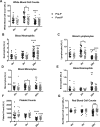
Pre- and post-pubertal whole blood cell counts during endotoxemia. Blood was obtained from pre- and post-pubertal (Pre-P, Post-P) C57Bl/6 mice via cardiac puncture and treated with K2EDTA to prevent clotting. All counts for total (A) white blood cells, (B) neutrophils, (C) lymphocytes, (D) monocytes, (E) eosinophils, (F) platelets, and (G) red blood cells and were obtained using the Hemavet 950 blood analyser. N ≥ 19/group at 0hr, N ≥ 35/group at 2 hr, and N ≥ 46/group at 20 hr. Significant changes in cell counts between 2 and 20 hours for either pre- or post-pubertal mice are labelled with ++++(p < 0.0001) or ++(p < 0.01). Significant differences in cell counts between pre- and post-pubertal mice are labelled with ****(p < 0.0001), **(p < 0.01), or *(p < 0.05). All comparisons were made using Two-way ANOVA followed by Tukey’s multiple comparisons test.
Figure 3
Figure 3
Serum cytokine expression suggests lack of resolution in post-pubertal animals. Serum samples obtained via cardiac puncture from pre- and post-pubertal C57Bl/6 mice (N ≥ 9/group) were subjected to a 32-plex cytokine assay. The 12 cytokines above show significant differences in expression between pre- and post-pubertal mice at either the (A) 2-hour time point or (B–K) 20-hour time point. For all graphs, pre- and post-pubertal data are shown in black and grey closed circles respectively. Data points above or below the detectable limit of the assay were not included. Significant age-associated differences in cytokine concentration are labelled with ****(p < 0.0001), ***(p < 0.001), **(p < 0.01), or *(p < 0.05). Significant changes in concentration between 2 and 20 hours for either pre- or post-pubertal mice are labelled with ++++(p < 0.0001), +++(p < 0.001), or ++(p < 0.01). All comparisons were made using Two-way ANOVA followed by Tukey’s multiple comparisons test.
Figure 4
Figure 4
Pre- and post-pubertal mice show differential recruitment of monocytic and neutrophilic cells into the peritoneal cavity. At 2 and 20 hours following endotoxemia, peritoneal cells from pre- and post-pubertal (Pre-P, Post-P) C57Bl/6 mice were collected through peritoneal lavage. Cells were blocked to prevent non-specific binding and treated with antibodies targeting the myeloid lineage marker CD11B, the neutrophilic marker Ly6G, and the monocytic marker Ly6C for subsequent immuno-phenotypic analysis by flow cytometry. (N = 4/group at 0 hr; N = 4/group at 2 hr; N = 8/group at 20 hr). Pre- and post-pubertal mice exhibited different temporal patterns in the influx of (A) neutrophilic and (B) monocytic cells. For each sample, a total of ten thousand cells were analysed using the gating strategy described in Figure S4. Significant differences in concentration between pre- and post-pubertal mice are labelled with ***(p < 0.001). All comparisons were made using Two-way ANOVA followed by Tukey’s multiple comparisons test.
Figure 5
Figure 5
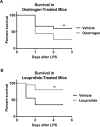
Delay or expedition of puberty by hormonal treatment alters mortality from endotoxemia. (A) Pre-pubertal CD-1 mice were pre-treated with daily subcutaneous injections of 17β-Oestradiol at 100 μg/ml or vehicle (0.4% DMSO) suspended in corn oil for three days prior and once on the day of intraperitoneal E. coli endotoxin injection. Per cent survival over three days was recorded (N = 45/group). (B) Pre-pubertal C57Bl/6 mice were treated with daily subcutaneous injections of leuprolide at 250 μg/ml or vehicle (0.9% sodium chloride) starting on PND 24 and continuing until post-puberty (PND 35) before S. enterica endotoxin injection (N = 20/group). Per cent survival over three days was recorded. Significant differences in survival between pre- and post-pubertal mice are labelled **(p < 0.01). Per cent survival was compared using a log rank Mantel Cox test.
Figure 6
Figure 6
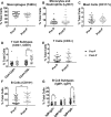
Pre- and post-pubertal (Pre-P, Post-P) mice show similar naïve peritoneal cell profiles. Naïve peritoneal cells from pre- and post-pubertal C57Bl/6 mice were collected through peritoneal lavage. Cells were blocked to prevent non-specific binding and then treated with antibodies for subsequent immuno-phenotypic analysis by flow cytometry. These included (A) F4/80+ Macrophages, (B) Ly6C+ Monocytes and Neutrophils, (C) CD117+ Mast Cells, (D) CD3+ T cells and associated (E) CD4+ and CD8+ subsets and (F) CD19+ B cells and associated (G) IgM+ and IgD+ subsets. Black closed circles signify pre-pubertal data. N ≥ 23/group. For each sample, a total of ten thousand cells were analysed using the gating strategy described in Figure S8. Significant differences in concentration between pre- and post-pubertal mice are labelled with **(p < 0.01). All comparisons were made using the Mann Whitney test.
Figure 7
Figure 7
Adoptive transfer of pre-pubertal cells improves post-pubertal survival. Naïve peritoneal cells were collected from pre-pubertal mice or post-pubertal mice by peritoneal lavage. Recipient post-pubertal mice were administered 1 mL of pre-pubertal (Pre-P) or post-pubertal (Post-P) peritoneal cell suspensions (2 million cells total), or 1 mL of the vehicle (PBS) via intraperitoneal injection. Following incubation of donor cells within the peritoneal cavity, the recipient mice were then subjected to endotoxemia with either S. enterica or E. coli endotoxin (N ≥ 37/group). Per cent survival was recorded over five days. Significant differences in survival between mice administered Pre-P cells vs. those receiving Post-P cells or Vehicle were ****(p < 0.0001). Per cent survival was compared using a log rank Mantel Cox test.

References

    1. Singer M, et al. The Third International Consensus Definitions for Sepsis and Septic Shock (Sepsis-3) JAMA. 2016;315:801–810. doi: 10.1001/jama.2016.0287. - DOI - PMC - PubMed
    1. Marshall JC. Why have clinical trials in sepsis failed? Trends Mol Med. 2014;20:195–203. doi: 10.1016/j.molmed.2014.01.007. - DOI - PubMed
    1. Mira JC, et al. Sepsis Pathophysiology, Chronic Critical Illness, and Persistent Inflammation-Immunosuppression and Catabolism Syndrome. Crit Care Med. 2017;45:253–262. doi: 10.1097/CCM.0000000000002074. - DOI - PMC - PubMed
    1. Ahmed R, Oldstone MB, Palese P. Protective immunity and susceptibility to infectious diseases: lessons from the 1918 influenza pandemic. Nat Immunol. 2007;8:1188–1193. doi: 10.1038/ni1530. - DOI - PMC - PubMed
    1. Linder, F. E. & Grove, R. D. Vital statistics rates in the United States 1900–1940. 248, 254 (US Government Printing Office, Washington, D.C., 1947).

Publication types

LinkOut - more resources