. 2017 Dec 26;56(51):6713-6725.
doi: 10.1021/acs.biochem.7b01066. Epub 2017 Dec 12.

Discovery and Biological Evaluation of Potent and Selective N-Methylene Saccharin-Derived Inhibitors for Rhomboid Intramembrane Proteases

Affiliations

Discovery and Biological Evaluation of Potent and Selective N-Methylene Saccharin-Derived Inhibitors for Rhomboid Intramembrane Proteases

Parul Goel et al. Biochemistry. .

Abstract

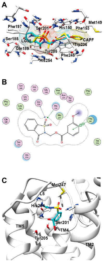
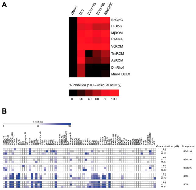
Rhomboids are intramembrane serine proteases and belong to the group of structurally and biochemically most comprehensively characterized membrane proteins. They are highly conserved and ubiquitously distributed in all kingdoms of life and function in a wide range of biological processes, including epidermal growth factor signaling, mitochondrial dynamics, and apoptosis. Importantly, rhomboids have been associated with multiple diseases, including Parkinson's disease, type 2 diabetes, and malaria. However, despite a thorough understanding of many structural and functional aspects of rhomboids, potent and selective inhibitors of these intramembrane proteases are still not available. In this study, we describe the computer-based rational design, chemical synthesis, and biological evaluation of novel N-methylene saccharin-based rhomboid protease inhibitors. Saccharin inhibitors displayed inhibitory potency in the submicromolar range, effectiveness against rhomboids both in vitro and in live Escherichia coli cells, and substantially improved selectivity against human serine hydrolases compared to those of previously known rhomboid inhibitors. Consequently, N-methylene saccharins are promising new templates for the development of rhomboid inhibitors, providing novel tools for probing rhomboid functions in physiology and disease.


Conflict of interest statement

Notes

The authors declare no competing financial interest.

Figures

Figure 1
Figure 1
(A) Workflow for the development of novel rhomboid inhibitors. (B) Proposed reaction mechanism of a saccharin-based inhibitor with the catalytic residues in the active site of the E. coli rhomboid protease GlpG. The mechanism involves the enzyme-induced formation of a Michael acceptor and results in an end product that is covalently cross-linked to the enzyme.
Figure 2
Figure 2
Docking of saccharin-based inhibitors into the active site of the rhomboid protease GlpG. (A) The model shows the initial interactions of a known rhomboid inhibitor (CAPF, yellow) and a saccharin-based inhibitor (with LG 38, turquoise) with amino acid side chains in the active site of GlpG. (B) Ligand interaction map of GlpG and the saccharin with LG38. The map shows the formation of two H-bonds of the sulfoxide moiety of the saccharin with His150 and Gly199. In addition, a ππ interaction between the aryl ring of the saccharin and Phe245 is visible. (C) Molecular model of N-methylated saccharin within the GlpG active site after the enzymatic reaction between the inhibitor (blue) and the rhomboid protease (gray). The model shows the end product based on the proposed reaction mechanism (see Figure 1B). Two covalent bonds have been formed between the saccharin and Ser201 and His254.
Figure 3
Figure 3
(A) Evaluation of saccharin inhibitors in the GlpG in vitro activity assay. The E. coli rhomboid GlpG was recombinantly expressed and preincubated with each saccharin inhibitor at concentrations ranging from 0.1 to 250 μM for 30 min at 37 °C prior to the addition of the Gurken substrate. The SDS–PAGE gel shows the reaction products at the end of a 90 min incubation period. The arrows indicate the full-length Gurken substrate (S, ~66 kDa) and the N-terminal Gurken cleavage product (P, ~55 kDa). Signal intensities of the N-terminal Gurken product were normalized to the DMSO control condition and plotted against log(inhibitor concentration). A nonlinear regression curve fit of the log(inhibitor concentration) vs percent N-terminal Gurken product was used to determine IC50 values. The SDS–PAGE gel was stained with Coomassie Brilliant Blue and quantified with ImageJ, and the values were plotted in GraphPad Prism. Two independent IC50 determinations were performed for each compound, and one representative experiment is shown. (B) In vivo inhibition of GlpG in live E. coli cells. Wild-type strain NR698 with endogenous GlpG activity or GlpG-deficient strain NR698 ΔglpG was transformed with the MBP-FLAG-LacYTM2-Trx substrate., Increasing concentrations of the indicated saccharin inhibitors from 5 nM to 100 μM were added to the culture media; substrate expression was induced with 1 mM L-rhamnose, and cells were grown in the presence of the inhibitors for 4 h. Cleavage fragments of the substrate (P, ~46 kDa) were quantified by SDS–PAGE and near-infrared Western blotting of cell lysates, and IC50 values were determined as described previously. Note that in the GlpG-deficient cells the substrate (S) was not processed and no cleavage fragment was produced (lane KO). Two independent IC50 determinations were performed for each compound, and one representative experiment is shown.
Figure 4
Figure 4
Mechanism of saccharin-based inhibitors. (A) The reaction of N-methylated saccharins with the rhomboid involves the release of the acidic LG. A saccharin derivative with anthranilic acid as the LG (BSc5193) was incubated with recombinantly expressed GlpG. According to the proposed mechanism, nucleophilic attack of Ser201 should lead to the formation of a Michael acceptor and release of the anthranilic acid LG, which becomes fluorescent. Incubation of GlpG with BSc5193 resulted in an increase in fluorescence as compared to those of reaction mixtures containing only the enzyme or the compound. However, no increase in fluorescence was observed for a different potent saccharin-based inhibitor (BSc5195) with a nonfluorescent LG. Average values of two independent experiments are shown. Error bars represent the standard deviation. (B) Purified wild-type GlpG was reacted in vitro with five different saccharins (BSc5205, BSc5156, BSc5195, BSc5196, and BSc5198), and the mass difference between free GlpG and the inhibitor–GlpG adduct was determined by electrospray mass spectrometry. The shift in the m/z value was the same after incubation with all five compounds. This indicated that all compounds inhibited GlpG via the same mechanism, which was expected because the covalently bound core structure was the same and only the LG differed between the compounds. Three independent experiments were performed. (C) Upon incubation of wild-type GlpG with the saccharin BSc5196, a shift in the m/z value corresponding to a covalent double-bonded end product after reaction with the protease was observed. Mutation of the catalytically active Ser201 to threonine or His254 to alanine abolished the difference in m/z observed with the wild-type protein. In contrast, mutation of His150 had no effect and produced a difference in m/z as seen with wild-type GlpG. Three independent experiments were performed, and one representative experiment is shown. (D) Reversibility of the saccharin reaction mechanism was investigated by the rapid dilution method. Saccharin BSc5195 (5 μM), irreversible isocoumarin inhibitor JLK-6 (10 μM), or reversible β-lactam inhibitor L29 (1 μM) was preincubated with GlpG for 1 h at 10 times their IC50 concentrations. The reaction mixtures were then rapidly diluted 100-fold, and a fluorogenic rhomboid substrate peptide (10 μM) was added. No recovery of enzyme activity was observed with JLK-6, while substantial recovery was seen with L29 as expected. With the saccharin inhibitor BSc5195, less but noticeable recovery of GlpG activity was observed over the time period of 100 min, demonstrating partial reversibility of the inhibition mechanism. No recovery of activity was apparent when the saccharin inhibitor concentration was maintained at 5 μM after the dilution step. Three independent experiments were performed, and one representative experiment is shown.
Figure 5
Figure 5
Selectivity of saccharin-based inhibitors. (A) Heat map representation of the activity-based probe (ABP) competition assay with three potent saccharin inhibitors against nine different rhomboid proteases. Recombinantly expressed rhomboids were preincubated with saccharins (50 μM), labeled with the ABP TAMRA-FP, and the labeled enzymes were visualized by SDS–PAGE and fluorescence scanning. The isocoumarin DCI was used as a positive control and inhibited around 80–100% of the activity of all nine rhomboids. Two independent experiments were performed, and average values for each experimental condition are shown. (B) A high-throughput activity-based probe competition assay was used to profile saccharins against human soluble serine hydrolases. Isocoumarins S006 and S016 inhibited multiple human serine hydrolases. In contrast, at concentrations sufficient to inhibit the rhomboid GlpG in the in vitro activity assay, saccharin-based compounds BSc5195, BSc5196, and BSc5205 showed far less activity against the panel of human serine hydrolases (no inhibition, white; weak inhibition, gray to light blue; strong inhibition, dark blue). Two independent experiments were performed, and one representative experiment is shown.

Similar articles

Cited by

References

    1. Puente XS, Sanchez LM, Overall CM, Lopez-Otin C. Human and mouse proteases: a comparative genomic approach. Nat Rev Genet. 2003;4:544–558. - PubMed
    1. Rawlings ND, Barrett AJ. Evolutionary families of peptidases. Biochem J. 1993;290(1):205–218. - PMC - PubMed
    1. Egeblad M, Werb Z. New functions for the matrix metalloproteinases in cancer progression. Nat Rev Cancer. 2002;2:161–174. - PubMed
    1. Krane SM. Elucidation of the potential roles of matrix metalloproteinases in skeletal biology. Arthritis Res Ther. 2003;5:2–4. - PMC - PubMed
    1. Lichtenthaler SF, Haass C, Steiner H. Regulated intramembrane proteolysis–lessons from amyloid precursor protein processing. J Neurochem. 2011;117:779–796. - PubMed

Publication types

LinkOut - more resources