. 2017 Dec 27;139(51):18576-18589.
doi: 10.1021/jacs.7b08870. Epub 2017 Dec 15.

Single-Molecule Fluorescence Reveals Commonalities and Distinctions among Natural and in Vitro-Selected RNA Tertiary Motifs in a Multistep Folding Pathway

Affiliations

Single-Molecule Fluorescence Reveals Commonalities and Distinctions among Natural and in Vitro-Selected RNA Tertiary Motifs in a Multistep Folding Pathway

Steve Bonilla et al. J Am Chem Soc. .

Abstract

Decades of study of the RNA folding problem have revealed that diverse and complex structured RNAs are built from a common set of recurring structural motifs, leading to the perspective that a generalizable model of RNA folding may be developed from understanding of the folding properties of individual structural motifs. We used single-molecule fluorescence to dissect the kinetic and thermodynamic properties of a set of variants of a common tertiary structural motif, the tetraloop/tetraloop-receptor (TL/TLR). Our results revealed a multistep TL/TLR folding pathway in which preorganization of the ubiquitous AA-platform submotif precedes the formation of the docking transition state and tertiary A-minor hydrogen bond interactions form after the docking transition state. Differences in ion dependences between TL/TLR variants indicated the occurrence of sequence-dependent conformational rearrangements prior to and after the formation of the docking transition state. Nevertheless, varying the junction connecting the TL/TLR produced a common kinetic and ionic effect for all variants, suggesting that the global conformational search and compaction electrostatics are energetically independent from the formation of the tertiary motif contacts. We also found that in vitro-selected variants, despite their similar stability at high Mg2+ concentrations, are considerably less stable than natural variants under near-physiological ionic conditions, and the occurrence of the TL/TLR sequence variants in Nature correlates with their thermodynamic stability in isolation. Overall, our findings are consistent with modular but complex energetic properties of RNA structural motifs and will aid in the eventual quantitative description of RNA folding from its secondary and tertiary structural elements.


Conflict of interest statement

The authors declare no competing financial interest.

Figures
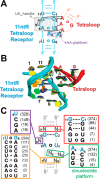
Figure 1
Figure 1
Structural modularity and sequence diversity of the GAAA/11ntR tertiary motif. (A) Secondary structure of the canonical 11ntR tetraloop-receptor (cyan) and GAAA tetraloop (red). Watson–Crick (WC) and noncanonical base-pairs are marked as colored dashed lines and circles, respectively. Tertiary interactions are marked in black. UA_handle and AA-platform submotifs are boxed. Residue numbering is used throughout. (B) Overlay of four crystal structures of the GAAA/11ntR motif. Backbone of tetraloop and tetraloop-receptor are colored as in (A). The structure of the TL/TLRs were extracted from crystal structures of the P4–P6 domain (PDB 1GID, blue), RNase P (PDB 1NBS, green), and Azoarcus group I intron (PDB 1ZZN, yellow and orange). PDB 1NBS contains a tetraloop-receptor variant in which the canonical AA-platform is replaced by an AC-platform. (C) Sequence variation in 515 11ntR-like tetraloop-receptors identified in the sequence and secondary structure database of aligned group I introns. Numbers in parentheses correspond to the total number of receptors containing the specified sequence variation. The complete sequences of the variants along with their observed frequencies are provided in Table S1. It is not known whether A–U and G–C at positions 6 and 7 (marked with *) form canonical WC base-pairs.
Figure 2
Figure 2
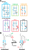
Natural and in vitro-selected TL/TLR variants were inserted into a minimal U7-tethered smFRET construct. (A) Sequence of the six tetraloop-receptor variants investigated. Residues that differ from the canonical 11ntR are boxed. Secondary structures of 11ntR and 11ntR_AC in the docked state are known experimentally., (B) Schematic of smFRET construct in which tetraloop-receptors in panel A were embedded. GAAA tetraloop (red) and residues in cyan are common to all of the TL/TLR variants investigated. This smFRET construct has been used in previous studies to characterize the canonical GAAA/11ntR., The complete sequence of all constructs is provided in Table S2.
Figure 3
Figure 3
smFRET reveals distinct kinetic behaviors and Mg2+ dependences of TL/TLR in the U7-tether minimal constructs (Figure 2). Sample smFRET traces of variants 11ntR (A) and C7.2 (B) at two Mg2+ concentrations. Green and red traces are donor and acceptor intensities respectively and black trace represents the probability of occupying the high FRET state as determined by a two-state hidden Markov model (HMM). The values of the fitted kdock and kundock for the sample traces are displayed above each trace. Traces were truncated at 6 s for ease of comparison. Sample traces for all variants and conditions are shown in Supporting Information (Figures S9–S275) and raw data are available to download. (C–D) Median kdock and kundock values (large circles) for a population of individual molecules (small circles) at a range of Mg2+ concentrations (colors) for 11ntR (C) and C7.2 (D). For clarity, only a subset of Mg2+ concentrations and 50 randomly selected molecules at each [Mg2+] are shown. Typically more than 100 molecules were measured at each Mg2+ concentration. The complete population of molecules at 0.25 mM Mg2+ is shown in the inset to better display the kinetic homogeneity. Data from measurements carried out by single-photon counting are marked with an internal “X”.
Figure 4
Figure 4
Docking kinetics, thermodynamics and Mg2+ dependence of TL/TLR variants in U7-tether minimal construct. (A–C) Median kdock, kundock, and Kobs values for each TL/TLR variant at a range of Mg2+ concentrations. All measurements were carried out in a background of 140 mM K+. Data and errors are summarized in Table S4. Standard errors calculated from bootstrapping were smaller than the size of the symbols. Data points marked with an internal “X” were determined using single-photon counting. Linear fits were determined by least-squares. Mg2+ uptake prior to and after formation of the docking transition state (D and E, respectively) and net Mg2+ uptake (F) were obtained from the linear fits in panels A–C as given by eq 1 and eqs 2A and 2B.
Figure 5
Figure 5
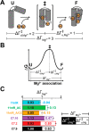
Physical interpretation of the sensitivity of Kobs, kdock, and kundock to Mg2+. (A) Schematic of a model RNA undergoing a folding transition from an extended unfolded (U) state, through a compact transition state (‡), to a final folded (F) state. Mg2+ is taken up to compensate for changes in charge density as the RNA folds. To maintain charge neutrality, Mg2+ uptake must be accompanied by the uptake of anions and/or release of K+ into ion atmosphere (not shown). The net number of Mg2+ taken up (ΔΓMg2+) is the sum of Mg2+ taken up prior to (ΔΓd,Mg2+) and after (ΔΓu,Mg2+) formation of the transition state (‡; ΔΓMg2+ = ΔΓd,Mg2+ + ΔΓu,Mg2+). (B) Folding process in panel A is represented in a free-energy diagram. (C) Mg2+ uptake of TL/TLR variants in U7 smFRET construct. Overall bar gives ΔΓMg2+ and is divided into contributions from ΔΓd,Mg2+ (filled) and ΔΓu,Mg2+ (open). Error bars are standard errors of ΔΓMg2+.
Figure 6
Figure 6
Comparison of TL/TLR docking kinetics in Mg2+ and Ba2+ reveals weak but consistent sequence-dependent discrimination for different types of divalent cations. Median kdock (A) and kundock (B) values over a range of Mg2+ (filled symbols) and Ba2+ (open symbols) for each TL/TLR variant. Mg2+ values are reproduced from Figure 4. Data and errors are summarized in Tables S4 and S5. Standard errors calculated from bootstrapping were smaller than the size of the symbols. Linear fits were determined by least-squares. All measurements were carried out in a background of 140 mM K+. Data collected with single-photon counting setup are marked with and internal “X”.
Figure 7
Figure 7
Conformational preferences and Mg2+-dependence of connecting tether produces a common kinetic and electrostatic effect on TL/TLR docking. (A,B) Median kdock (A) and kundock (B) values of four TL/TLR variants as a function of Mg2+ in a background of 140 mM K+ with an A7 (circles) or U7 (triangles) tether connecting the tertiary contact partners. Data and errors are summarized in Tables S4 and S6. Standard errors calculated from bootstrapping were smaller than the size of the symbols. Data collected with single-photon counting setup are marked with an internal “X”. Linear fits were determined by least-squares. (C–D) Ratio of the kdock (C) and kundock (D) values with the U7 and A7 tethers. Open symbols were obtained from interpolation using the linear fits in panels A and B. Dashed lines at a constant value of one are shown for reference.
Figure 8
Figure 8
Docking kinetics and thermodynamics of TL/TLR variants in KCl. To account for nonideal electrolyte interactions, the data are plotted as a function of the mean activity (a±) of the salt solution. (A–C) Median kdock, kundock, and Kobs values for each of the TL/TLR variants over a range of KCl concentrations. Coloring scheme is shown in panel A. Data and errors are summarized in Table S7. Standard errors determined from bootstrapping were smaller than the size of the symbols. Data marked with an internal “X” were determined by single-photon counting. Linear fits were determined by least-squares.
Figure 9
Figure 9
Relative effects of Mg2+ vs K+ on the docking kinetics and thermodynamics of the TL/TLR variants. kdock (A), kundock (B), and Kobs (C) in 140 mM K+ were compared with and without 1 mM Mg2+. (kdock)rel is defined as kdock at 140 mM K+ and 1 mM Mg2+ divided by kdock at 140 mM K+; (kundock)rel and (Kobs)rel are similarly defined. Values for variants that were too weak to measure in 140 mM K+ (a±,KCl = 103 mM) were extrapolated from the linear fits in Figure 8.
Figure 10
Figure 10
Conformational rearrangements of canonical 11ntR tetraloop-receptor and kinetic effect of sequence variations relative to 11ntR. (A) Schematic of the experimentally determined conformations of the undocked (left) and docked (right) canonical 11ntR tetraloop-receptor.,, The GAAA tetraloop does not undergo significant conformational changes upon docking., For the 11ntR tetraloop-receptor, in the undocked state A4, A8, and A5 form a series of stacking interactions (A-zipper) that break upon formation of the AA-platform present in the docked state. Base-stacking and hydrogen-bond interactions in the undocked state that need to break prior to attaining the docked state are colored in red, and interactions that form in the docked state are colored in green. Residues that were mutated to U in a previous study are colored in purple. kdock (B) and kundock (C) of variants relative to the canonical 11ntR at 1 mM Mg2+ and 140 mM K+ (except for single point mutants marked with ‘*’, which were measured previously in 1 mM Ba2+ and 140 mM K+; measurements across a range of [M2+] suggest that TL/TLRs display only minor kinetic differences in Mg2+ vs Ba2+ (Figure 6)). The sequences of the residues that comprise the putative platforms are shown in small boxes on the top of left panel. Variants that are identical to the 11ntR except for a single mutation to the AA-platform are marked with a red star.
Figure 11
Figure 11
Schematic model for TL/TLR folding for the GAAA/11ntR. The canonical 11ntR is shown starting with a schematic of the experimentally determined unbound form of the tetraloop-receptor and ending with the know structure of the docked GAAA/11ntR., The features of this pathway are supported by mutational and ionic effects described in the text. Nevertheless, this model simplifies the many rearrangements that must occur in folding, and the experimental data distinguish only between effects that occur prior and subsequent to the rate-limiting transition state (‡). The U·U base pair in the unbound state is expected to be weak and in the simplest model it breaks to allow rearrangement of the A residues to form the dinucleotide platform.
Figure 12
Figure 12
Thermodynamic stability of TL/TLR variants in isolated smFRET construct at near physiological ionic conditions correlates with observed frequency in group I introns. (A) Stability of TL/TLR variants in smFRET construct relative to the canonical 11ntR at high and near-physiological Mg2+ concentrations (values with “*” were measured previously in Ba2+). Values at 20 mM Mg2+ (left) for TL/TLR variants studied here were extrapolated from linear fits in Figure 4C. All measurements were carried out in a background of 140 mM K+. (B) Relative stability of TL/TLR variants in 1 mM Mg2+ and 140 mM K+ (except for variants with “*” in figure legend which were measured at 1 mM Ba2+ and 140 mM K+ previously) plotted against their observed frequency in the Group I Intron Sequence and Structure Database (GISSD).

References

    1. Wan R.; Yan C.; Bai R.; Huang G.; Shi Y. Science 2016, 353 (6302), 895–904. 10.1126/science.aag2235. - DOI - PubMed
    1. Ban N.; Nissen P.; Hansen J.; Moore P. B.; Steitz T. A. Science 2000, 289 (5481), 905–920. 10.1126/science.289.5481.905. - DOI - PubMed
    1. Steitz T. A. Nat. Rev. Mol. Cell Biol. 2008, 9 (3), 242–253. 10.1038/nrm2352. - DOI - PubMed
    1. Stagno J. R.; Liu Y.; Bhandari Y. R.; Conrad C. E.; Panja S.; Swain M.; Fan L.; Nelson G.; Li C.; Wendel D. R.; White T. A.; Coe J. D.; Wiedorn M. O.; Knoska J.; Oberthuer D.; Tuckey R. A.; Yu P.; Dyba M.; Tarasov S. G.; Weierstall U.; Grant T. D.; Schwieters C. D.; Zhang J.; Ferré-D’Amaré A. R.; Fromme P.; Draper D. E.; Liang M.; Hunter M. S.; Boutet S.; Tan K.; Zuo X.; Ji X.; Barty A.; Zatsepin N. A.; Chapman H. N.; Spence J. C. H.; Woodson S. A.; Wang Y.-X. Nature 2016, 541 (7636), 242–246. 10.1038/nature20599. - DOI - PMC - PubMed
    1. Herschlag D. J. Biol. Chem. 1995, 270 (36), 20871–20874. 10.1074/jbc.270.36.20871. - DOI - PubMed

Publication types