. 2017 Nov 29;6(4):31.
doi: 10.3390/antibiotics6040031.

Antimicrobial Activity of Bee Venom and Melittin against Borrelia burgdorferi

Affiliations

Antimicrobial Activity of Bee Venom and Melittin against Borrelia burgdorferi

Kayla M Socarras et al. Antibiotics (Basel). .

Abstract

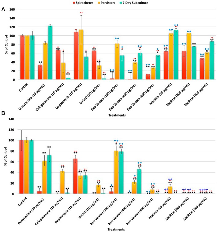
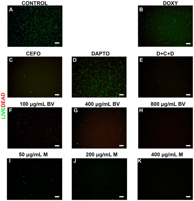
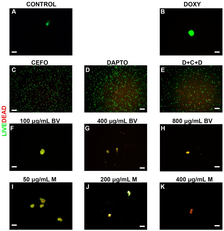
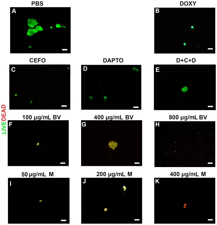
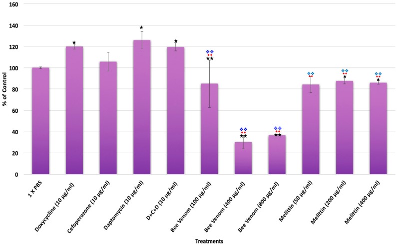
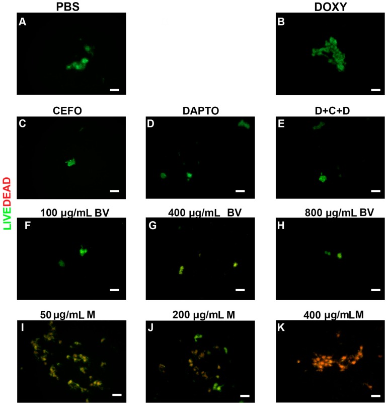
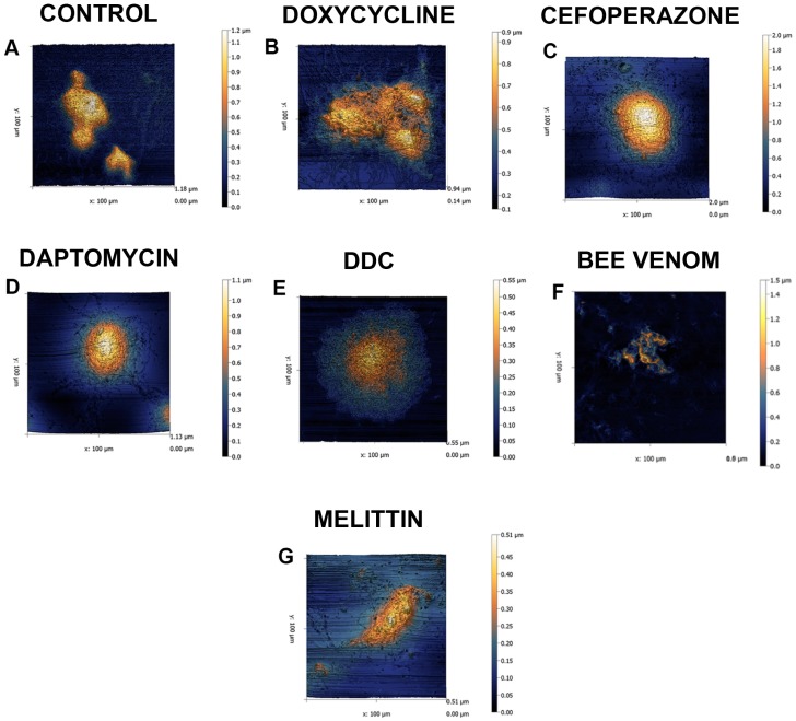
Lyme disease is a tick-borne, multi-systemic disease, caused by the bacterium Borrelia burgdorferi. Though antibiotics are used as a primary treatment, relapse often occurs after the discontinuation of antimicrobial agents. The reason for relapse remains unknown, however previous studies suggest the possible presence of antibiotic resistant Borrelia round bodies, persisters and attached biofilm forms. Thus, there is an urgent need to find antimicrobial agents suitable to eliminate all known forms of B. burgdorferi. In this study, natural antimicrobial agents such as Apis mellifera venom and a known component, melittin, were tested using SYBR Green I/PI, direct cell counting, biofilm assays combined with LIVE/DEAD and atomic force microscopy methods. The obtained results were compared to standalone and combinations of antibiotics such as Doxycycline, Cefoperazone, Daptomycin, which were recently found to be effective against Borrelia persisters. Our findings showed that both bee venom and melittin had significant effects on all the tested forms of B. burgdorferi. In contrast, the control antibiotics when used individually or even in combinations had limited effects on the attached biofilm form. These findings strongly suggest that whole bee venom or melittin could be effective antimicrobial agents for B. burgdorferi; however, further research is necessary to evaluate their effectiveness in vivo, as well as their safe and effective delivery method for their therapeutic use.

Keywords: Lyme disease; antibiotic resistance; bee venom; biofilms; melittin; persisters.


Conflict of interest statement

The authors declare no conflicts of interest.

Figures

Figure 1
Figure 1
The effects of various antimicrobial agents on B. burgdorferi as determined by SYBR Green I/PI assay Panel (A) or direct counting assay Panel (B). Doxycycline, Cefoperazone, Daptomycin and their combination (D + C + D) as well as different concentrations of bee venom and melittin were tested on B. burgdorferi logarithmic phase (spirochetes) culture and stationary phase (persisters) cultures as well as in 7-day recovery subculture as described previously [6,7,8]. Significance against sterile PBS buffer (control vehicle) with the p value of <0.05 and <0.01 are indicated in * and ** respectively. Significance against Doxycycline with the p value of <0.05 and <0.01 are indicated in ♦ and ♦♦ respectively. Significance against the three-antibiotic combination (D + C + D) with the p value of <0.05 and <0.01 are indicated in ❖ and ❖❖ respectively. n = 9.
Figure 2
Figure 2
Representative Live/Dead staining images of B. burgdorferi log phase spirochetal cultures treated with different antimicrobial agents. Cells were stained with SYBR Green I/PI as outlined in the Methods and representative images were taken at 100× magnification. Panel (A) Borrelia culture treated only with PBS was used as a negative control. Panel (B) Doxycycline (DOXY) treated; Panel (C) Cefoperazone (CEFO) treated; Panel (D) Daptomycin (DAPTO) treated and Panel (E) Three-antibiotic combination (D + C + D). Panels (FH) Bee venom (BV) was used in increasing concentrations while Panels (IK) depicts melittin (M) treated cells. Live cells are stained with green color while dead cells are stained red. Scale bar: 100 μm.
Figure 3
Figure 3
Representative Live/Dead staining images of B. burgdorferi stationary phase persister cultures following treatment with different antimicrobial agents. Cells were stained with SYBR Green I/PI as outlined in the Methods and representative images were taken at 100× magnification. Panel (A) Borrelia culture treated only with PBS was used as a negative control. Panel (B) Doxycycline (DOXY) treated, Panel (C) Cefoperazone (CEFO) treated, Panel (D) Daptomycin (DAPTO) treated and Panel (E) Three-antibiotic combination (D + C + D). Panels (FH) Bee venom (BV) was used in increasing concentrations while Panels (IK) depicts melittin (M) treated cells. Live cells are stained with green color while dead cells are stained red. Scale bar: 100 μm.
Figure 4
Figure 4
Representative Live/Dead staining images of B. burgdorferi 7-day recovery cultures following treatment with different antimicrobial agents. Cells were stained with SYBR Green I/PI as outlined in the Material and Methods and representative images were taken at 100× magnification. Panel (A) Borrelia culture treated only with PBS was used as a negative control. Panel (B) Doxycycline (DOXY) treated, Panel (C) Cefoperazone (CEFO) treated, Panel (D) Daptomycin (DAPTO) treated and Panel (E) Three-antibiotic combination (D + C + D). Panels (FH) Bee venom (BV) was used in increasing concentrations while Panels (IK) depict melittin (M) treated cells at different concentrations. Live cells are stained with green color while dead cells are stained red. Scale bar: 100 μm.
Figure 5
Figure 5
Effect of different antimicrobial agents on attached B. burgdorferi biofilms. Susceptibility of attached B. burgdorferi biofilms to antimicrobial agents after a three-day treatment was analyzed by crystal violet method as described in Material and Methods. Doxycycline, Cefoperazone, Daptomycin and their combination (D + C + D) as well as different concentration of bee venom and melittin were tested on attached Borrelia biofilms. Significance against PBS buffer (negative control vehicle) with the p value of <0.05 and <0.01 are indicated in * and ** respectively. Significance against Doxycycline with the p value of <0.05 and <0.01 are respectively indicated in ♦ and ♦♦ Significance against the three-antibiotic combination (D + C + D) with the p-value of <0.05 and <0.01 are indicated in ❖ and ❖❖ respectively. n = 9.
Figure 6
Figure 6
Representative Live/Dead images of the viability of attached Borrelia biofilms following treatment with different antimicrobial agents. Biofilms were stained with SYBR Green I and PI as outlined in the Material and Methods and representative images were taken at 100× magnification. Panel (A) Borrelia culture treated only with PBS was used as a negative control. Panel (B) Doxycycline (DOXY) treated, Panel (C) Cefoperazone (CEFO) treated, Panel (D) Daptomycin (DAPTO) treated and Panel (E) Three-antibiotic combination (D + C + D). Panels (FH) Bee venom (BV) was used in increasing concentrations while Panels (IK) depict melittin treated cells at different concentration. Live cells are stained with green color while dead cells are stained red. Scale bar: 100 μm.
Figure 7
Figure 7
Representative atomic force microscopy images showing the ultrastructural details of Borrelia biofilm before and after treatment with antimicrobial agents. The preparations of B. burgdorferi strain B31 biofilms on chamber slides are described in Methods section. All biofilms were scanned at 0.4 Hz using contact mode and the individual Z ranges (height) are indicated next to each panel by means of a scale. The images were scanned using the Nanosurf Easyscan 2 software and the images were processed using Gwyddion software. Scale bar located on the side of corresponding AFM scan indicate the height changes of the topography of the biofilm. Darker colors (black and blue) indicate the surface of the slide while lighter colors (yellow to white) indicate high points of attached biofilms. Attached Borrelia biofilms treated with Panel (A) PBS (control), Panel (B) Doxycycline (10 μg/mL), Panel (C) Cefoperazone (10 μg/mL), Panel (D) Daptomycin (10 μg/mL), Panel (E) Three antibiotic combination: Doxycycline + Daptomycin + Cefoperazone (DCC, 10 μg/mL/each), Panel (F) Bee venom (400 μg/mL) and Panel (G) Melittin (200 μg/mL).

References

    1. Barbour A.G., Hayes S.F. Biology of Borrelia species. Microbiol. Rev. 1986;50:381–400. - PMC - PubMed
    1. Rudenko N., Golovchenko M., Grubhoffer L., Oiver J.H., Jr. Updates on Borrelia burgdorferi sensu lato complex with respect to public health. Ticks Tick-borne Dis. 2011;2:123–128. doi: 10.1016/j.ttbdis.2011.04.002. - DOI - PMC - PubMed
    1. Brisson D., Vandermause M.F., Meece J.K., Reed K.D., Dykhyizen D.E. Evolution of Northeastern and Midwestern Borrelia burgdorferi, United States. Emerg. Infect. Dis. 2010;16:911–917. doi: 10.3201/eid1606.090329. - DOI - PMC - PubMed
    1. Center of Disease Control and Prevention Lyme Disease. [(accessed on 13 September 2017)];2016 Available online: http://www.cdc.gov/lyme/
    1. Wormser G.P., Nadelman R.B., Dattwyler R.J., Dennis D.T., Shapiro E.D., Steere A.C., Rush T.J., Rahn D.W., Coyle P.K., Persing D.H., et al. Practice guidelines for the treatment of Lyme disease. Clin. Infect. Dis. 2000;31:S1–S14. doi: 10.1086/314053. - DOI - PubMed

LinkOut - more resources