. 2017 Nov 29;22(12):2089.
doi: 10.3390/molecules22122089.

HPLC-PDA-MS/MS Characterization of Bioactive Secondary Metabolites from Turraea fischeri Bark Extract and Its Antioxidant and Hepatoprotective Activities In Vivo

Affiliations

HPLC-PDA-MS/MS Characterization of Bioactive Secondary Metabolites from Turraea fischeri Bark Extract and Its Antioxidant and Hepatoprotective Activities In Vivo

Mansour Sobeh et al. Molecules. .

Erratum in

Abstract

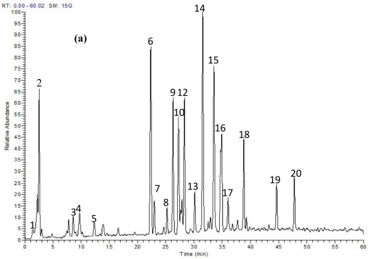
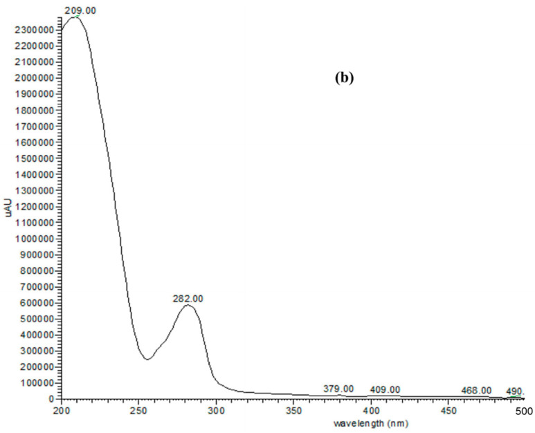
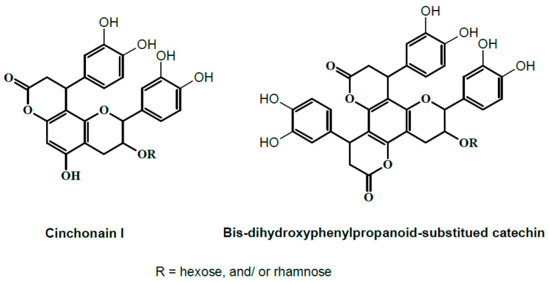
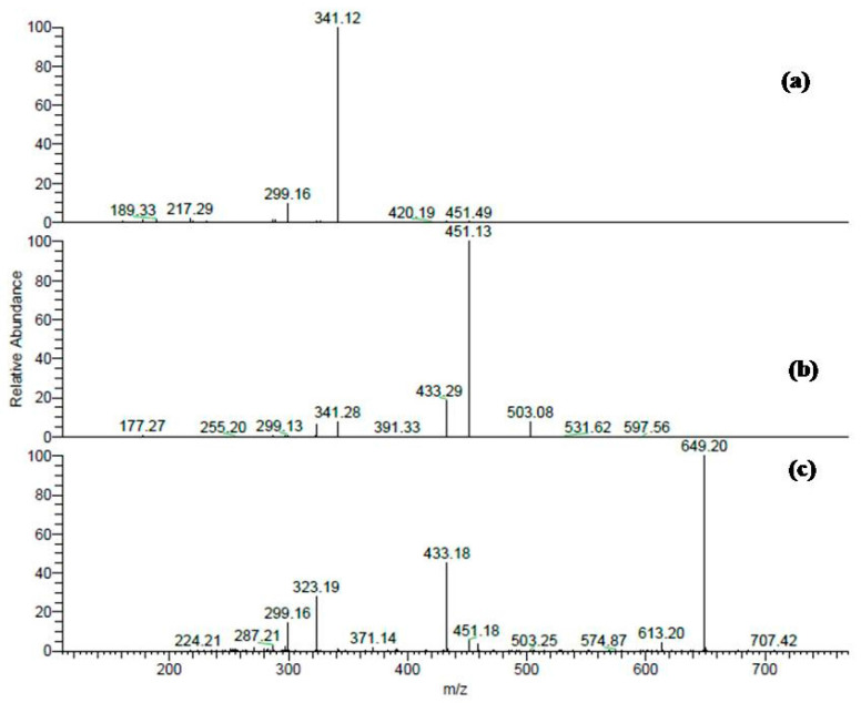
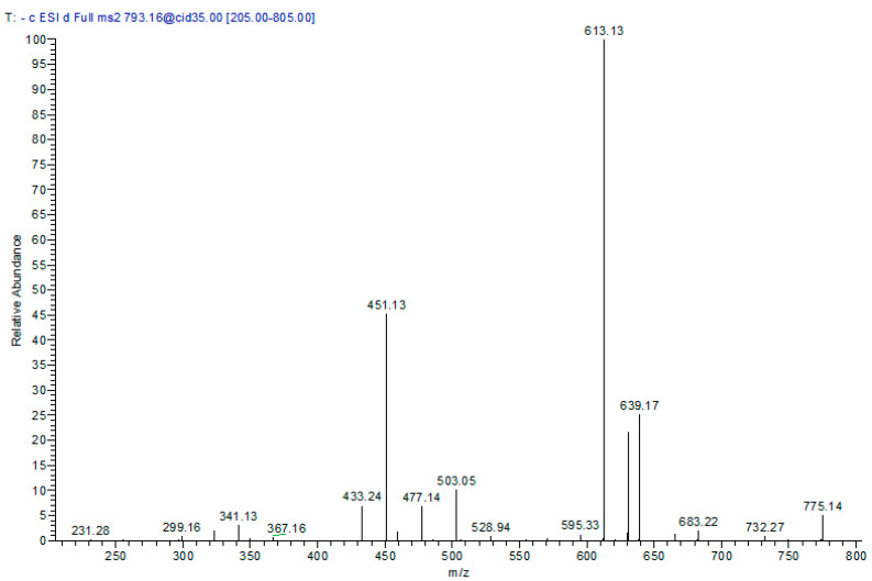
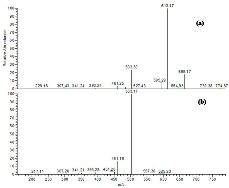
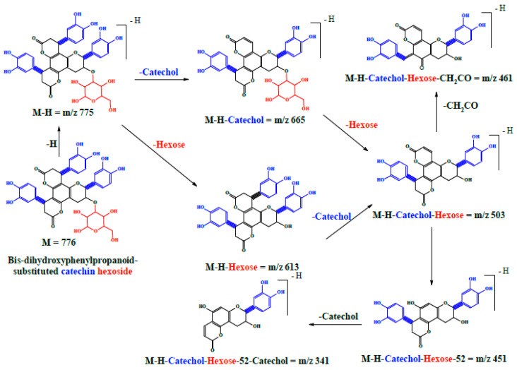
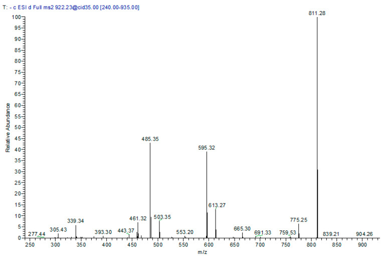
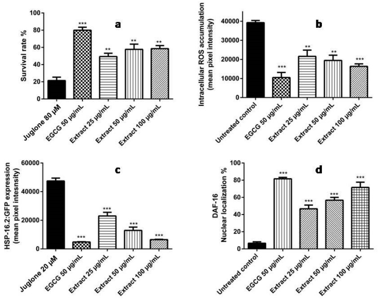
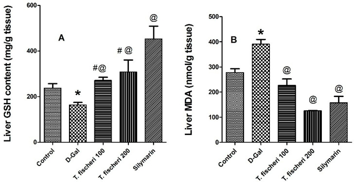
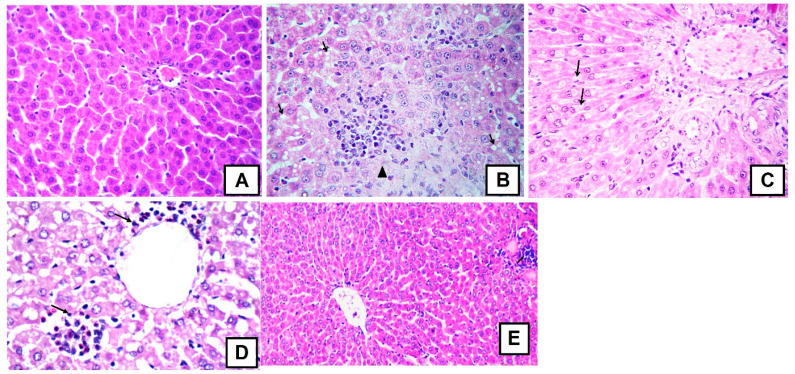
Turraea fischeri is an East African traditional herb, which is widely used in traditional medicine. In this study, we profiled the secondary metabolites in the methanol extract of T. fischeri bark using HPLC-PDA-ESI-MS/MS, and 20 compounds were tentatively identified. Several isomers of the flavonolignan cinchonain-I and bis-dihydroxyphenylpropanoid-substituted catechin hexosides dominated the extract. Robust in vitro and in vivo antioxidant properties were observed in 1,1-diphenyl-2-picrylhydrazyl radical scavenging assay (DPPH) and ferric reducing antioxidant power (FRAP) assay, and in the model organism Caenorhabditis elegans. Additionally, the extract exhibited promising hepatoprotective activities in D-galactosamine (D-GaIN) treated rats. A significant reduction in the elevated levels of aspartate aminotransferase (AST), total bilirubin, gamma-glutamyltransferase (GGT), and malondialdehyde (MDA) and increase of glutathione (GSH) was observed in rats treated with the bark extract in addition to D-galactosamine when compared with rats treated with D-galactosamine alone. In conclusion, T. fischeri is apromising candidate for health-promoting and for pharmaceutical applications.

Keywords: HPLC-PDA-ESI-MS/MS; Turraea fischeri; antioxidant; cinchonains; flavonolignan; hepatoprotection.


Conflict of interest statement

The authors declare no conflict of interest.

Figures

Figure 1
Figure 1
(a) HPLC-ESI (−)-MS/MS profile of a methanol extract of T. fischeri bark; (b) A sample spectra (UV) of compounds (320) at 280 nm.
Figure 1
Figure 1
(a) HPLC-ESI (−)-MS/MS profile of a methanol extract of T. fischeri bark; (b) A sample spectra (UV) of compounds (320) at 280 nm.
Figure 2
Figure 2
Tentative structures of some compounds from Table 1.
Figure 3
Figure 3
(a) MS/MS fragmentation of cinchonain-I at [M − H] (m/z) at 451; (b) MS/MS fragments of cinchonain-I hexoside at [M − H] (m/z) at 613; (c) MS/MS spectra of cinchonain-I rhamnosyl-hexoside at [M − H] m/z 759.
Figure 4
Figure 4
MS/MS spectra of cinchonain-I syringyl-hexoside at [M − H] m/z 793.
Figure 5
Figure 5
Negative ion ESI-MS/MS spectra of bis-dihydroxyphenylpropanoid-substituted catechin–hexoside; (a) MS2 of [M − H] m/z 775; (b) MS3 of main daughter ion at m/z 613.
Figure 6
Figure 6
A proposed fragmentation pattern of bis-dihydroxyphenylpropanoid-substituted catechin hexoside at [M − H] m/z 775.
Figure 7
Figure 7
Negative ion ESI-MS/MS spectra of bis-dihydroxyphenylpropanoid-substituted catechin rhamnosyl-hexoside at [M − H] m/z 921.
Figure 8
Figure 8
(a) Influence of T. fischeri extract on the survival rate in N2 worms against the deleterious effects of juglone (80 µM). The extract improved the survival rate to 49.39%, 57.73% and 58.54% at concentrations of 25, 50, and 100 µg/mL, respectively, when compared to the juglone-alone control (21.49%), (mean ± SEM, n = 3); (b) Influence on intracellular ROS accumulation in N2 nematodes evidenced by H2DCF-DA dye. A significant reduction was observed in ROS levels by 44.96%, 50.44% and 58.32% when the worms were treated with 25, 50, and 100 µg/mL extract, respectively; the control was set 100%. ROS levels were measured by fluorescence microscopy. Data are expressed as relative fluorescent intensity compared to control group (mean ± SEM, n = 3); (c) Influence of the extract on Phsp-16.2::GFP expression in mutant strains TJ375. Phsp-16.2::GFP levels were significantly decreased by 51.49%, 72.73%, 86.36% after pre-treatment of the nematodes with 25, 50, and 100 μg/mL extract followed by 20 µM juglone; (d) Influence of the extract on the localisation of the transcription factor DAF-16 in mutant TJ356 strains. The extract induced nuclear localization to 46.67%, 56.67%, and 71.67% at concentrations of 25, 50, and 100 µg/mL extract, respectively. DAF-16::GFP localization was determined using fluorescence microscopy. The worms were assigned into three groups: cytosolic, intermediate, and nuclear according to their phenotype. ** p < 0.01, *** p < 0.001 related to control was analysed by one-way ANOVA. Interestingly, the extract showed comparable activities with the reference compound epigallocatechin gallate (EGCG) in all assays.
Figure 9
Figure 9
Influence of a single oral dose of d-galactosamine—induced liver injury (800 mg/kg) and oral administration of two doses (100 mg/kg and 200 mg/kg b.w.) of T. fischeri extract and the positive control silymarin (100 mg/kg) on serum enzyme activities (A) alanine aminotransferase (ALT); (B) aspartate aminotransferase (AST); (C) gamma-glutamyltransferase (GGT); (D) Total bilirubin level. Results are expressed as mean ± SEM. * Significant difference compared to normal control group; @ Significant difference compared to D-GalN treated group at p < 0.05. n = 6; by One Way ANOVA and Tukey post hoc test.
Figure 10
Figure 10
Influence of a single oral dose of D-galactosamine-induced liver injury (800 mg/kg) and oral administration of two doses (100 mg/kg and 200 mg/kg b.w.) of T. fischeri extract and the positive control silymarin (100 mg/kg) on (A) Reduced glutathione content (GSH, mg/g liver tissue); (B) Malondialdehyde content (MDA, nmol/g liver tissue). Results are expressed as mean ± SEM. * Significant difference compared to normal control group; @ Significant difference when compared to D-GalN treated group at p < 0.05; # significant difference compared to silymarin treated group at p < 0.05, n = 6; by One Way ANOVA and Tukey post hoc test.
Figure 11
Figure 11
Representative photomicrographs of cross sections from six rat livers (staining with hematoxylin and eosin, 400×); (A) Liver of healthy rats with normal hepatocytes arranged in hepatic cords radiating from the central vein with normal portal area, central vein and normal ducts; (B) Liver of D-galactosamine treated rats showing focal hepatic necrosis infiltrated by mononuclear cells (arrow head) with microsteatosis of the adjacent hepatocytes (arrows); (C) Liver of extract (100 mg/kg) treated rats with minute fibrous strands in portal area and interlobular tissue with reversible degenerative changes mainly vacuolar degeneration in the hepatic cells (arrows); (D) Liver of extract (200 mg/kg) treated rats showing portal inflammation (arrows) but no steatosisornecrosis; and, (E) Liver of silymarin extract (100 mg/kg) treated rats showing partial improvement but some monocellular infiltration. Photomicrographs from control, D-galactosamine and silymarin groups were published before in [23], where the experiments were carried out in parallel.

References

    1. Adly A.A. Oxidative stress and disease: An updated review. Res. J. Immunol. 2010;3:129–145.
    1. Van Wyk B.-E., Wink M. Phytomedicines, Herbal Drugs and Poisons. University of Chicago Press; Chicago, IL, USA: 2015.
    1. Van Wyk B.-E., Wink M. Medicinal Plants of the World. 2nd ed. CABI; Wallingford, UK: 2017.
    1. Mabberley D.J. Mabberley’s Plant-Book: A Portable Dictionary of Plants, Their Classifications and Uses. 3rd ed. Cambridge University Press; Cambridge, UK: 2008.
    1. Yuan C.-M., Tang G.-H., Zhang Y., Wang X.-Y., Cao M.-M., Guo F., Li Y., Di Y.-T., Li S.-L., Hua H.-M. Bioactive limonoid and triterpenoid constituents of Turraea pubescens. J. Nat. Prod. 2013;76:1166–1174. doi: 10.1021/np400276q. - DOI - PubMed

MeSH terms

LinkOut - more resources