. 2017 Dec 7;552(7683):101-105.
doi: 10.1038/nature24994. Epub 2017 Nov 29.

Immune evasion of Plasmodium falciparum by RIFIN via inhibitory receptors

Affiliations

Immune evasion of Plasmodium falciparum by RIFIN via inhibitory receptors

Fumiji Saito et al. Nature. .

Erratum in

Abstract

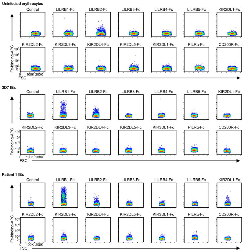
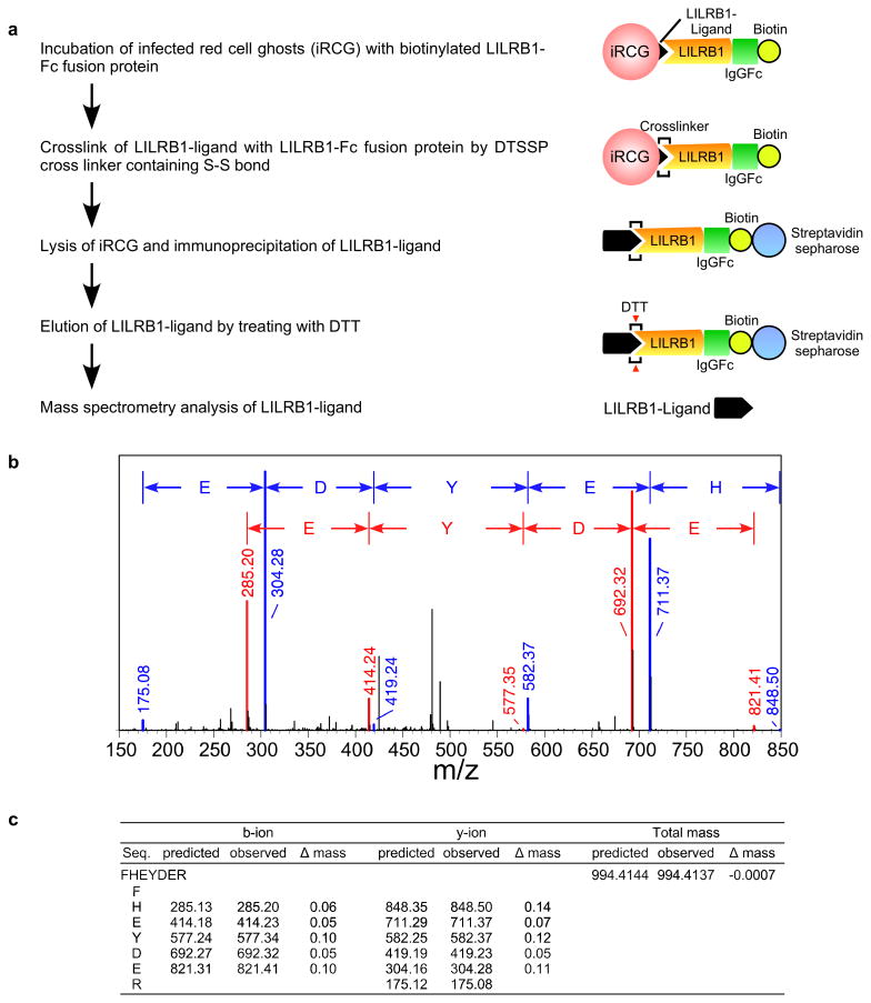
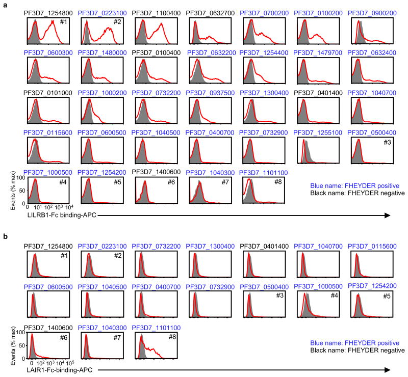
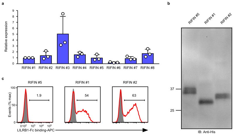
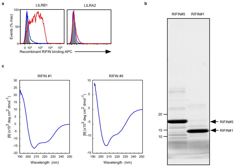
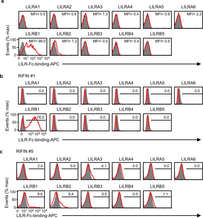
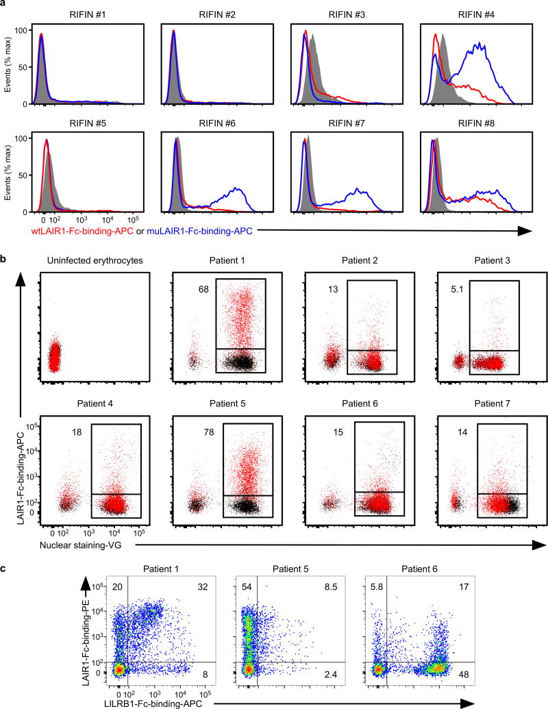
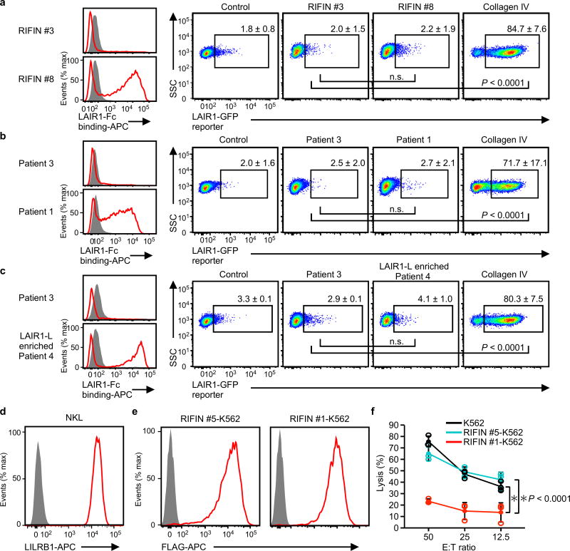
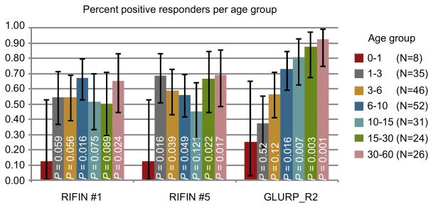
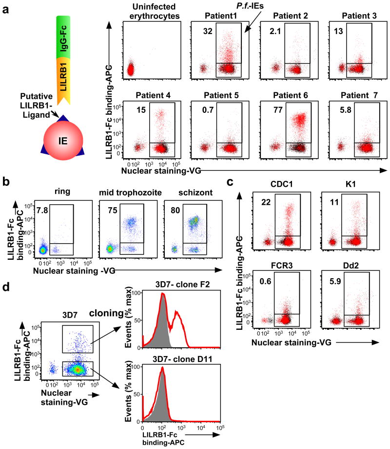
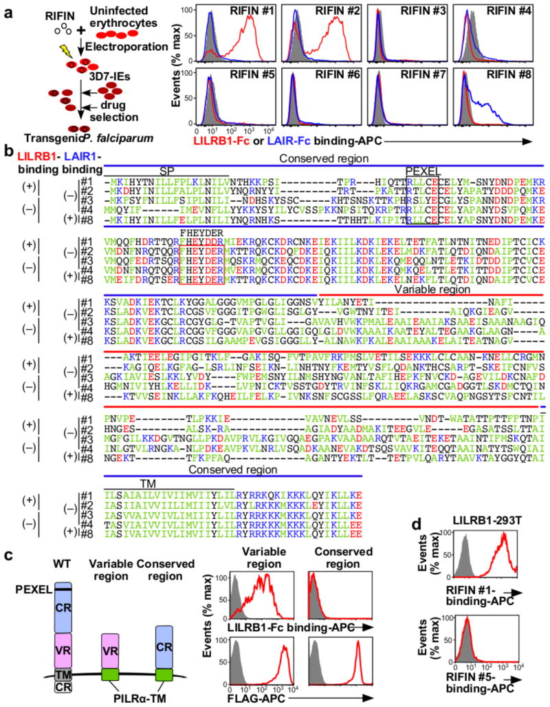
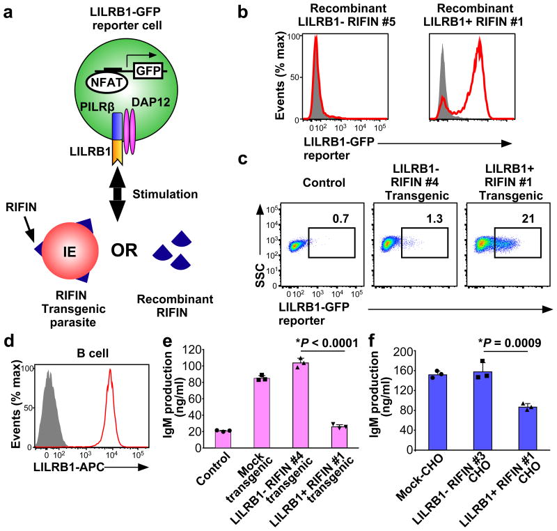
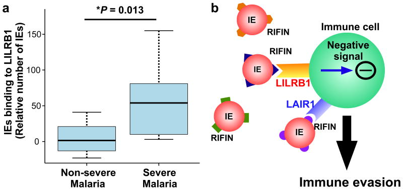
Malaria is among the most serious infectious diseases affecting humans, accounting for approximately half a million deaths each year. Plasmodium falciparum causes most life-threatening cases of malaria. Acquired immunity to malaria is inefficient, even after repeated exposure to P. falciparum, but the immune regulatory mechanisms used by P. falciparum remain largely unknown. Here we show that P. falciparum uses immune inhibitory receptors to achieve immune evasion. RIFIN proteins are products of a polymorphic multigene family comprising approximately 150-200 genes per parasite genome that are expressed on the surface of infected erythrocytes. We found that a subset of RIFINs binds to either leucocyte immunoglobulin-like receptor B1 (LILRB1) or leucocyte-associated immunoglobulin-like receptor 1 (LAIR1). LILRB1-binding RIFINs inhibit activation of LILRB1-expressing B cells and natural killer (NK) cells. Furthermore, P. falciparum-infected erythrocytes isolated from patients with severe malaria were more likely to interact with LILRB1 than erythrocytes from patients with non-severe malaria, although an extended study with larger sample sizes is required to confirm this finding. Our results suggest that P. falciparum has acquired multiple RIFINs to evade the host immune system by targeting immune inhibitory receptors.


Figures

Extended Data Figure 1
Extended Data Figure 1. Binding of Fc fusion proteins of inhibitory receptors to IEs
Erythrocytes infected with 3D7 (3D7 IEs), P. falciparum obtained from patient 1 (Patient 1 IEs), or uninfected erythrocytes were stained with Fc fusion proteins of inhibitory receptors. As a control, IEs were stained with APC-labelled anti-human IgG Fc Abalone (Control). FSC: Forward-scattered light. The experiments were replicated twice.
Extended Data Figure 2
Extended Data Figure 2. Variability and stability of LILRB1 binding to IEs and LILRB1 reporter activity
a, LILRB1 binding to schizont-stage P. falciparum-infected erythrocytes from patients with malaria in Figure 1a. b, LILRB1 binding to P. falciparum-infected erythrocytes at the ring, mid-trophozoite and schizont stages of P. falciparum-infected erythrocytes derived from patient 6 in Figure 1b. c, LILRB1 binding to schizont-stage P. falciparum-infected erythrocytes from the laboratory strains CDC1, K1, FCR3 and Dd2 shown in Figure 1c. d, GFP expression in LILRB1-expressing reporter cells upon stimulation with P. falciparum-infected erythrocytes in Figure 3c. Data represent the mean ± s.d. of three independent experiments. e, Proportions of LILRB1-Fc- binding erythrocytes infected with clone 3D7-F2 were analysed during 5 weeks of culture. The experiment was performed once.
Extended Data Figure 3
Extended Data Figure 3. Identification of the LILRB1-ligand
a, Diagram of LILRB1-ligand identification. A putative LILRB1 ligand was immunoprecipitated from IE ghosts using an LILRB1-Fc fusion protein and was identified using mass spectrometry analysis. b, Mass spectrometry of LILRB1-Fc immunoprecipitates. The observed m/z values of b-ions (red) and y-ions (blue) in the MS/MS spectra of the peptide FHEYDER present in RP-HPLC fractions of trypsin-digests of LILRB1 precipitates from IEs infected with F2 clones. The experiments were replicated twice. c, The observed m/z values of b-ions and y-ions in the MS/MS spectra of the peptide FHEYDER present in RP-HPLC fractions of trypsin-digests of LILRB1 precipitates. The predicted m/z values are shown for comparison. The differences between the m/z values for observed ions and the predicted values are shown.
Extended Data Figure 4
Extended Data Figure 4. Screening of RIFINs that bound the LILRB1-Fc or LAIR1-Fc fusion protein
a, IEs of 3D7 carrying RIFIN-transgenes were stained with the LILRB1-Fc fusion protein. RIFIN-transgenes are indicated in the figure. Red and shaded histograms indicate staining with LILRB1-Fc and control-Fc fusion proteins, respectively. b, IEs of 3D7 carrying RIFIN-transgenes were stained with the LAIR1-Fc fusion protein. RIFIN-transgenes are indicated in the figure. Red and shaded histograms indicate staining with LAIR1-Fc and control-Fc fusion proteins, respectively. Presence of the FHEYDER sequence in each RIFIN is indicated in the figure. Representative data from independent analyses are shown. Therefore, the proportions of IEs bound to Fc-fusion proteins and the levels of Fc-fusion protein binding IEs may not be comparable among different RIFINs. All experiments were replicated twice.
Extended Data Figure 5
Extended Data Figure 5. Expression of RIFINs in transgenic malaria parasites
a, The rif transgene transcript levels normalised to the internal control gene. The average of RIFIN #1 transcript levels was defined as 1. Data represent the mean ± s.d. (n = 3 technically independent samples). RIFIN #6 expression was lowest among the transgenes. However, cell surface expression of RIFIN #6 was detected using a mutated LAIR1-Fc fusion protein (Extended Data Fig. 8a), indicating that all the transgenes were sufficiently expressed at the transcript level. b, Western blot analysis of the expression of transfected C-terminally His-tagged RIFINs transfected in malaria parasites using an anti-His-tag mAb. The His-tagged RIFINs were detected at approximately equal levels (Supplementary Data). The expected molecular masses are 31.7 (RIFIN #1), 32.9 (RIFIN #2) and 34.8 (RIFIN #5) kDa. The experiment was performed once. c, P. falciparum-infected erythrocytes expressing C-terminally His-tagged RIFIN-transgenes RIFIN #1: PF3D7_1254800, RIFIN #2: PF3D7_0223100 and RIFIN #5: PF3D7_1254200 were stained with LILRB1-Fc (red) and control-Fc (shaded). The experiments were replicated at least twice.
Extended Data Figure 6
Extended Data Figure 6. Recombinant RIFINs
a, Binding of recombinant RIFINs to LILRB1 that was produced using a wheat germ cell-free protein expression system. 293T cells expressing transfected LILRB1 or LILRA2 were stained with recombinant His-tagged RIFINs that were produced using a wheat germ cell-free protein expression system. LILRA2 is an activating counterpart of LILRB1 and was used as a control. Red and blue histograms indicate binding of LILRB1+RIFIN #1 and LILRB1−RIFIN #5, respectively. The shaded histogram represents an unstained control. The experiments were replicated at least twice. b, Production of recombinant RIFINs in Escherichia coli.N-terminal-His-tagged variable regions of RIFINs were expressed in E. coli and purified using TALON metal-affinity chromatography. Recombinant RIFINs were analysed using SDS-PAGE and Oriole staining. LILRB1+RIFIN #1 and LILRB1−RIFIN #5 are shown on the right and left, respectively. The experiments were replicated at least twice. c, Circular dichroism (CD) spectra of recombinant RIFINs. Refolded and purified recombinant RIFIN #1 and #5 were subjected to CD spectral analysis. The spectra are shown as the mean residue ellipticity after subtracting the solvent background. RIFINs #1 and #5 exhibited CD spectra typical for well-folded proteins with α-helix (208 nm +222 nm) and β-sheet (215 nm) structures. Prediction of the secondary structures of each RIFIN using the BeStSel server (http://bestsel.elte.hu/index.php) yielded α/β values of ∼30%/∼10% and ∼30%/∼20% for RIFINs #1 and #5, respectively. The experiment was performed once.
Extended Data Figure 7
Extended Data Figure 7. LILRB1-binding RIFIN did not bind to other LILRs
a, The sequence encoding the variable region of LILRB1+ RIFIN #1 was transfected into 293T cells, and the transfectants were stained with LILR-Fc fusion proteins. The levels of LILR-Fc-binding are indicated in the figureas mean fluorescence intensities (MFIs). Control indicates fluorescence of cells reacted only with the secondary antibody. The experiment was performed once. b and c, Binding of LILR-Fc fusion proteins to IEs. RIFIN #1- and RIFIN #5-transgenic IEs were stained with 11 LILR-Fc fusion proteins and analysed using flow cytometry. Control indicates fluorescence of cells reacted only with the secondary antibody. The proportions of IEs stained with LILR-Fc fusion proteins are shown. The experiment in b was replicated twice and experiment in c was performed once.
Extended Data Figure 8
Extended Data Figure 8. Binding of wild-type and mutated LAIR1 to IEs
a, IEs from the RIFIN-transgenic parasites RIFIN #1: PF3D7_1254800, RIFIN #2: PF3D7_0223100, RIFIN #3: PF3D7_0500400, RIFIN #4: PF3D7_1000500, RIFIN #5: PF3D7_1254200, RIFIN #6: PF3D7_1400600, RIFIN #7: PF3D7_1040300 and RIFIN #8: PF3D7_1101100 were stained with wild-type LAIR1-Fc (wtLAIR1-Fc, red), mutated LAIR1-Fc (muLAIR1-Fc, blue), and control-Fc (shaded histogram) fusion proteins. The experiments were replicated twice. b, LAIR1-Fc bound to erythrocytes infected with P. falciparum derived from Thai patients with malaria. Schizont-stage erythrocytes infected with P. falciparum from patients with malaria and uninfected erythrocytes were stained with LAIR1-Fc (red dot) and control-Fc (black dot) fusion proteins, followed by Vybrant Green(VG) staining. Percentages of LAIR1-ligand-expressing IEs are shown. The experiments were replicated at least twice. c, Patterns of LILRB1 and LAIR1 binding to IEs derived from Thai patients with malaria. Schizont-stage IEs derived from Thai patients with malaria were stained with LAIR1-Fc (vertical) and LILRB1-Fc (horizontal) fusion proteins, followed by VG staining. VG-positive cells were analysed. The percentages of LAIR1-ligand single-positive, LILRB1-ligand single-positive and LAIR- and LILRB1-ligand double-positive IEs. The experiments were replicated twice.
Extended Data Figure 9
Extended Data Figure 9. Functional analysis of cells expressing LAIR1 and LILRB1
a–c, Erythrocytes infected with LAIR1-Fc-binding parasites (RIFIN #8 transgenic parasites [a], parasites from Thai patient 1 [b], LAIR1-Fc-binding parasites enriched by cell sorting from Thai patient isolate 4 [LAIR1-L enriched patient 4, c]) or erythrocytes infected with parasites that did not bind LAIR1-Fc (RIFIN #3 transgenic parasites [a], parasites from Thai malaria patient 3 [b, c]) were prepared (left histogram). LAIR1-reporter cells were co-cultured with these IEs and their expression levels in reporter cells were analysed using flow cytometry (right dot plots). Immobilised collagen IV served as a positive control for LAIR1-reporter activation. Proportions of GFP-expressing cells are shown as the mean ± s.d. (n = 3 biologically independent samples, one-way ANOVA with Tukey's post hoc test).d, LILRB1 expressed by the NK cell line NKL. e, FLAG-tagged RIFINs were expressed by K562 cells stably transfected with LILRB1+ RIFIN #1 or LILRB1− RIFIN #5. f, RIFIN-K562 cells or parental K562 cells were used as targets for NKL. The E:T ratio indicates the Effector: Target ratio. Data represent the mean ± s.d. (n = 3 technically independent samples). *P < 0.05, two-way ANOVA with Tukey's post hoc test. SSC: Side-scattered light. The experiments in d and e were replicated at least twice.
Extended Data Figure 10
Extended Data Figure 10. Age dependence of the antibody response to LILRB1+ and LILRB1− RIFINs
Plasma IgG positivity for the recombinant proteins comprising the variable regions of LILRB1+ RIFIN #1 and LILRB1− RIFIN #5 as well as GLURP_R2 in 222 Tanzanian individuals divided into age groups. Error bars represent 95% CI.P values were calculated using logistic regression comparing percent responders among children aged 0-1 years to children of the other age groups.
Figure 1
Figure 1. Plasmodium falciparum-infected erythrocytes (IEs) are recognised by LILRB1
a, Analysis of IEs with an LILRB1-Fc fusion protein. Diagram of LILRB1-Fc binding to IEs. Schizont-stage P. falciparum IEs and uninfected erythrocytes were stained with LILRB1-Fc (red dot) and control-Fc (black dot), followed by Vybrant Green (VG) staining. Percentages of LILRB1-binding IEs are shown. b, Different stages of P. falciparum IEs derived from patient 6 were stained with LILRB1-Fc, followed by VG staining. Percentages of LILRB1-positive IEs are shown. c, Schizont-stage IEs from four P. falciparum laboratory strains were stained with LILRB1-Fc (red) and control-Fc (shaded), followed by VG staining. Percentages of LILRB1-binding IEs are shown. d, LILRB1-binding clone 3D7 (F2) and non-binding clone 3D7 (D11). Red and shaded histograms indicate staining with LILRB1-Fc and control-Fc, respectively. Data represent at least three independent experiments and the variabilities of data shown a, b and c are shown in Extended Data Figure 2a, 2b and 2c, respectively.
Figure 2
Figure 2. RIFINs are ligands for LILRB1 and LAIR1
a, Diagram of the method used to generate transgenic P. falciparum. LILRB1-Fc and LAIR1-Fc bind IEs expressing RIFIN-transgenes. RIFIN-transgenic parasites are RIFIN #1: PF3D7_1254800, RIFIN #2: PF3D7_0223100, RIFIN #3: PF3D7_0500400, RIFIN #4: PF3D7_1000500, RIFIN #5: PF3D7_1254200, RIFIN #6: PF3D7_1400600, RIFIN #7: PF3D7_1040300 and RIFIN #8: PF3D7_1101100. Red, blue and shaded histograms indicate staining with LILRB1-Fc, LAIR1-Fc and control-Fc, respectively. b, Amino acid sequence alignments of RIFINs that bind LILRB1, LAIR1 or neither. SP and TM indicate the signal peptide and transmembrane domains, respectively. Green, blue, red and black characters indicate hydrophobic, basic, acidic and neutral amino acid residues, respectively. The FHEYDER sequence is contained in the red box. c, N-terminally FLAG-tagged conserved or variable regions of LILRB1+ RIFIN #1 fused to the PILRα transmembrane region (TM) expressed by 293T cells were tested for LILRB1-Fc binding (upper panel). RIFIN expression was detected using an anti-FLAG antibody (lower panel). Red and shaded histograms indicate RIFIN and mock (GFP) transfectants, respectively. d, LILRB1-transfected 293T cells were stained with His-tagged LILRB1+ RIFIN #1 or LILRB1− RIFIN #5, followed by staining with an anti-His antibody. Data represent at least three independent experiments.
Figure 3
Figure 3. Inhibition of LILRB1-expressing B cells by RIFIN
a, Diagram of the LILRB1 NFAT-GFP reporter assay. b, GFP expression in LILRB1-reporter cells upon treatment with recombinant RIFIN. Red histograms indicate treatment with recombinant LILRB1+ RIFIN#1 or LILRB1− RIFIN#5. Shaded histogram areas represent medium alone. c, GFP expression in LILRB1-reporter cells upon treatment with IEs expressing the LILRB1+ RIFIN #1 or LILRB1− RIFIN #4 transgene. Percentages of GFP-expressing cells are shown. SSC: Side-scattered light. d, Red and shaded histograms indicate staining of primary human B cells from a healthy donor with an anti-LILRB1 antibody and control, respectively. e, Inhibition of human immunoglobulin M (IgM) production in PBMCs by IEs. Human PBMCs were co-cultured with IEs, and IgM was measured in culture supernatants (mean ± s.d.). Transgenic malarial parasites expressing LILRB1+ RIFIN #1, LILRB1− RIFIN #4 or mock (GFP) are shown. Control indicates PBMCs alone. n = 3 technically independent samples. f, Inhibition of human IgM production by RIFIN-transfected CHO cells. PBMCs were co-cultured with CHO cells expressing LILRB1+ RIFIN #1, LILRB1− RIFIN #3 and an unrelated gene (MDA5). Data represent the mean ± s.d. (n = 3 technically independent samples). *P< 0.05 (one-way ANOVA with Tukey's post hoc test). Data represent at least three independent experiments, and the variability of data presented in c is shown in Extended Data Fig.2d.
Figure 4
Figure 4. Binding of Plasmodium falciparum-infected erythrocytes to LILRB1 is associated with severe malaria
a, Binding of IEs from Tanzanian patients with malaria diagnosed with cerebral malaria, severe anaemia or both (severe malaria, n = 9) or non-cerebral or non-severe anaemia (non-severe malaria, n = 30) to LILRB1-Fc, relative to control-Fc (median, 75 and 95 percentiles and two-sided Mann–Whitney U test). b, Diagram of the suggested mechanism of immune evasion. P. falciparum induces the expression of RIFINs on the surface of IEs. Individual RIFINs may have evolved to target host inhibitory receptors, thus facilitating escape from host immune systems, which may lead to inefficient development of immunity.

References

    1. World Health Organization. World malaria report 2016. World Health Organization; 2016.
    1. Langhorne J, Ndungu FM, Sponaas AM, Marsh K. Immunity to malaria: more questions than answers. Nat Immonol. 2008;9:725–732. - PubMed
    1. Joannin N, Abhiman S, Sonnhammer EL, Wahlgren M. Sub-grouping and sub-functionalization of the RIFIN multi-copy protein family. BMC Genomics. 2008;9:19. - PMC - PubMed
    1. Gomes PS, Bhardwaj J, Rivera-Correa J, Freire-De-Lima CG, Morrot A. Immune Escape Strategies of Malaria Parasites. Front Microbiol. 2016;7:1617. - PMC - PubMed
    1. Renia L, Goh YS. Malaria Parasites: The Great Escape. Front Immunol. 2016;7:463. - PMC - PubMed

Publication types

MeSH terms

Substances