

Histopathological diagnosis of small melanocytic lesions suspicious for malignant melanoma
- PMID: 29186251
- PMCID: PMC5514579
- DOI: 10.1590/abd1806-4841.20175169
Histopathological diagnosis of small melanocytic lesions suspicious for malignant melanoma
Abstract
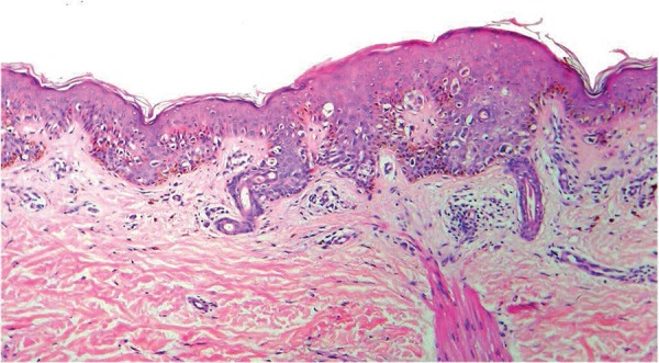
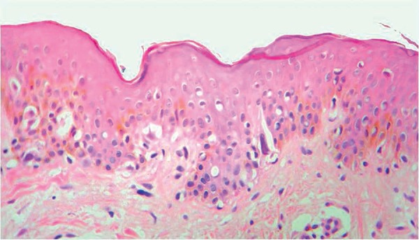
The concern about malignant skin neoplasms leads to the excision of smaller lesions. This study on small melanocytic lesions aims to evaluate the range of possible histopathological diagnoses, describe histopathological aspects, and assess the usefulness of serial histological sections. We performed a cross-sectional descriptive histopathological study examining 76 pigmented skin lesions up to 6 mm in diameter. Histopathological diagnoses included atypical melanocytic nevi (n=38), common melanocytic nevi (n=18), atypical lentiginous melanocytic hyperplasia with architectural features of atypical melanocytic nevi (n=7), lentigo simplex (n=2), and malignant melanoma (n=1). Ten cases were non-diagnostic. Cytological atypia was not an exclusive finding of atypical lesions. Examination of serial sections did not change histopathological impression. Early detection of malignant melanoma is important, but clinical and dermoscopy exams may be leading to the resection of a great number of benign lesions. Strict attention to histopathological criteria results in a large number of non-diagnostic cases.
Conflict of interest statement
Conflict of interest: None
Figures
Similar articles
-
Role of In Vivo Reflectance Confocal Microscopy in the Analysis of Melanocytic Lesions.Acta Dermatovenerol Croat. 2018 Apr;26(1):64-67. Acta Dermatovenerol Croat. 2018. PMID: 29782304 Review.
-
Atypical melanocytic nevi of the genital type with a discussion of reciprocal parenchymal-stromal interactions in the biology of neoplasia.Hum Pathol. 1998 Jan;29(1 Suppl 1):S1-24. doi: 10.1016/s0046-8177(98)80028-2. Hum Pathol. 1998. PMID: 9445124
-
Correlation between dermoscopic and histopathological diagnoses of atypical nevi in a dermatology outpatient clinic of the Medical School of São José do Rio Preto, SP, Brazil.An Bras Dermatol. 2013 Mar-Apr;88(2):199-203. doi: 10.1590/S0365-05962013000200002. An Bras Dermatol. 2013. PMID: 23739709 Free PMC article.
-
Benign melanocytic lesions: risk markers or precursors of cutaneous melanoma?J Am Acad Dermatol. 1995 Dec;33(6):1000-7. doi: 10.1016/0190-9622(95)90294-5. J Am Acad Dermatol. 1995. PMID: 7490345
-
Melanocytic aggregation in the skin: diagnostic clues from lentigines to melanoma.Dermatol Clin. 2007 Jul;25(3):303-20, vii-viii. doi: 10.1016/j.det.2007.04.007. Dermatol Clin. 2007. PMID: 17662896 Review.
Cited by
-
Melanocytic lesions ≤ 6mm: Prospective series of 481 melanocytic trunk and limb lesions in Brazil.PLoS One. 2021 Jun 8;16(6):e0252162. doi: 10.1371/journal.pone.0252162. eCollection 2021. PLoS One. 2021. PMID: 34101726 Free PMC article.
-
The "Virtual Biopsy" of Cancerous Lesions in 3D: Non-Invasive Differentiation between Melanoma and Other Lesions Using Vibrational Optical Coherence Tomography.Dermatopathology (Basel). 2021 Dec 13;8(4):539-551. doi: 10.3390/dermatopathology8040058. Dermatopathology (Basel). 2021. PMID: 34940035 Free PMC article.
References
-
- Tran KT, Wright NA, Cockerell CJ. Biopsy of the pigmented lesion--when and how. J Am Acad Dermatol. 2008;59:852–871. - PubMed
-
- Cohen DE, Sangueza OP, Fass E, Stiller MJ. In vivo cutaneous surface microscopy: revised nomenclature. Int J Dermatol. 1993;32:257–258. - PubMed
-
- Bafounta ML, Beauchet A, Aegerter P, Saiag P. Is dermoscopy (epiluminescence microscopy) useful for the diagnosis of melanoma? Results of a meta-analysis using techniques adapted to the evaluation of diagnostic tests. Arch Dermatol. 2001;137:1343–1350. - PubMed
-
- Kittler H, Pehamberger H, Wolff K, Binder M. Diagnostic accuracy of dermoscopy. Lancet Oncol. 2002;3:159–165. - PubMed
-
- Carli P, De Giorgi V, Chiarugi A, Nardini P, Mannone F, Stante M, et al. Effect of lesion size on the diagnostic performance of dermoscopy in melanoma detection. Dermatology. 2003;206:292–296. - PubMed
MeSH terms
LinkOut - more resources
Full Text Sources
Other Literature Sources
Medical