

doi: 10.1590/abd1806-4841.20176018.
White piedra, black piedra, tinea versicolor, and tinea nigra: contribution to the diagnosis of superficial mycosis
Affiliations
- PMID: 29186263
- PMCID: PMC5514591
- DOI: 10.1590/abd1806-4841.20176018
Item in Clipboard
White piedra, black piedra, tinea versicolor, and tinea nigra: contribution to the diagnosis of superficial mycosis
An Bras Dermatol.
2017 May-Jun.
Abstract
Superficial mycoses are fungal infections restricted to the stratum corneum and to the hair shafts, with no penetration in the epidermis; they are: white piedra, black piedra, tinea versicolor, and tinea nigra. This study presents images of mycological tests performed in the laboratory, as well as exams performed at the authors office, in order to improve the dermatologist's knowledge about the diagnosis of these dermatoses, which are common in many countries.
Conflict of interest statement
Conflict of interest: None
Figures
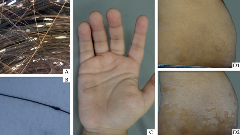
Clinical appearance of actual superficial mycoses. (A) White
piedra: whitish nodule attached to the hair shaft. (B) Black
piedra: darkened nodule attached to the hair shaft. (C) Tinea
nigra: brownish macula on children’s palms. (D) Pityriasis
versicolor: scattered maculas on the abdomen (D1), which become
more evident after skin stretching (Zireli’s sign – D2)
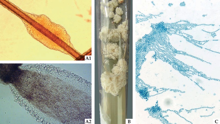
Mycological examinations of white piedra: (A1) Optical
microscopy (x40) offering a detailed illustration of the light color nodule
attached to the pillar shaft. (A2) Optical microscopy (x100)
illustrates the yeasts the make up the structure on the edge of the nodule.
(B) Culture Mycosel medium (Difco, USA) with yeast-like
colony, with the cerebriform filamentous appearance. (C)
Microgrowth demonstrates yeasts with blasto-arthrospores, typical of
Trichosporon sp
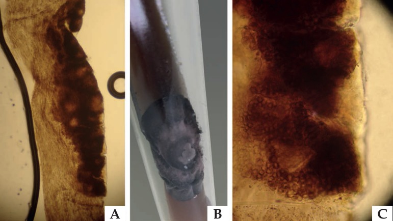
Mycological examinations of black piedra: (A) Optical microscopy
(x40) offering a detailed illustration of the dark nodule attached to the
pillar shaft. (B) Culture Mycosel medium (Difco, USA) with
dematiaceous colony. (C) Optical microscopy (x100) identifying
the ascus, round structures typical of parasitism caused by Piedraia
hortae
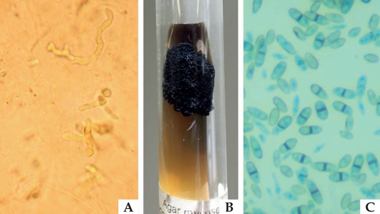
Mycological examinations for tinea nigra: (A) Direct mycological
examination of a sample collected through skin lesion scraping, clarified
with KOH 10%, illustrating dematiaceous septate hyphae. (B)
Culture Mycosel medium (Difco, USA) with dematiaceous colony with a waxy
appearance. (C) Microgrowth revealing dematiaceous yeasts with
binary fission, typical of Hortaea werneckii
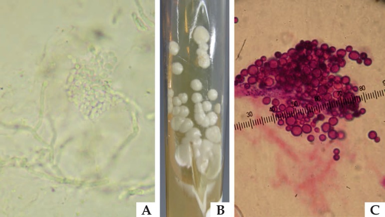
Mycological examinations for pityriasis versicolor: (A) Direct mycological
examination of a sample collected through skin lesion scraping, clarified
with KOH 10%, illustrating yeasts grouped in a “grape bunch” format, and of
short and thick pseudo-hyphae. (B) Sabouraud agar culture, enriched with
olive oil, with beige yeast-like colony. (C) Yeasts grouped with short base
single budding, with “bowling pin” appearance, stained by the hematoxylin
eosin method, typical of Malassezia sp microgrowth
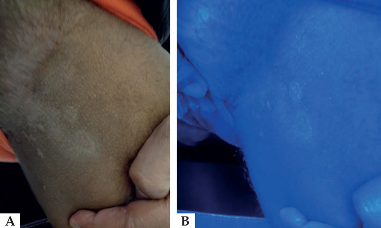
Patient exhibiting pityriasis versicolor lesions in the inguinal region
(A), under Wood lamp, reveals silver fluorescence
(B)
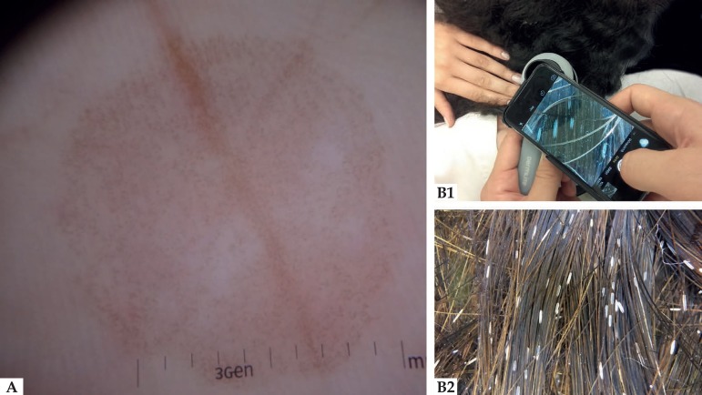
Dermatoscopy of tinea nigra palmar lesion under polarized light
(A) and polarized light dermatoscopy performed on child
with whitish nodules attached to the hair (B1), illustrating
structures that are similar to those viewed in optical microscopy
(B2)
References
-
- Sociedade Brasileira de Dermatologia Perfil nosológico das consultas dermatológicas no Brasil. An Bras Dermatol. 2006;81:549–558.
-
- Zaitz C, Campbell I, Marques SA, Ruiz LRB, Framil VMS. Compêndio de Micologia Médica. 2.ed. Rio de Janeiro: Guanabara Koogan; 2010.
-
- Odds FC, Arai T, Disalvo AF, Evans EG, Hay RJ, Randhawa HS, et al. Nomenclature of fungal diseases: a report and recomendations from a Sub-Committee of the International Society for Human and Animal Mycology (ISHAM) J Med Vet Mycol. 1992;30:1–10. - PubMed
-
- Bonifaz A, Gómez-Daza F, Paredes V, Ponce RM. Tinea versicolor, tinea nigra, white piedra, and black piedra. Clin Dermatol. 2010;28:140–145. - PubMed
-
- Schwartz RA. Superficial fungal infections. Lancet. 2004;364:1173–1182. - PubMed
MeSH terms
LinkOut - more resources
Full Text Sources
Other Literature Sources
Medical