. 2018 Apr 1;59(4):756-764.
doi: 10.1093/pcp/pcx187.

Type-B ARRs Control Carpel Regeneration Through Mediating AGAMOUS Expression in Arabidopsis

Affiliations

Type-B ARRs Control Carpel Regeneration Through Mediating AGAMOUS Expression in Arabidopsis

Xiao Fei Rong et al. Plant Cell Physiol. .

Erratum in

  • Erratum.
    [No authors listed] [No authors listed] Plant Cell Physiol. 2018 Apr 1;59(4):876. doi: 10.1093/pcp/pcy075. Plant Cell Physiol. 2018. PMID: 29718476 Free PMC article. No abstract available.

Abstract

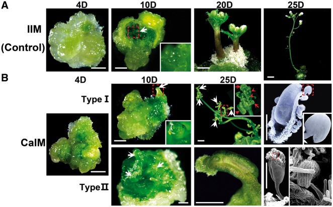
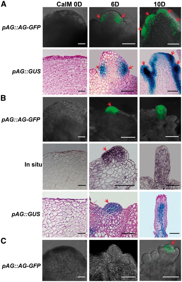
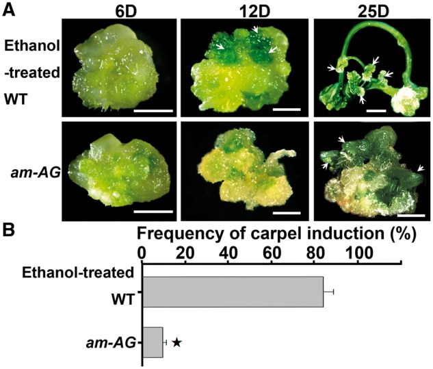
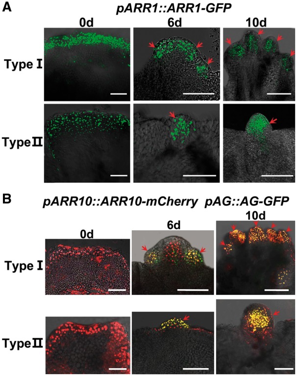
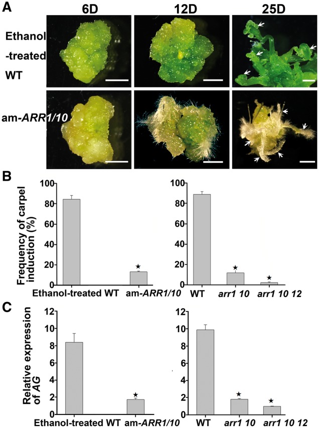
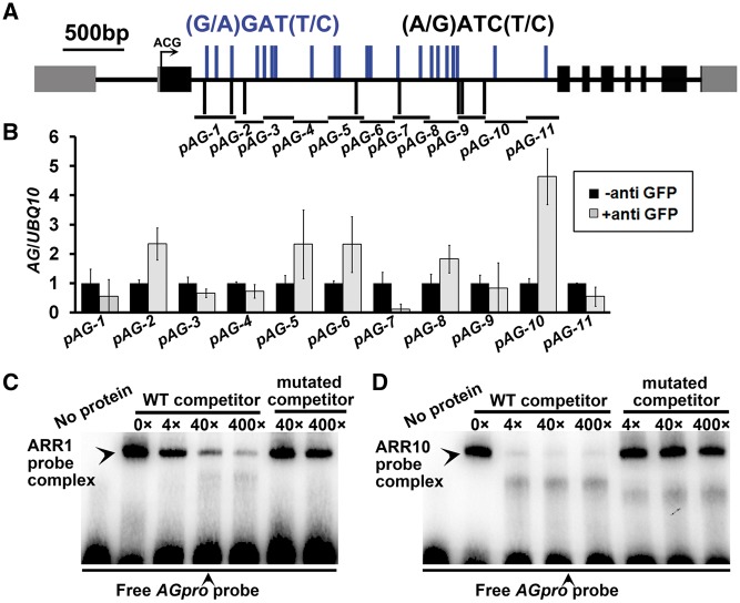
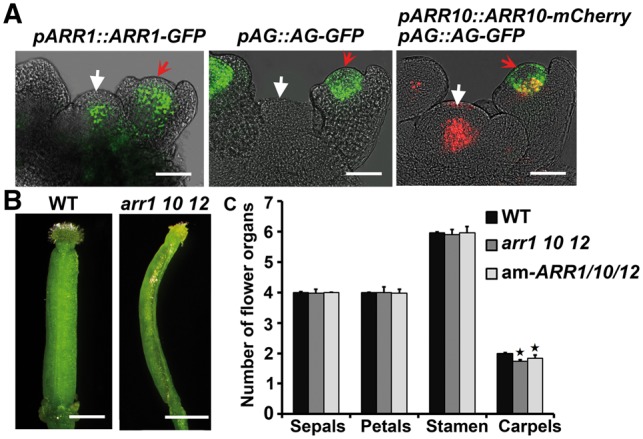
Plants are known for their capacity to regenerate organs, such as shoot, root and floral organs. Recently, a number of studies contributed to understanding the mechanisms of shoot and root regeneration. However, the mechanisms underlying floral organ regeneration are largely unknown. In this study, we established a carpel regeneration system in which two types of carpels were induced by exogenous cytokinin. For type I, all the floral organs in the regenerated inflorescence were transformed into carpels. For type II, carpels were generated directly from callus. The transcript level of AGAMOUS (AG), the carpel identity gene, was up-regulated during carpel induction. The expression signals of AG were detected in the initiating carpel primordia and regenerating carpels, and co-localized with those of two Type-B ARABIDOPSIS RESPONSE REGULATORs (ARRs), ARR1 and ARR10. Repression of either AG or type-B ARRs reduced carpel regeneration. Binding analyses showed that ARR1 and ARR10 directly bound to transcriptional regulatory regions of AG and positively regulated its expression. In addition, the expression of type-B ARRs overlapped with that of AG in the floral primordia in planta. Defects in type-B ARRs reduced the number of carpels. The results indicate that type-B ARRs control carpel regeneration through activating AG expression. Our results provide new information for understanding the mechanism of carpel formation.


Figures

Fig. 1
Fig. 1
Morphology of regenerated carpels induced by cytokinin. (A) Inflorescence regeneration procedure as control. Calli were cultured on inflorescence induction medium (IIM) for 4, 10, 20 and 25 d. The white arrow indicates the inflorescence meristem initiation site. (B) Carpel regeneration procedures. Calli were cultured on carpel induction medium (CaIM) for 4, 10, 20 and 25 d. For type I regeneration (upper), inflorescence regeneration initiated at 10 CaIM. Carpels were generated at floral organ primordia of regenerated inflorescences. For type IIregeneration (lower), carpels initiated directly from calli. White arrows indicate regenerated carpels; red arrows indicate formation of ovules. Scanning electron microscope images show structures of ovules (upper) and stigma (lower) of regenerated carpels. Dotted boxes indicate regions magnified in insets. Scale bars = 2 mm. ‘D’ indicates the number of days of culture on IIM or CaIM.
Fig. 2
Fig. 2
AG expression patterns during carpel regeneration. (A) Expression patterns of pAG::AG-GFP and pAG::GUS during type I carpel regeneration. Calli were cultured in CaIM for 0, 6 and 10 d. Red arrows indicate carpel primordial and regenerating carpels. (B) Distribution pattern of pAG::AG-GFP and pAG::GUS, and in situ hybridization of AG during type II regeneration. Calli were cultured on CaIM for 0, 6 and 10 d. Red arrows indicate carpel primordia. (C) Expression pattern of pAG::AG-GFP during inflorescence induction as control. The red arrow indicates GFP signals in the central region of the floral primordium. Calli were cultured on IIM for 0, 6 and 10 d. Scale bars = 100 μm. ‘D’ indicates the number of days of culture on IIM or CaIM.
Fig. 3
Fig. 3
AG is required for carpel regeneration. (A) Carpel induction from wild-type and am-AG transgenic calli. Calli were cultured on CaIM for 6, 12 and 25 d. Arrows indicate initiation of carpel regeneration and regenerating carpels from wild-type callus, and regeneration of leaf-like structures from am-AG transgenic callus. Scale bars = 2 mm. ‘D’ indicates the number of days of culture on CaIM. (B) Frequency of carpel regeneration in (A). Error bars indicate tjhe SDs of three biological replicates. The star indicates P < 0.01 (Student’s t-test).
Fig. 4
Fig. 4
Overlapping expression patterns of type-B ARRs and AG during carpel regeneration. (A) Expression of pARR1::ARR1-GFP during carpel regeneration. (B) Expression patterns of pARR10::ARR10-mCherry (red) and pAG::AG-GFP (green) in the double reporter line. Expression signals of ARR10 overlapped with that of AG in carpel primordia and regenerating carpels (yellow). Calli were cultured on CaIM for 0, 6 and 10 d. Red arrows indicate carpel primordial and regenerating carpels. Scale bars = 100 μm. ‘D’ indicates the number of days of culture on CaIM.
Fig. 5
Fig. 5
Type-B ARRs are involved in carpel regeneration. (A) Carpel induction from wild-type and am-ARR1/10 transgenic calli. Calli were cultured on CaIM for 6, 12 and 25 d. Arrows indicate initiation of carpel regeneration and regenerating carpels from wild-type callus, and root regeneration from am-ARR1/10 transgenic callus. Scale bars = 2 mm. ‘D’ indicates the number of days of culture on CaIM. (B) Compared with the wild type, am-ARR1/10 transgenic lines as well as arr1 10 double and arr1 10 12 triple mutants showed reduced frequencies of carpel regeneration. (C) Compared with the wild type, am-ARR1/10 transgenic lines as well as arr1 10 double and arr1 10 12 triple mutants showed reduced transcriptional levels of AG. (B, C) Error bars indicate the SDs of three biological replicates. The star indicates P < 0.01 (Student’s t-test).
Fig. 6
Fig. 6
ARR1 and ARR10 bind to the transcriptional regulatory region of AG. (A) Diagram of the AG genomic region. The arrow marks the translation start site, black boxes indicate coding sequences, gray boxes indicate untranslated regions and black bold lines indicate intron/intergenic regions. pAG-1 to pAG-10 indicate positions of fragments used in ChIP-qPCR analyses. Blue and the black bars indicate type-B ARR-binding elements (G/A)GAT(T/C) and (A/G)ATC(T/C), respectively. (B) ChIP analyses showing an association between ARR1 and the transcriptional regulatory region of AG. (C) EMSAs confirming binding of ARR1 and ARR10 to the transcriptional regulatory region of AG. Arrowheads above indicate band shifts (complexes of ARR proteins and probe DNA). Arrows below indicate free probes. Non-labeled oligonucleotides were used as competitors. Mutated competitors were generated by replacing two base pairs in ARR-binding elements (GATC/T to CTTC/T).
Fig. 7
Fig. 7
ARRs regulate carpel initiation in planta. (A) Expression pattern of pARR1::ARR1-GFP (green), pAG::AG-GFP (green), pARR10::ARR10-mCherry (red) and pAG::AG-GFP (green) double reporters in inflorescence meristems and floral primordia. White arrows indicate inflorescence meristems and red arrows indicate floral primordia at stage 3. ARR10 and AG signals overlapped in the central regions of the floral primordium (yellow). Scale bars = 50 μm. (B) Pistil (15.71%) from the arr1 10 12 triple mutant carpels formed only one carpel. Scale bars = 1 mm. (C) Compared with the wild type, arr1 10 12 triple mutant and am-ARR1/10 transgenic lines showed a reduced number of carpels, but not other floral organs. Error bars indicate the SDs of three biological replicates. The star indicates P < 0.01 (Student’s t-test).

References

    1. Alvarez-Buylla E.R., Benítez M., Corvera-Poiré A., Chaos Cador A., de Folter S., Gamboa, de Buen A., et al. (2010) Flower development. Arabidopsis Book 8: e0127. - PMC - PubMed
    1. An Y.R., Li X.G., Su H.Y., Zhang X.S. (2004) Pistil induction by hormones from the callus of Oryza sativa in vitro. Plant Cell Rep. 23: 448–452. - PubMed
    1. Bartrina I., Otto E., Strnad M., Werner T., Schmülling T. (2011) Cytokinin regulates the activity of reproductive meristems, flower organ size, ovule formation, and thus seed yield in Arabidopsis thaliana. Plant Cell 23: 69–80. - PMC - PubMed
    1. Bertrand C., Bergounioux C., Domenichini S., Delarue M., Zhou D.X. (2003) Arabidopsis histone acetyltransferase AtGCN5 regulates the floral meristem activity through the WUSCHEL/AGAMOUS pathway. J. Biol. Chem. 278: 28246–2851. - PubMed
    1. Bowman J.L., Drews G.N., Meyerowitz E.M. (1991) Expression of the Arabidopsis floral homeotic gene AGAMOUS is restricted to specific cell types late in flower development. Plant Cell 3: 749–758. - PMC - PubMed

MeSH terms

LinkOut - more resources