. 2017 Nov 28;21(9):2487-2499.
doi: 10.1016/j.celrep.2017.10.110.

Testosterone Attenuates Group 2 Innate Lymphoid Cell-Mediated Airway Inflammation

Affiliations

Testosterone Attenuates Group 2 Innate Lymphoid Cell-Mediated Airway Inflammation

Jacqueline-Yvonne Cephus et al. Cell Rep. .

Abstract

Sex hormones regulate many autoimmune and inflammatory diseases, including asthma. As adults, asthma prevalence is 2-fold greater in women compared to men. The number of group 2 innate lymphoid cells (ILC2) is increased in patients with asthma, and we investigate how testosterone attenuates ILC2 function. In patients with moderate to severe asthma, we determine that women have an increased number of circulating ILC2 compared to men. ILC2 from adult female mice have increased IL-2-mediated ILC2 proliferation versus ILC2 from adult male mice, as well as pre-pubescent females and males. Further, 5α-dihydrotestosterone, a hormone downstream of testosterone, decreases lung ILC2 numbers and IL-5 and IL-13 expression from ILC2. In vivo, testosterone attenuated Alternaria-extract-induced IL-5+ and IL-13+ ILC2 numbers and lung eosinophils by intrinsically decreasing lung ILC2 numbers, as well as by decreasing expression of IL-33 and thymic stromal lymphopoietin (TSLP), ILC2-stimulating cytokines. Collectively, these findings provide a foundational understanding of sexual dimorphism in ILC2 function.

Keywords: asthma; innate lymphoid cells; sex hormones; testosterone.


Figures

Figure 1
Figure 1. Circulating ILC2 are increased in women compared to men with asthma
Blood was collected from men and women with moderate to severe asthma or healthy controls. (A) Representative dot plots from a woman and man with asthma showing flow cytometry gating strategy for circulating ILC2, defined as Lin-, CD45+, CD127+, CD161+, CD25+, and CRTH2+ cells. (B) ILC2 as a percentage of PBMCs/ml of blood. (C) Representative histograms of Gata3+ ILC2 from woman with asthma (red outline) compared to man with asthma (blue outline). Gray peak represents isotype control. (D–E). Gata3+ ILC2 as a percentage of PBMCs/ml of blood and GATA3 MFI. (F–H). ILC2 were restimulated with PMA, ionomycin and golgi-stop to determine IL-5 production. (F) Representative dot plots from a woman and man with asthma. (G). Representative histograms of IL-5+ ILC2, gray peak represents isotype control. (H). IL-5+ ILC2 as a percentage of PBMCs/ml of blood. Data are mean ± SEM, n=4 healthy women or men, n=6 women with asthma and n=7 men with asthma; * p<0.05, n.s., not significant, Kruskal-Wallis test.
Figure 2
Figure 2. Lung ILC numbers and CD25 expression are decreased in male mice compared to female mice
ILCs were sorted from the lungs of naïve adult and pre-pubescent male and female mice. (A–B) representative dot plot and quantification of sorted ILCs. (C–E) Representative histograms and quantification of CD25+ or CD127+ staining and MFI on lung ILCs. (F–I) Sorted ILCs were stimulated with IL-2 and IL-33 for 6 days. IL-5 and IL-13 protein expression as well as Gata3 and Rorα mRNA relative expression normalized to GAPDH was determined. Data are mean ± SEM n=6–9 mice per group, * p<0.05, ANOVA (B, D, E) or t-test (F–I).
Figure 3
Figure 3. IL-2-mediated ILC2 proliferation is decreased in male mice compared to female mice
(A) Representative histogram and quantitation of ILC2 proliferation over 3 days from IL-33 and IL-2 stimulated ILC2 from adult male and female mice. Gray peak represents baseline of cell tracer dye, added immediately before flow cytometry analysis. (B) Ki-67+ ILC2 from adult male and female mice measured after 3 days of stimulation with IL-2 PLUS IL-33 or IL-7 PLUS IL-33. (C) Phospho-STAT5 in ILC2 24 hours after stimulation. (D–E) Representative dot plots and quantification of ILC2 cultured for 6 days with IL-2 PLUS IL-33 or IL-7 PLUS IL-33 and restimulated to measure IL-5. Data are mean ± SEM. n=6 wells from 2 combined experiments; * p<0.05, t-test (A–B) or ANOVA with Tukey post-hoc analysis (C–F).
Figure 4
Figure 4. 5α-DHT inhibited IL-5 and IL-13 protein expression by ILC2 cells
(A–I) ILCs were sorted from the lungs of sham-operated or gonadectomized male and female mice were administered hormone or vehicle pellets for 21 days. (A) Total number of sorted ILCs. (B–D) Representative histograms and quantification of CD25+ or CD127+ staining and MFI on lung ILCs. (E–H) ILC2 were stimulated with IL-33 and IL-2 for 6 days. IL-5 and IL-13 protein expression as well as Gata3 and Rorα mRNA relative expression normalized to GAPDH was determined. (I). AR mRNA expression normalized to GAPDH on ILC2. (H–I) 5α-DHT or vehicle was added concurrently with IL-33 and IL-2 stimulation of ILCs for 6 days. IL-5 and IL-13 protein expression as well as Gata3 and Rorα mRNA relative expression normalized to GAPDH was determined. Data are mean ± SEM, n=4–5 wells from 3 combined experiments; * p<0.05, n.s., not significant, ANOVA with Tukey post-hoc analysis.
Figure 5
Figure 5. Testosterone decreased and ovarian hormones increased Alt Ext-induced airway inflammation
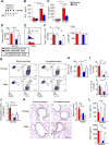
(A) Timeline for Alt Ext protocol on sham-operated and gonadectomized male and female mice. (B–C) IL-5 and IL-13 protein expression in lung homogenates of male and female mice. Data are mean ± SEM, n=5 mice; * p<0.05, ANOVA with Tukey post-hoc analysis. (D) Total number of ILC2 in the lungs 24 hours after last Alt Ext challenge. (E) Representative histogram of CD25 staining in ILC2, pregated on viable, Lin- CD45+ CD127+ ST2+ cells. Gray peak represents isotype control. (F). Quantification of CD25+ ILC2 and CD25 MFI. (G) Representative dot plots of IL-5 and IL-13 producing ILC2. (H). Quantification of ST2+ ILC2. (I). Quantification of IL-5+ ILC2 (top quadrants) and IL-13+ ILC2 (right quadrants). (J) Eosinophils in BAL fluid. (K) Representative sections at 20× magnification and quantification of PAS staining to detect mucus in lung sections on 48 hours after final challenge. Bar represents 100µm. (L) IL-33 was measured in BAL fluid of mice 1 hour following the last Alt Ext challenge and TSLP protein expression was measured in whole lung homogenates 6 hours following the last Alt Ext challenge. (D–L) Data are mean ± SEM, n=5–9 mice per group; * p<0.05, ANOVA with Tukey post-hoc analysis.
Figure 6
Figure 6. Testosterone intrinsically attenuated Alt Ext-induced lung ILC2
(A) Experimental design of mixed bone marrow chimera experiment with a 1:1 bone marrow mixture from WT (CD90.1) or ARtfm (CD90.2) adult male mice being transferred into lethally irradiated heterozygous CD90.1+ CD90.2+ WT adult male recipient mice. After 6 weeks of reconstitution, recipient mice underwent the Alt Ext protocol. (B–C) Representative gating strategy and quantification of the total number of IL-5 and IL-13+ ILC2 from WT (CD90.1) and ARtfm (CD90.2) mice. (D–F). Histogram and quantification of CD25+ ILC2 and CD25 MFI on WT and ARtfm ILC2. Gray peak represents isotype control. Data are mean ± SEM, n=6–10 mice per group; * p<0.05, n.s., not significant, ANOVA with Tukey post-hoc analysis.

References

    1. Agarwal R. Severe asthma with fungal sensitization. Curr Allergy Asthma Rep. 2011;11:403–413. - PubMed
    1. American Lung Association. Trends in Asthma Morbidity and Mortality 2012
    1. Barlow JL, Bellosi A, Hardman CS, Drynan LF, Wong SH, Cruickshank JP, McKenzie AN. Innate IL-13-producing nuocytes arise during allergic lung inflammation and contribute to airways hyperreactivity. J Allergy Clin Immunol. 2012;129:191–198. e191–194. - PubMed
    1. Bartemes KR, Kephart GM, Fox SJ, Kita H. Enhanced innate type 2 immune response in peripheral blood from patients with asthma. J Allergy Clin Immunol. 2014;134:671–678. e674. - PMC - PubMed
    1. Busse WW, Israel E, Nelson HS, Baker JW, Charous BL, Young DY, Vexler V, Shames RS Daclizumab Asthma Study, G. Daclizumab improves asthma control in patients with moderate to severe persistent asthma: a randomized, controlled trial. Am J Respir Crit Care Med. 2008;178:1002–1008. - PubMed

MeSH terms

LinkOut - more resources