Review
. 2017 Nov 24;9(12):359.
doi: 10.3390/v9120359.

Autophagy in Measles Virus Infection

Affiliations
Review

Autophagy in Measles Virus Infection

Aurore Rozières et al. Viruses. .

Abstract

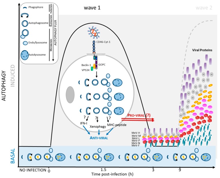
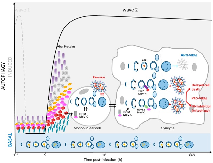
Autophagy is a biological process that helps cells to recycle obsolete cellular components and which greatly contributes to maintaining cellular integrity in response to environmental stress factors. Autophagy is also among the first lines of cellular defense against invading microorganisms, including viruses. The autophagic destruction of invading pathogens, a process referred to as xenophagy, involves cytosolic autophagy receptors, such as p62/SQSTM1 (Sequestosome 1) or NDP52/CALCOCO2 (Nuclear Dot 52 KDa Protein/Calcium Binding And Coiled-Coil Domain 2), which bind to microbial components and target them towards growing autophagosomes for degradation. However, most, if not all, infectious viruses have evolved molecular tricks to escape from xenophagy. Many viruses even use autophagy, part of the autophagy pathway or some autophagy-associated proteins, to improve their infectious potential. In this regard, the measles virus, responsible for epidemic measles, has a unique interface with autophagy as the virus can induce multiple rounds of autophagy in the course of infection. These successive waves of autophagy result from distinct molecular pathways and seem associated with anti- and/or pro-measles virus consequences. In this review, we describe what the autophagy-measles virus interplay has taught us about both the biology of the virus and the mechanistic orchestration of autophagy.

Keywords: CD46; IRGM; NDP52; OPTN; T6BP; autophagy; autophagy receptors; measles virus; selective autophagy.


Conflict of interest statement

The authors declare no conflict of interest.

Figures

Figure 1
Figure 1
Early induction of autophagy upon MeV infection. Attenuated MeV strains induce autophagy as soon as 1.5 h after infection via a CD46/GOPC (Golgi Associated PDZ and Coiled-Coil Motif Containing)-dependent pathway. This wave of autophagy could contribute to protecting the infected cells and surrounding cells from further infections due to its anti-viral potential. On the other hand, infectious MeV could use such an autophagy induction to facilitate its own replication (pro-viral). Before induction of a second wave of autophagy, which correlates with the accumulation of viral proteins, the first transient wave of autophagy ceases. Basal autophagy remains possibly efficient during the all cycles of MeV infection. Note that virulent/clinical strains of MeV, which do not bind to CD46, do not induce such an early autophagy wave. Upper left panel: schematic representation of the main steps of the complete autophagy flux process. (blue arrows = anti-viral effect; red arrows = pro-viral effect; dotted line = wave 2).
Figure 2
Figure 2
Second wave of autophagy induction upon MeV infection. After nine hours of MeV infection, a second autophagy wave is induced, which correlates with the accumulation of MeV proteins. Several successive signals seem associated with this late autophagy wave, including IRGM engagement and events associated with syncytia formation. Autophagosome maturation is required for an efficient MeV replication (pro-viral effect). This effect could involve the two autophagy adaptors NDP52 (Nuclear Dot 52 KDa Protein) and T6BP (TRAF6-Binding Protein), which are both targeted by MeV proteins. In contrast, p62 appears to oppose MeV replication (anti-viral effect). The pro-viral function of autophagy has been shown to involve a delayed death of infected cells and a decreased innate anti-viral response. (blue arrows = anti-viral effect; red arrows = pro-viral effect; dotted line = wave 1).

References

    1. Moss W.J., Griffin D.E. Measles. Lancet. 2012;379:153–164. doi: 10.1016/S0140-6736(10)62352-5. - DOI - PubMed
    1. Griffin D.E. Measles virus-induced suppression of immune responses. Immunol. Rev. 2010;236:176–189. doi: 10.1111/j.1600-065X.2010.00925.x. - DOI - PMC - PubMed
    1. Noyce R.S., Bondre D.G., Ha M.N., Lin L.T., Sisson G., Tsao M.S., Richardson C.D. Tumor cell marker pvrl4 (nectin 4) is an epithelial cell receptor for measles virus. PLoS Pathog. 2011;7:e1002240. doi: 10.1371/journal.ppat.1002240. - DOI - PMC - PubMed
    1. Tatsuo H., Ono N., Tanaka K., Yanagi Y. Slam (CDw150) is a cellular receptor for measles virus. Nature. 2000;406:893–897. - PubMed
    1. Muhlebach M.D., Mateo M., Sinn P.L., Prufer S., Uhlig K.M., Leonard V.H., Navaratnarajah C.K., Frenzke M., Wong X.X., Sawatsky B., et al. Adherens junction protein nectin-4 is the epithelial receptor for measles virus. Nature. 2011;480:530–533. doi: 10.1038/nature10639. - DOI - PMC - PubMed

MeSH terms

LinkOut - more resources