. 2017 Nov 24;18(12):2522.
doi: 10.3390/ijms18122522.

Impact of Antibiotics on the Proliferation and Differentiation of Human Adipose-Derived Mesenchymal Stem Cells

Affiliations

Impact of Antibiotics on the Proliferation and Differentiation of Human Adipose-Derived Mesenchymal Stem Cells

Aleksanra Skubis et al. Int J Mol Sci. .

Abstract

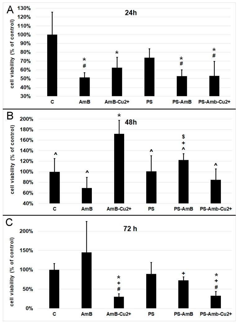
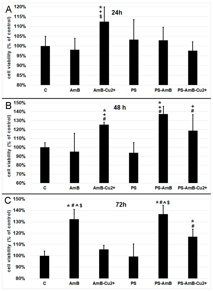
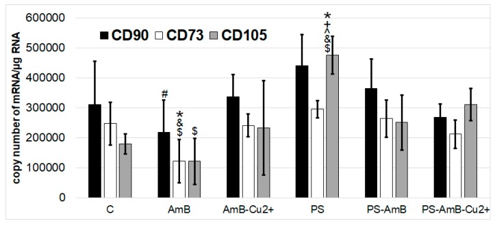
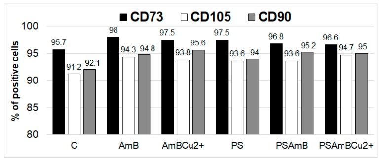
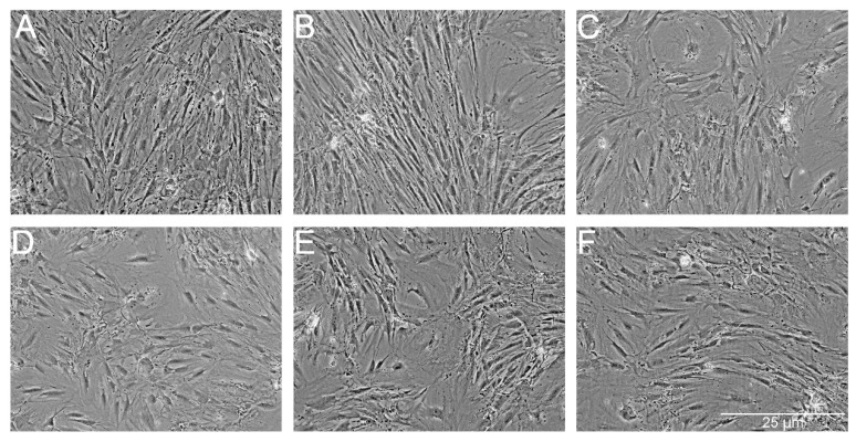
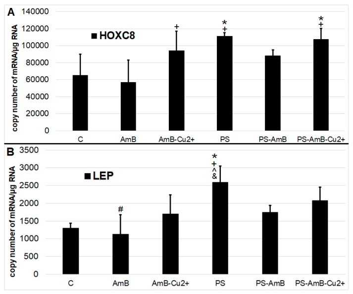
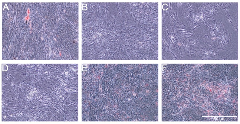
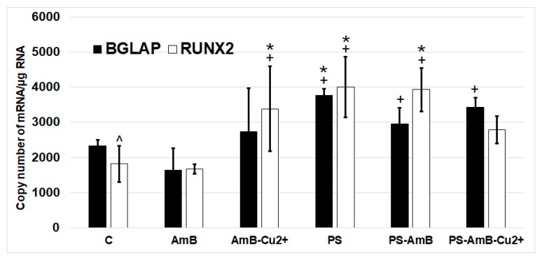
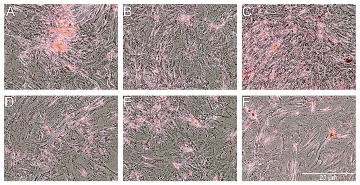
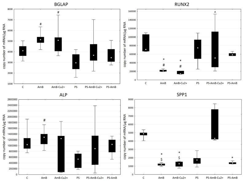
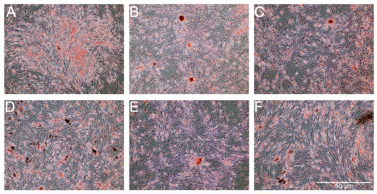
Adipose tissue is a promising source of mesenchymal stem cells. Their potential to differentiate and regenerate other types of tissues may be affected by several factors. This may be due to in vitro cell-culture conditions, especially the supplementation with antibiotics. The aim of our study was to evaluate the effects of a penicillin-streptomycin mixture (PS), amphotericin B (AmB), a complex of AmB with copper (II) ions (AmB-Cu2+) and various combinations of these antibiotics on the proliferation and differentiation of adipose-derived stem cells in vitro. Normal human adipose-derived stem cells (ADSC, Lonza) were routinely maintained in a Dulbecco's Modified Eagle Medium (DMEM) that was either supplemented with selected antibiotics or without antibiotics. The ADSC that were used for the experiment were at the second passage. The effect of antibiotics on proliferation was analyzed using the 3-[4,5-dimethylthiazol-2-yl]-2,5-diphenyltetrazolium bromide (MTT) and sulforhodamine-B (SRB) tests. Differentiation was evaluated based on Alizarin Red staining, Oil Red O staining and determination of the expression of ADSC, osteoblast and adipocyte markers by real-time RT-qPCR. The obtained results indicate that the influence of antibiotics on adipose-derived stem cells depends on the duration of exposure and on the combination of applied compounds. We show that antibiotics alter the proliferation of cells and also promote natural osteogenesis, and adipogenesis, and that this effect is also noticeable in stimulated osteogenesis.

Keywords: adipose-derived stem cells; amphotericin B; copper (II) ions; osteogenesis; penicillin; streptomycin.


Conflict of interest statement

The authors declare no conflicts of interest.

Figures

Figure 1
Figure 1
Cell viability based on the measurement of total cellular protein content after exposure to the tested antibiotics and their combinations for 24 (A), 48 (B) and 72 h (C). The bars represent the means ± standard deviation (SD) of the percentages of the control cell viability (100%); analysis of variance (ANOVA) with the Tukey post hoc test, * p < 0.05 vs. control, # p < 0.05 vs. PS, ^ p < 0.05 vs. AmB-Cu2+, + p < 0.05 vs. AmB, $ p < 0.05 vs. PS-AmB-Cu2+.
Figure 2
Figure 2
Cell viability based on the measurement of mitochondrial oxidative activity after exposure to the tested antibiotics and their combinations for 24 (A), 48 (B) and 72 h (C). The bars represent the means ± standard deviation (SD) of the percentages of the control cell viability (100%); ANOVA with the Tukey post hoc test, * p < 0.05 vs. the control, # p < 0.05 vs. PS, ^ p < 0.05 vs. AmB-Cu2+, + p < 0.05 vs. AmB, $ p < 0.05 vs. PS-AmB-Cu2+.
Figure 3
Figure 3
The mRNA levels of MSC markers CD73, CD90 and CD105 in the ADSC after exposure to the tested antibiotic combinations (RT-qPCR analysis). The bars represent the means ± standard deviation (SD) of the copy numbers per 1 µg of total RNA; ANOVA with the Tukey post hoc test, * p < 0.05 vs. control, # p < 0.05 vs. PS, ^ p < 0.05 vs. AmB-Cu2+, + p < 0.05 vs. AmB, & p < 0.05 vs. PS-AmB, $ p < 0.05 vs. PS-AmB-Cu2+.
Figure 4
Figure 4
The percentage of positive cells for CD73, CD90 and CD105 markers after treatment with tested antibiotic and their combinations (FACS analysis).
Figure 5
Figure 5
The morphology of the ADSC after exposure to the tested combinations of antibiotics. ADSC were exposed for 14 days to AmB (B), AmB-Cu2+ (C), PS (D), PS-AmB (E) and PS-AmB-Cu2+ (F), and compared to the control cells (A).
Figure 6
Figure 6
The mRNA levels of adipose tissue markers ((A)—HOXC5; (B)—LEP). The bars represent the means ± standard deviation (SD) of the copy numbers per 1 µg of total RNA; ANOVA with the Tukey post hoc test, * p < 0.05 vs. control, # p < 0.05 vs. PS, ^ p < 0.05 vs. AmB-Cu2+, + p < 0.05 vs. AmB, & p < 0.05 vs. PS-AmB.
Figure 7
Figure 7
Oil red staining of lipid droplets after exposure of adipose-derived stem cells (ADSC) to the tested combinations of antibiotics. ADSC were exposed for 14 days to AmB (B), AmB-Cu2+ (C), PS (D), PS-AmB (E) and PS-AmB-Cu2+ (F), and compared to the control cells (A).
Figure 8
Figure 8
The mRNA levels of osteoblastic markers BGLAP and RUNX2 in the ADSC after exposure to the tested antibiotic combinations. The bars represent the means ± standard deviation (SD) of the copy numbers per 1 µg of total RNA; ANOVA with the Tukey post hoc test, * p < 0.05 vs. control, + p < 0.05 vs. AmB, ^ p < 0.05 vs. AmB-Cu2+.
Figure 9
Figure 9
Alizarin Red staining of extracellular calcium after exposure of ADSC to the tested combinations of antibiotics. ADSC were exposed for 14 days to AmB (B), AmB-Cu2+ (C), PS (D), PS-AmB (E) and PS-AmB-Cu2+ (F), and compared to the control cells (A).
Figure 10
Figure 10
The mRNA levels of osteogenic markers BGLAP, RUNX2, SPP1 and ALP in the ADSC after exposure of ADSC to the tested combinations of antibiotics. The bars represent the (Me) with the 25th and 75th quartiles and the minimum and maximum of the copy numbers per 1 µg of total RNA; the Kruskal–Wallis test with post hoc was applied to assess any differences in the expression of the genes, * p < 0.05 vs. control, ^ p < 0.05 vs. AmB-Cu2+, # p < 0.05 vs. PS, $ p < 0.05 vs. PS-AmB-Cu2+.
Figure 11
Figure 11
The Alizarin Red staining in the ADSC after exposure to the osteogenesis medium with the addition of AmB (B), AmB-Cu2+ (C), PS (D), PS-AmB (E) and PS-AmB-Cu2+ (F) compared to the control cells (A).

Similar articles

Cited by

References

    1. Cieslar-Pobuda A., Knoflach V., Ringh M.V., Stark J., Likus W., Siemianowicz K., Ghavami S., Hudecki A., Green J.L., Los M.J. Transdifferentiation and reprogramming: Overview of the processes, their similarities and differences. Biochim. Biophys. Acta. 2017;1864:1359–1369. doi: 10.1016/j.bbamcr.2017.04.017. - DOI - PubMed
    1. Baglioni S., Francalanci M., Squecco R., Lombardi A., Cantini G., Angeli R., Gelmini S., Guasti D., Benvenuti S., Annunziato F., et al. Characterization of human adult stem-cell populations isolated from visceral and subcutaneous adipose tissue. FASEB J. 2009;23:3494–3505. doi: 10.1096/fj.08-126946. - DOI - PubMed
    1. Mohammadi Z., Afshari J.T., Keramati M.R., Alamdari D.H., Ganjibakhsh M., Zarmehri A.M., Jangjoo A., Sadeghian M.H., Ameri M.A., Moinzadeh L. Differentiation of adipocytes and osteocytes from human adipose and placental mesenchymal stem cells. Iran. J. Basic Med. Sci. 2015;18:259. - PMC - PubMed
    1. Romanov Y.A., Darevskaya A.N., Merzlikina N.V., Buravkova L.B. Mesenchymal stem cells from human bone marrow and adipose tissue: Isolation, characterization, and differentiation potentialities. Bull. Exp. Biol. Med. 2005;140:138–143. doi: 10.1007/s10517-005-0430-z. - DOI - PubMed
    1. Bunnell B.A., Flaat M., Gagliardi C., Patel B., Ripoll C. Adipose-derived stem cells: Isolation, expansion and differentiation. Methods. 2008;45:115–120. doi: 10.1016/j.ymeth.2008.03.006. - DOI - PMC - PubMed