

How do PrPSc Prions Spread between Host Species, and within Hosts?
- PMID: 29186791
- PMCID: PMC5750584
- DOI: 10.3390/pathogens6040060
How do PrPSc Prions Spread between Host Species, and within Hosts?
Abstract
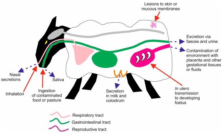
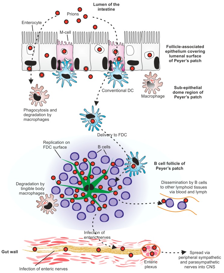
Prion diseases are sub-acute neurodegenerative diseases that affect humans and some domestic and free-ranging animals. Infectious prion agents are considered to comprise solely of abnormally folded isoforms of the cellular prion protein known as PrPSc. Pathology during prion disease is restricted to the central nervous system where it causes extensive neurodegeneration and ultimately leads to the death of the host. The first half of this review provides a thorough account of our understanding of the various ways in which PrPSc prions may spread between individuals within a population, both horizontally and vertically. Many natural prion diseases are acquired peripherally, such as by oral exposure, lesions to skin or mucous membranes, and possibly also via the nasal cavity. Following peripheral exposure, some prions accumulate to high levels within the secondary lymphoid organs as they make their journey from the site of infection to the brain, a process termed neuroinvasion. The replication of PrPSc prions within secondary lymphoid organs is important for their efficient spread to the brain. The second half of this review describes the key tissues, cells and molecules which are involved in the propagation of PrPSc prions from peripheral sites of exposure (such as the lumen of the intestine) to the brain. This section also considers how additional factors such as inflammation and aging might influence prion disease susceptibility.
Keywords: PrPSc; central nervous system; horizontal transmission; intestine; prion protein; prions; secondary lymphoid organs; vertical transmission.
Conflict of interest statement
The author declares no conflict of interest. The sponsors had no role in the writing of the manuscript, and in the decision to publish the results.
Figures
References
Publication types
Grants and funding
- BB/F019726/1/BB_/Biotechnology and Biological Sciences Research Council/United Kingdom
- BB/J014672/1/BB_/Biotechnology and Biological Sciences Research Council/United Kingdom
- BB/M024288/1/BB_/Biotechnology and Biological Sciences Research Council/United Kingdom
- BB/G003947/1/BB_/Biotechnology and Biological Sciences Research Council/United Kingdom
- BBS/E/D/20251968/BB_/Biotechnology and Biological Sciences Research Council/United Kingdom
LinkOut - more resources
Full Text Sources
Other Literature Sources
Research Materials
