Review
. 2017 Nov 25;22(12):2064.
doi: 10.3390/molecules22122064.

Upconversion Luminescence Sensitized pH-Nanoprobes

Affiliations
Review

Upconversion Luminescence Sensitized pH-Nanoprobes

Manoj Kumar Mahata et al. Molecules. .

Abstract

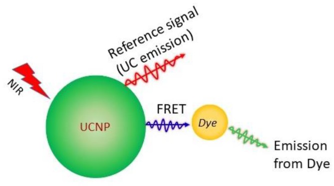
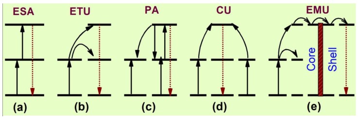
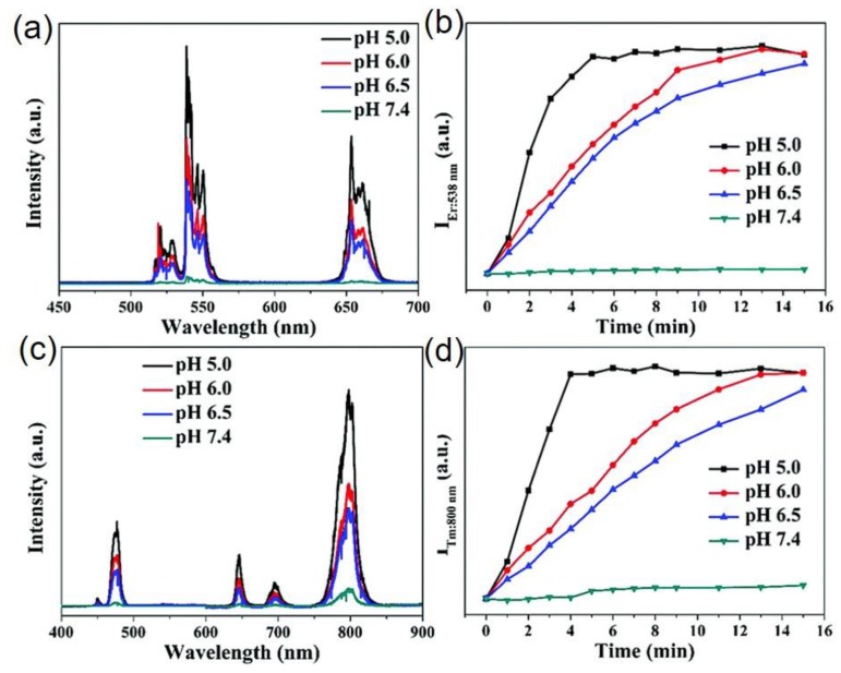
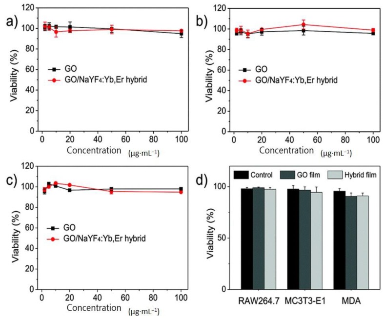
Photon upconversion materials, featuring excellent photophysical properties, are promising for bio-medical research due to their low autofluorescence, non-cytotoxicity, low photobleaching and high photostability. Upconversion based pH-nanoprobes are attracting considerable interest due to their superiority over pH-sensitive molecular indicators and metal nanoparticles. Herein, we review the advances in upconversion based pH-nanoprobes, the first time in the seven years since their discovery in 2009. With a brief discussion on the upconversion materials and upconversion processes, the progress in this field has been overviewed, along with the toxicity and biodistribution of upconversion materials for intracellular application. We strongly believe that this survey will encourage the further pursuit of intense research for designing molecular pH-sensors.

Keywords: molecular probes; optical sensors; pH-sensors; photoluminescence; upconversion.


Conflict of interest statement

There is no conflict of interests.

Figures

Figure 1
Figure 1
Schematic representation of upconversion sensitized pH-sensor. The pH-sensitive dye emission is analyzed with respect to the UC emission. The Förster resonance energy transfer (FRET) from UCNP to the pH-sensitive dye is shown.
Figure 2
Figure 2
Schematic representation of different upconversion processes [47].
Figure 3
Figure 3
(a) pH-dependent upconversion emission spectra of bromothymal blue conjugated NaYF4:Er3+/Yb3+ film upon 980 nm excitation (Reproduced from Ref. [78] with permission from The Royal Society of Chemistry). (b) The upconversion spectrum of Y2O2S:Er3+/Yb3+ UCNPs (red line, left y-axis) and the extinction spectra (right y-axis) of bromocresol green-doped silica films in pH 3.0 (orange) and buffer 7.0 (green). Inset shows the red luminescence with higher resolution. (c) The calibration curves of bromocresol green conjugated Y2O2S:Er3+/Yb3+ films, without (black dots) and with (red points) passing through porcine tissue. Left inset shows the photograph of the sensor film sandwiched between two porcine tissues for pH calibration. The inset in the right side shows the schematic representation of the pH sensor. (d) pH determination in real-time with the sensor film passing through porcine tissue. The red dots exhibit the variation of pH due to the bacterial development at the interface of the TSA plate and pH-sensor; the black dots show the variation of pH of the controlled sample with same volume of phosphate buffer solution. The corresponding pH values, calculated according to the calibration curve, are shown in the right y-axis. (e) The photograph at the left side shows the pH sensor film by the end of the real-time experiment with S. epidermidis and at the right side shows the film of the control sample by the end of acquisition (Figure 3b–d: Reproduced from Ref. [34] with permission of John Wiley & Sons Ltd.).
Figure 4
Figure 4
(a) The upconversion spectra of the graphene oxide–PEI-NaYF4; Er3+/Yb3+ film in buffer solutions upon excitation at 980 nm; (b) ratio of green emission intensity at pH 5 to green emission intensity at other pH values; (c) repetitive cycles of the pH-sensor between pH 6.00 and pH 8.00, showing response time; and (d) upconversion spectra of graphene oxide–PEI-NaYF4; Er3+/Yb3+ in urine of mice. Inset shows the ratio of green emission intensity at pH 5 to green emission intensity at other pH values plotted against the pH values in diluted urine (reproduced from Ref. [32] with permission from the PCCP Owner Societies).
Figure 5
Figure 5
(a) Imaging calibration row (RGB) of the membrane. The graph at the middle of (a) shows the ratio G/R values versus the pH for calibration point. At the bottom, four sensor spots were impregnated with serum specimens. The first three samples have adjusted pH values at pH 6.5, pH 7.0, and pH 7.5 and the fourth one is original. Autofluorescence from background is seen under UV illumination (first row). IR excitation and RGB imaging of the real serum samples remains unaffected from the background (second row). The G/R ratio is shown in pseudocolor (bottom row) and converted into corresponding pH values (Adapted with permission from Ref. [76]. Copyright (2014) American Chemical Society.). (b) The 980 nm excited upconversion spectra of the pH-sensor film containing UCNPs (NaYF4; Er3+/Yb3+) and chromoionophore (ETH 5418), within pH values 6–11. Inset shows intensity ratio of 656 nm to 542 nm emission band as a function of pH; (c) Upconversion spectra of the pH-sensor film containing UCNPs and ETH 5418 in diluted blood. Inset shows the intensity variation of 656 nm emission band of sensor film in buffer and diluted blood as a function of pH (Figure 5b,c: Adapted with permission from Ref. [84]. Copyright (2012) American Chemical Society).
Figure 6
Figure 6
(a) Upconversion luminescence spectrum of dye conjugated-UCNPs in pH 7.62 and 3.23. The excitation spectrum of pHrodo™ Red (red dots, right y-axis) is overlapped with the upconversion emission spectrum. (b) Upconversion-sensitized luminescence emission spectrum of pHrodo™ Red in pH 7.65, 5.13 and 2.95. (c) Ratio of intensities of the sensitized luminescence emission of pHrodo™ Red at 590 nm and the upconversion emission at 550 nm with (black) and without (red) a physiological salt concentration. (d) Fluorescence microscopy images of the green luminescence of pHrodo™-conjugated-UCNPs at different pHs (Reproduced from Ref. [85] with permission from The Royal Society of Chemistry).
Figure 7
Figure 7
(a) Red emission normalized luminescence spectra of QBC939 cells (incubated with UCNPs) under 980 nm excitation at different pH. (b) Variation of the relative luminescence intensity ratio of 475 nm to 645 nm at different pH values: (c,d) images of the QBC939 cell’s nucleus under 405 nm excitation; (e,f) the upconversion images of different sites in single cell upon 980 nm light excitation; and (g,h) merged images of (c&e) and (d&f), respectively, in 10 µm scale bar. (i) The upconversion emission spectra of (e,f) sites of the cell, normalized at 645 nm (Reproduced from Ref. [30], Creative Commons license: http://creativecommons.org/licenses/by/4.0/).
Figure 8
Figure 8
Upconversion response of: (a) NaYF4:Er3+/Yb3+@NaYF4@Ni; and (c) NaYF4:Tm3+/Yb3+@NaYF4@Ni nanoparticles in different pH solution. The plot of upconversion emission intensity a:t (b) 538 nm of Er3+; and (d) 800 nm of Tm3+ ions doped with Ni-modified UCNPs in different pH buffers versus of reaction time (Reproduced from Ref. [33] with permission from The Royal Society of Chemistry).
Figure 9
Figure 9
(a) Serum indicators of mice injected with PAA-conjugated NaYF4:Tm3+/Yb3+ UCNPs and mice receiving no injection (control); and (b) observation of heart function and heart size of zebrafish upon treatment with quantum dots and NaYF4 UCNPs (Figure 9a,b was reproduced from Ref. [94,97], Copyright (2010), (2014), respectively, with permission from Elsevier).
Figure 10
Figure 10
Biocompatibility of graphene oxide-PEI-NaYF4:Er3+/Yb3+ hybrid and hybrid film. (ac) In vitro relative cellular viability of three types of cells (RAW264.7, MC3T3-E1 and MDA) treated with graphene oxide and the graphene oxide–NaYF4:Er3+/Yb3+ hybrid, respectively. (d) Cytotoxicity of pure graphene oxide and graphene oxide–NaYF4:Er3+/Yb3+ films (reproduced from Ref. [32] with permission from the PCCP Owner Societies).
Figure 11
Figure 11
Upconversion imaging of mice with injection of PAA-coated NaYF4:Tm3+/Yb3+ UCNPs at various time points (Reproduced from Ref. [94], Copyright (2010), with permission from Elsevier).

References

    1. Drees C., Raj A.N., Kurre R., Busch K.B., Haase M., Piehler J. Engineered upconversion nanoparticles for resolving protein interactions inside living cells. Angew. Chem. Int. Ed. 2016;55:11668–11672. doi: 10.1002/anie.201603028. - DOI - PubMed
    1. Ong L.C., Ang L.Y., Alonso S., Zhang Y. Bacterial imaging with photostable upconversion fluorescent nanoparticles. Biomaterials. 2014;35:2987–2998. doi: 10.1016/j.biomaterials.2013.12.060. - DOI - PubMed
    1. Schäferling M. Nanoparticle-based luminescent probes for intracellular sensing and imaging of pH. WIREs Nanomed. Nanobiotechnol. 2016;8:378–413. doi: 10.1002/wnan.1366. - DOI - PubMed
    1. Michalet X., Pinaud F.F., Bentolila L.A., Tsay J.M., Doose S.J.J.L., Li J.J., Sundaresan G., Wu A.M., Gambhir S.S., Weiss S. Quantum dots for live cells, in vivo imaging, and diagnostics. Science. 2005;307:538–544. doi: 10.1126/science.1104274. - DOI - PMC - PubMed
    1. Mader H.S., Wolfeis O.S. Optical Ammonia Sensor Based on Upconverting Luminescent Nanoparticles. Anal. Chem. 2010;82:5002–5004. doi: 10.1021/ac1007283. - DOI - PubMed

Substances

LinkOut - more resources