Review
. 2017 Nov 25;18(12):2528.
doi: 10.3390/ijms18122528.

Translational Aspects of Sphingolipid Metabolism in Renal Disorders

Affiliations
Review

Translational Aspects of Sphingolipid Metabolism in Renal Disorders

Alaa Abou Daher et al. Int J Mol Sci. .

Abstract

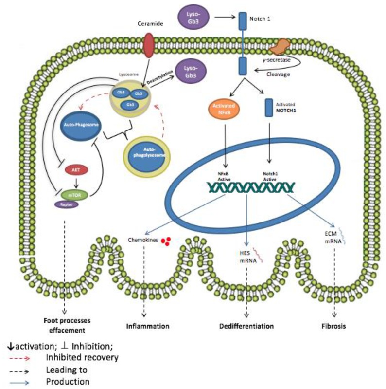
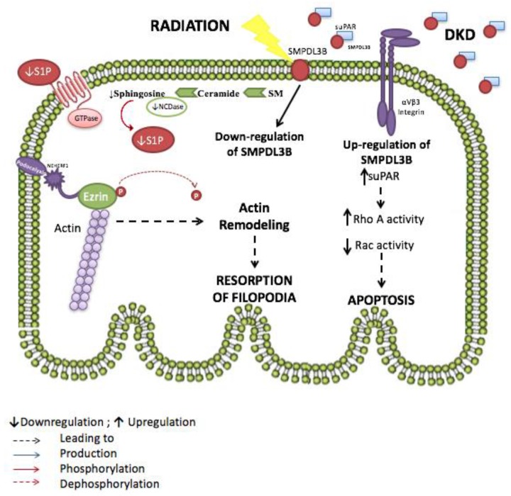
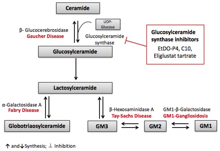
Sphingolipids, long thought to be passive components of biological membranes with merely a structural role, have proved throughout the past decade to be major players in the pathogenesis of many human diseases. The study and characterization of several genetic disorders like Fabry's and Tay Sachs, where sphingolipid metabolism is disrupted, leading to a systemic array of clinical symptoms, have indeed helped elucidate and appreciate the importance of sphingolipids and their metabolites as active signaling molecules. In addition to being involved in dynamic cellular processes like apoptosis, senescence and differentiation, sphingolipids are implicated in critical physiological functions such as immune responses and pathophysiological conditions like inflammation and insulin resistance. Interestingly, the kidneys are among the most sensitive organ systems to sphingolipid alterations, rendering these molecules and the enzymes involved in their metabolism, promising therapeutic targets for numerous nephropathic complications that stand behind podocyte injury and renal failure.

Keywords: Fabry’s disease; podocytes; renal failure; renal injury; sphingolipid metabolism; sphingolipids.


Conflict of interest statement

The authors declare no conflict of interest.

Figures

Figure 1
Figure 1
Potential mechanism of action of Globotriaosylceramide (Gb3) and Globotriaosylsphingosine (Lyso-Gb3) in Fabry’s disease podocyte. The accumulation of Gb3 in lysosomes inhibits AKT and mTOR pathway leading to the dysregulation of autophagy signaling in the podocyte. The inhibition of mTOR prevents the recovery of autophagosomes and lysosomes from autophagolysosomes by negative regulation. The formation of the autophagolysosomes causes podocyte foot processes effacement thus injury. Lyso-Gb3, the deacetylated form of Gb3, also plays a role in podocyte injury by promoting inflammation, fibrosis and dedifferentiation of podocytes. Lyso-Gb3 activates both the NOTCH signaling pathway, through γ-secretase, and nuclear factor κB (NFκB) leading to the release of chemokines. NOTCH-1 activation also promotes the transcription of HES genes and genes coding for extracellular matrix (ECM) proteins thus inducing, respectively, the dedifferentiation and fibrosis of the podocyte.
Figure 2
Figure 2
Proposed role for sphingomyelinase phosphodiesterase acid-like 3b (SMPDL3b) in radiation and diabetic kidney disease-induced podocytopathy. Radiation causes the downregulation of SMPDL3B, altering the sphingolipid metabolism. Ceramide levels increase while sphingosine and sphingosine 1 phosphate levels decrease. This leads to the dephosphorylation of ezrin and subsequent actin remodeling and resorption of filopodia. However, in diabetic kidney disease (DKD) SMPDL3B levels are elevated and bind to increased circulating soluble urokinase plasminogen activator receptor (suPAR), thus inhibiting the activation of αVβ3, but enabling RhoA activation and increasing podocyte apoptosis.
Figure 3
Figure 3
Substrate reduction theory: Glucosylceramide synthase inhibitors are therapeutic targets for lipid storing diseases. Glucosylceramide synthase inhibitors (EtDO-P4, C10 and Eliglustat tartrate) halt the synthesis of glucosylceramide, thereby preventing the downstream synthesis and accumulation of different sphingolipids involved in diseases such as Gaucher, Fabry’s, Tay-Sachs and GM1-Gangliosidosis.

References

    1. Pavenstadt H., Kriz W., Kretzler M. Cell biology of the glomerular podocyte. Physiol. Rev. 2003;83:253–307. doi: 10.1152/physrev.00020.2002. - DOI - PubMed
    1. Merscher S., Fornoni A. Podocyte pathology and nephropathy—Sphingolipids in glomerular diseases. Front. Endocrinol. 2014;5:127. doi: 10.3389/fendo.2014.00127. - DOI - PMC - PubMed
    1. Cellesi F., Li M., Rastaldi M.P. Podocyte injury and repair mechanisms. Curr. Opin. Nephrol. Hypertens. 2015;24:239–244. doi: 10.1097/MNH.0000000000000124. - DOI - PubMed
    1. Nagata M. Podocyte injury and its consequences. Kidney Int. 2016;89:1221–1230. doi: 10.1016/j.kint.2016.01.012. - DOI - PubMed
    1. Futerman A.H., Hannun Y.A. The complex life of simple sphingolipids. EMBO Rep. 2004;5:777–782. doi: 10.1038/sj.embor.7400208. - DOI - PMC - PubMed