. 2018 Jan 15;78(2):558-571.
doi: 10.1158/0008-5472.CAN-17-1700. Epub 2017 Nov 29.

Photodynamic Priming Mitigates Chemotherapeutic Selection Pressures and Improves Drug Delivery

Affiliations

Photodynamic Priming Mitigates Chemotherapeutic Selection Pressures and Improves Drug Delivery

Huang-Chiao Huang et al. Cancer Res. .

Abstract

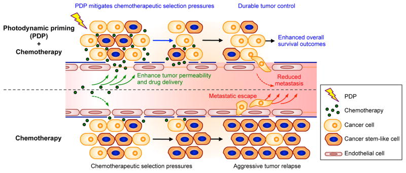
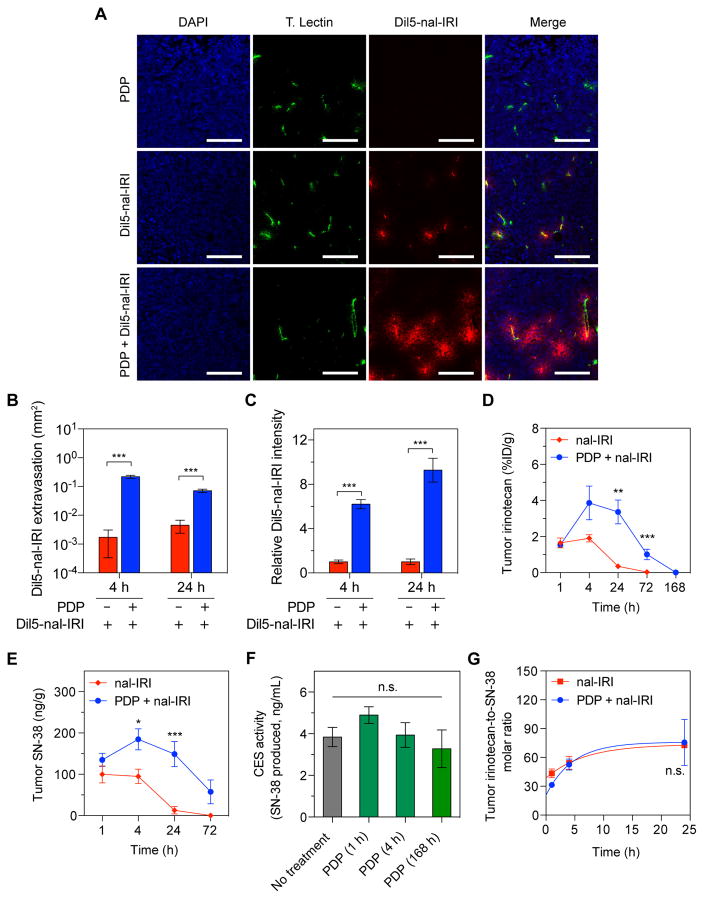
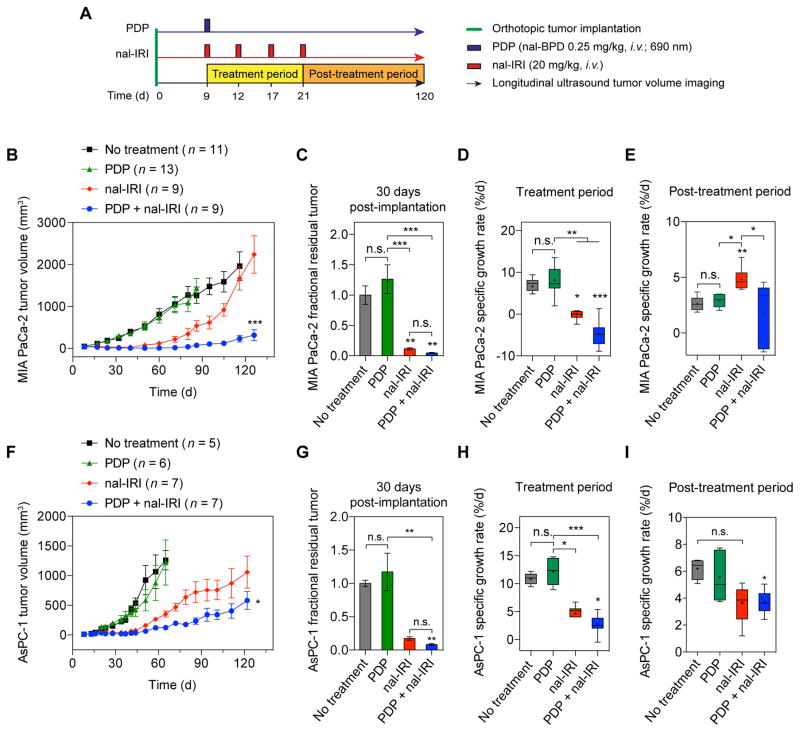
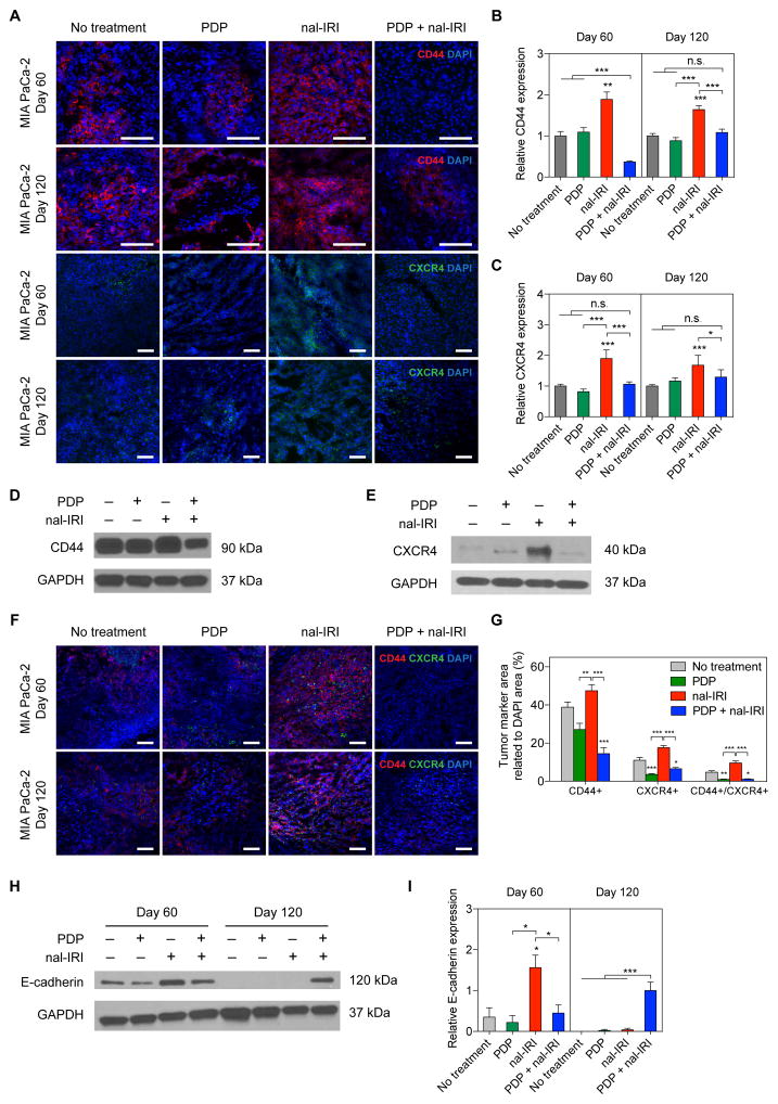
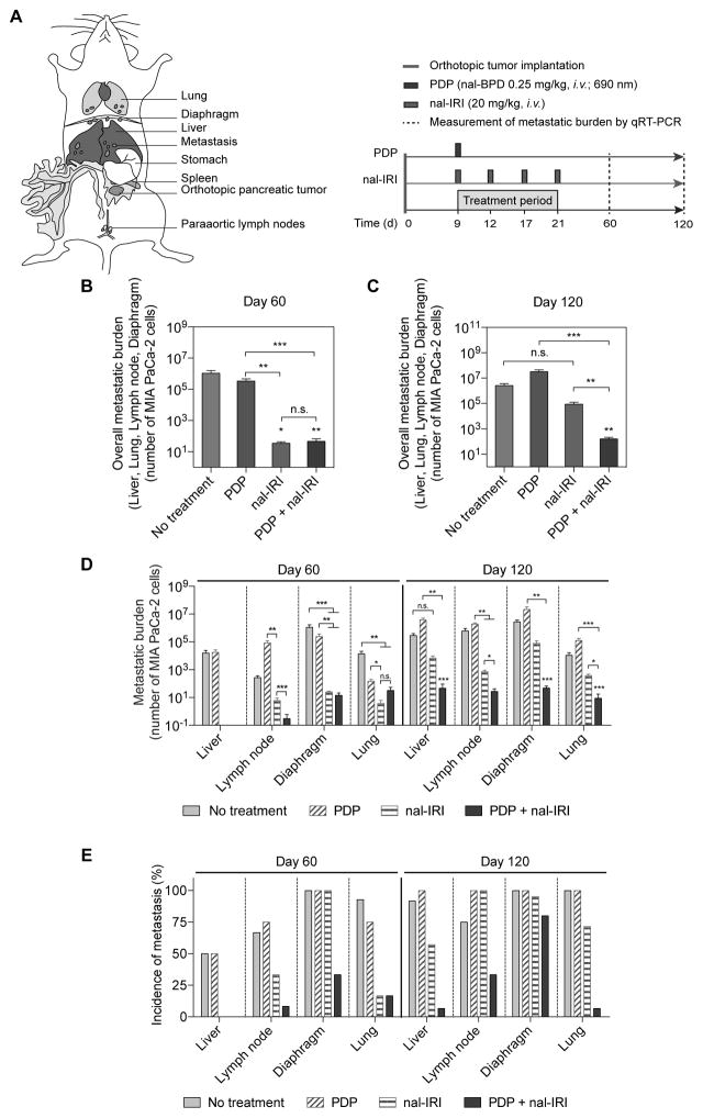
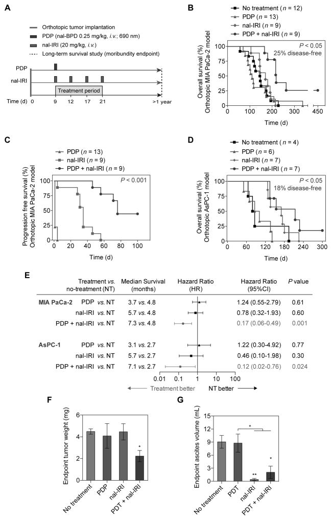
Physiologic barriers to drug delivery and selection for drug resistance limit survival outcomes in cancer patients. In this study, we present preclinical evidence that a subtumoricidal photodynamic priming (PDP) strategy can relieve drug delivery barriers in the tumor microenvironment to safely widen the therapeutic window of a nanoformulated cytotoxic drug. In orthotopic xenograft models of pancreatic cancer, combining PDP with nanoliposomal irinotecan (nal-IRI) prevented tumor relapse, reduced metastasis, and increased both progression-free survival and 1-year disease-free survival. PDP enabled these durable improvements by targeting multiple tumor compartments to (i) increase intratumoral drug accumulation by >10-fold, (ii) increase the duration of drug exposure above a critical therapeutic threshold, and (iii) attenuate surges in CD44 and CXCR4 expression, which mediate chemoresistance often observed after multicycle chemotherapy. Overall, our results offer preclinical proof of concept for the effectiveness of PDP to minimize risks of tumor relapse, progression, and drug resistance and to extend patient survival.Significance: A biophysical priming approach overcomes key treatment barriers, significantly reduces metastases, and prolongs survival in orthotopic models of human pancreatic cancer. Cancer Res; 78(2); 558-71. ©2017 AACR.


Figures

Fig. 1
Fig. 1. Concept of sub-tumoricidal photodynamic priming
Spatiotemporally controlled photodynamic priming (PDP) of tumor microvasculature and parenchyma simultaneously improves therapeutic agent accessibility and overcomes chemotherapeutic selection pressures. Subtumoricidal PDP increases tumor permeability to enhance intratumoral accumulation of chemotherapeutic agents for a prolonged period of time. In addition, it attenuates the insidious surge of stemness marker expression that is typically observed after multiple cycles of chemotherapy. Combining subtumoricidal PDP with cytotoxic chemotherapeutic agents prevents aggressive tumor regrowth, reduces metastatic burden, and enhances survival outcomes.
Fig. 2
Fig. 2. PDP increases tumor vascular permeability to enhance nal-IRI delivery in an orthotopic PDAC model
Orthotopic MIA PaCa-2 tumors were exposed to 75 J/cm2 of light (100 mW/cm2) one-hour following intravenous injection of nal-BPD (0.25 mg/kg) and a single dose of Dil5-nal-IRI (20 mg/kg). Control tumors were only injected with Dil5-nal-IRI (20 mg/kg) without light treatment. (A) Representative confocal fluorescence microscopy images of control tumors (top row) and PDP-treated tumors (bottom row) obtained 4 hours after intravenous injection Dil5-nal-IRI. In presence of PDP, Dil5-nal-IRI (red) was widely distributed throughout the tumor tissue and extravasated from the blood vessels (tomato lectin staining; green), whereas the signals arising from Dil5-nal-IRI in control tumors were confined to the immediate vicinity of the tumor blood vessels. No Dil5-nal-IRI signal was detected in the tumors treated with PDP alone. Nuclear staining (blue-fluorescence, DAPI); Scale bar 200 μm. (B, C) Quantitative analyses of Dil5-nal-IRI fluorescence intensity (B) and distribution (C) showing PDP significantly enhanced Dil5-nal-IRI accumulation and extravasation within MIA PaCa-2 tumors 4 and 24 hours after Dil5-nal-IRI injection (n ≥ 3 animals per group; n ≥ 19 images per group; ***P<0.001, Bonferroni-corrected Mann-Whitney U test). (D, E) PDP mediated changes in tumor pharmacokinetic profile of nal-IRI. Swiss nude mice bearing orthotopic MIA PaCa-2 tumors were treated with a single cycle of nal-IRI (nal-IRI, 20 mg/kg; IV) (red line; solid square) or a combination of PDP and single cycle nal-IRI (20 mg/kg; IV) (blue line; solid circle). Tumors were collected at various intervals and the irinotecan (d) and SN-38 (e) levels were measured by HPLC analysis (n ≥ 5 per time point; ***P<0.01, **P=0.022, *P<0.05, Bonferroni-corrected Mann-Whitney U test). (F) Carboxylesterase (CES) activities in MIA PaCa-2 tumors were not modulated by PDP at various time posts after treatment (n = 3–9 animals per condition; ns, non-significant, Kruskal–Wallis one-way ANOVA). (G) Comparison of tumoral irinotecan to SN-38 molar ratio at various time-points between ‘single cycle nal-IRI’ monotherapy and ‘PDP + single cycle nal-IRI’ arm. (n ≥ 5 per time point; Solid lines are nonlinear fits; n.s., non-significant, P=0.79, two-way ANOVA PDP·time interaction term). Results are mean ± standard error of the mean (SEM).
Fig. 3
Fig. 3
PDP of tumors extends the efficacy of multi-cycle nal-IRI chemotherapy for durable tumor control in two orthotopic PDAC models. (A) In vivo treatment schedule: Treatments were initiated nine days after MIA PaCa-2 or AsPC-1 cancer cell implantation when tumor volumes reached approximately 50 mm3 (see Methods). Mice were randomized into groups that received (i) no-treatment, (ii) PDP (nal-BPD 0.25 mg/kg; 690 nm light at 100 mW/cm2 to achieve 75 J/cm2), (iii) nal-IRI (four doses, each at 20 mg/kg irinotecan hydrochloride salt, on days 9, 12, 17, and 21 after tumor implantation), and (iv) combination of PDP and nal-IRI (PDP+nal-IRI). Injections of nal-BPD (for PDP) and nal-IRI were done intravenously. (B–E) Orthotopic MIA PaCa-2 and (FI) AsPC-1 tumor volumes were longitudinally monitored by non-invasive ultrasound imaging. A combination of PDP and nal-IRI prolonged and enhanced tumor growth inhibition in both MIA PaCa-2 and AsPC-1 animal models compared to nal-IRI alone. (n = 9–13 for MIA PaCa-2 model; n = 5–7 for AsPC-1 model; *P<0.05, ***P<0.001, one-way ANOVA with Tukey’s post hoc test for ‘nal-IRI’ vs. ‘PDP+nal-IRI’ groups). (C, G) Gross tumor volume changes were quantified between day 8 (one day prior to treatment) and approximately day 30 (21 days after treatment initiation) in (C) MIA PaCa-2 and (G) AsPC-1 orthotopic xenograft models. Approximately, a 90% reduction in mean tumor volume was observed in mice treated with ‘nal-IRI’ and ‘PDP+nal-IRI’ compared to the ‘no-treatment’ control animals. Asterisks denote significance compared with no-treatment group or amongst the indicated groups at each time point. (*P<0.05, **P<0.01, ***P<0.001, n.s., non-significant, Kruskal–Wallis one-way ANOVA with Dunn’s post hoc test) The specific growth rate (SGR) of tumors during the treatment period (D, H) and post-treatment period (E, I) were determined using the following formula: SGR = (1/V)(dV/dt), where V is tumor volume and t is time. In the MIA PaCa-2 mouse model, shortly following the termination of treatment and up to 120 days, nal-IRI-treated animals experienced a rapid tumor regrowth at a significantly higher SGR (4.8±0.3 %/d), compared to the ‘no-treatment’ control tumors. In contrast, the combination of PDP and nal-IRI continued to suppress tumor growth to a low SGR (1.7±0.9 %/d) for a prolonged period of time up to 120 days. (*P<0.05, **P<0.01, ***P<0.001, n.s., non-significant, Kruskal–Wallis one-way ANOVA with Dunn’s post hoc test). (n = 9–13 mice per group for MIA PaCa-2 model; n = 5–7 for AsPC-1 model). Results are mean ± standard error of the mean (SEM).
Fig. 4
Fig. 4. PDP suppresses chemotherapy-induced enrichment of CD44 and CXCR4 expression in PDAC tumors
(A) Representative immunofluorescence images of CD44 and CXCR4 in MIA PaCa-2 tumors subjected to (1) no-treatment; (2) PDP (nal-BPD 0.25 mg/kg; 690 nm light at 100 mW/cm2 to achieve 75 J/cm2); (3) four cycles of nal-IRI (nal-IRI; at 20 mg/kg each, on days 9, 12, 17 and 21); and (4) PDP+nal-IRI. Significant increases in CD44 and CXCR4 expression were observed in tumors treated with nal-IRI alone at days 60 and 120 post-implantation; Blue: DAPI (nuclei), Red: CD44, Green: CXCR4. Scale bar, 100 μm. (B, C) To quantify immunofluorescence intensities, at least 25 images, evenly distributed across the entire tumor cross-section, were collected from at least three tumor samples for each condition. CD44 and CXCR4 fluorescence intensities were normalized to DAPI area per image. Relative CD44 and CXCR4 levels were found to be significantly higher in the ‘nal-IRI’ groups compared to other groups. (*P<0.05, **P<0.01, ***P<0.001, Kruskal–Wallis one-way ANOVA with Dunn’s post hoc test) Asterisks denote significance compared to the no-treatment group or amongst the indicated groups at each time point. (D, E) Representative immunoblotting of CD44 and CXCR4 in tumors collected at day 120 confirmed that the enhanced protein expression of CD44 and CXCR4 after nal-IRI treatment could be effectively mitigated by PDP. (F) Representative CD44/CXCR4 double-stained images of MIA PaCa-2 tumors subjected to (1) no-treatment; (2) PDP (nal-BPD 0.25 mg/kg; 690 nm light at 100 mW/cm2 to achieve 75 J/cm2); (3) four cycles of nal-IRI (nal-IRI; at 20 mg/kg each, on days 9, 12, 17 and 21); and (4) PDP+nal-IRI. (G) The CD44+, CXCR4+, and CD44+/CXCR4+ areas of tumors (collected at day 120) were quantified and normalized to DAPI area using ImageJ software. At least 12 images, evenly distributed across the entire tumor cross-section, were collected from at least four tumor samples for each condition. (*P<0.05, **P<0.01, ***P<0.001, Kruskal–Wallis one-way ANOVA with Dunn’s post hoc test) Asterisks denote significance compared to the no-treatment group or amongst the indicated groups at each time point. (H) Immunoblot analysis of E-cadherin in MIA PaCa-2 primary tumor tissues at days 60 and 120. Expression of E-cadherin (relative to ‘PDP+nal-IRI’ at day 120) was normalized to glyceraldehyde 3-phosphate dehydrogenase (GAPDH) (n = 2–3; *P<0.05, **P<0.01, ***P<0.001, One-way ANOVA with Tukey’s post hoc test). (I) Representative Immunoblotting showed that the no-treatment, PDP, nal-IRI and treated tumors exhibited a complete loss of E-Cadherin expression, suggesting that ‘PDP+nal-IRI’ may help reduce the dedifferentiation of cancer cells. Results in B, C, D, E and I are mean ± standard error of the mean (SEM).
Fig. 5
Fig. 5. PDP enhances the anti-metastatic effects of multi-cycle nal-IRI in vivo
(A) To assess the efficacy of PDP and nal-IRI in controlling metastases, treatments were initiated nine days after MIA PaCa-2 tumor implantation in mice randomized to the following groups: (i) no-treatment; (ii) PDP (nal-BPD 0.25 mg/kg; 690 nm light at 100 mW/cm2 to achieve 75 J/cm2); (iii) nal-IRI (four doses at 20 mg/kg each, on days 9, 12, 17 and 21); and (iv) PDP+nal-IRI. (B–D) The number of metastases to the liver, retroperitoneal lymph nodes, diaphragm and lung were quantified by qRT-PCR (see Methods) on day 60 and day 120 after tumor implantation. (n > 11 per group; *P<0.05, **P<0.01, ***P<0.001, Kruskal–Wallis one-way ANOVA with Dunn’s post hoc test). Asterisks denote significance compared with no-treatment group or amongst the indicated groups at each time point. (B, C) The overall metastatic burden includes liver, lung, retroperitoneal lymph nodes, and diaphragm metastases. (D) Metastases to individual organs are presented. At day 60, both ‘nal-IRI’ and ‘PDP+nal-IRI’ completely inhibited liver metastasis and significantly reduced distant organ metastases to less than 50 cancer cells, as compared to the ‘no-treatment’ group (>1 million cancer cells at lung, diaphragm, lymph node). At day 120, the combination treatment of PDP and nal-IRI significantly reduced liver and distant organ metastases by ~16,000-fold and ~40,000-fold (P<0.001), respectively, compared to the ‘no-treatment’ control. (E) The incidence of metastases in mice bearing orthotopic MIA PaCa-2 tumors were significantly reduced by the combination treatment on days 60 and 120 (n > 11 per group). At day 120, the combination of PDP and nal-IRI effectively reduced the incidence of metastases to a range from 6.7 to 33.3%, while the incidence of metastases ranged from 60 to 100% in the ‘no-treatment’, ‘PDP’, and ‘nal-IRI’ groups.
Fig. 6
Fig. 6. PDP and multi-cycle nal-IRI achieve durable and significant survival enhancement and reduce endpoint disease burden in two orthotopic PDAC models
(A) Swiss nude mice were orthotopically inoculated with MIA PaCa-2 or AsPC-1 cells, divided into four groups, and subjected to (1) no-treatment; (2) PDP (day 9 post-implantation; nal-BPD 0.25 mg/kg; 690 nm light at 100 mW/cm2 to achieve 75 J/cm2); (3) multiple cycles of nal-IRI (nal-IRI; four doses at 20 mg/kg each, on days 9, 12, 17 and 21 post-implantation); and (4) PDP+nal-IRI. Moribundity was used as the endpoint for the survival study with proper justification and special approval by the MGH IACUC. Animals were monitored for up to 450 days (15 months). (B, C) Kaplan-Meier plot of overall animal survival (B) and progression-free survival (C) in MIA PACa-2 model. (n = 9–13 animals per group). (D) Kaplan-Meier plot of animal overall survival in the AsPC-1 model. (n = 4–7 animals per group). (E) Median survival time, hazard ratio forest plot, and differences in survival were evaluated by the log-rank test. A global test demonstrated a difference exists among the groups. Specifically, pairwise comparisons were performed to evaluate the advantage of treatment over no-treatment. Animals treated with PDP+nal-IRI were found to be significantly less likely to die by the next time point (hazard ratio < 1). No advantage to monotherapies (compared to no-treatment) were observed. Primary tumor weight, metastatic burden, and ascites volume were evaluated at animal death or day 450. (F) The combination of PDP+nal-IRI significantly reduced the endpoint primary tumor weight by half compared to the monotherapies and the no-treatment group. (n = 3–5 animals per group, *P<0.05, Unpaired t test). (G) The ascites formation in moribund animals were significantly reduced after ‘nal-IRI’ and ‘PDP+nal-IRI’ treatments, compared to the ‘no-treatment’ arm. (n = 3–6 animals per group; (*P<0.05, **P<0.01, Unpaired t test). Asterisks denote significance compared with no-treatment group or amongst the indicated groups at each time point. Results are mean ± standard error of the mean (SEM).

References

    1. Holohan C, Van Schaeybroeck S, Longley DB, Johnston PG. Cancer drug resistance: an evolving paradigm. Nat Rev Cancer. 2013;13:714–26. - PubMed
    1. Adiseshaiah PP, Crist RM, Hook SS, McNeil SE. Nanomedicine strategies to overcome the pathophysiological barriers of pancreatic cancer. Nature reviews Clinical oncology. 2016;13:750–65. - PubMed
    1. Garrido-Laguna I, Hidalgo M. Pancreatic cancer: from state-of-the-art treatments to promising novel therapies. Nature reviews Clinical oncology. 2015;12:319–34. - PubMed
    1. Minchinton AI, Tannock IF. Drug penetration in solid tumours. Nat Rev Cancer. 2006;6:583–92. - PubMed
    1. Greaves M, Maley CC. Clonal evolution in cancer. Nature. 2012;481:306–13. - PMC - PubMed

Publication types

MeSH terms