. 2018 Jan 30;92(4):e01776-17.
doi: 10.1128/JVI.01776-17. Print 2018 Feb 15.

Inflammasome Antagonism by Human Parainfluenza Virus Type 3 C Protein

Affiliations

Inflammasome Antagonism by Human Parainfluenza Virus Type 3 C Protein

Niraj K Shil et al. J Virol. .

Abstract

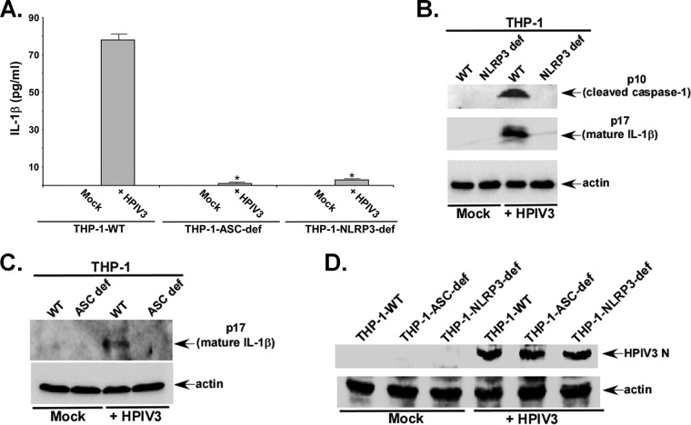
Human parainfluenza virus type 3 (HPIV3) is a negative-sense single-stranded RNA virus belonging to the Paramyxoviridae family. HPIV3 is a lung-tropic virus causing airway diseases, including pneumonia, croup, and bronchiolitis, during infancy and childhood. The activation of the inflammasome by pathogens results in the production of proinflammatory cytokines such as interleukin-1β (IL-1β) during infection. Thus, the inflammasome-mediated proinflammatory response plays a critical role in regulating the immune response and virus clearance. The inflammasome is a multimeric protein complex triggering caspase-1 activation. Activated caspase-1 cleaves pro-IL-1β into its mature (and active) secretory form. Our study revealed inflammasome activation in macrophages following HPIV3 infection. Specifically, the activation of the NLRP3/ASC inflammasome resulted in the production of mature IL-1β from HPIV3-infected cells. Furthermore, Toll-like receptor 2 (TLR2) activation (first signal) and potassium efflux (second signal) constituted two cellular events mediating inflammasome activation following HPIV3 infection. During our studies, we surprisingly identified the HPIV3 C protein as an antagonist of inflammasome activation. The HPIV3 C protein is an accessory protein encoded by the open reading frame of the viral phosphoprotein (P) gene. The HPIV3 C protein interacted with the NLRP3 protein and blocked inflammasome activation by promoting the proteasomal degradation of the NLRP3 protein. Thus, our studies report NLRP3/ASC inflammasome activation by HPIV3 via TLR2 signaling and potassium efflux. Furthermore, we have identified HPIV3 C as a viral component involved in antagonizing inflammasome activation.IMPORTANCE Human parainfluenza virus type 3 (HPIV3) is a paramyxovirus that causes respiratory tract diseases during infancy and childhood. Currently, there is no effective vaccine or antiviral therapy for HPIV3. Therefore, in order to develop anti-HPIV3 agents (therapeutics and vaccines), it is important to study the HPIV3-host interaction during the immune response. Inflammasomes play an important role in the immune response. Inflammasome activation by HPIV3 has not been previously reported. Our studies demonstrated inflammasome activation by HPIV3 in macrophages. Specifically, HPIV3 activated the NLRP3/ASC inflammasome by TLR2 activation and potassium efflux. C proteins of paramyxoviruses are accessory proteins encoded by the viral phosphoprotein gene. The role of the C protein in inflammasome regulation was unknown. Surprisingly, our studies revealed that the HPIV3 C protein antagonizes inflammasome activation. In addition, we highlighted for the first time a mechanism utilized by paramyxovirus accessory proteins to block inflammasome activation. The HPIV3 C protein interacted with the NLRP3 protein to trigger the proteasomal degradation of the NLRP3 protein.

Keywords: inflammasome; innate immunity.


Figures

FIG 1
FIG 1
Inflammasome activation by HPIV3. (A) THP-1 cells were infected with HPIV3. At 6 h, 12 h, and 24 h postinfection, the medium supernatant was assessed for IL-1β by an ELISA. (B) The TCA-precipitated supernatants derived from HPIV3-infected (8 h and 16 h) THP-1 cells were subjected to Western blotting with either IL-1β (p17) or caspase-1 (p10) antibody. Actin served as a loading control. (C) THP-1 cells were infected with HPIV3 for 6 h in the presence of either water (vehicle control) or a caspase-1 inhibitor (10 μM Ac-YVAD-CHO). The IL-1β level in the supernatant was assessed by an ELISA. (D) The IL-1β level was measured in the medium supernatant of THP-1 cells infected with either UV-irradiated HPIV3 (+ UV) or non-UV-irradiated HPIV3 (− UV). The ELISA values (A, C, and D) represent the means ± standard deviations (n = 8). *, P ≤ 0.05 by using Student's t test. The immunoblot (B) is representative of data from two independent experiments with similar results. UT, untreated.
FIG 2
FIG 2
HPIV3 activates the NLRP3/ASC inflammasome. (A) THP-1-WT (control), NLRP3-deficient THP-1 (THP-1-NLRP3-def), and ASC-deficient THP-1 (THP-1-ASC-def) cells were infected with HPIV3 for 6 h. IL-1β levels in the supernatant were assessed by an ELISA. (B) Detection of the cleaved caspase-1 p10 subunit and the mature p17 subunit of IL-1β in the supernatant of HPIV3-infected THP-1-WT and THP-1-NLRP3-def cells by performing Western blotting with p10- and p17-specific antibodies. Actin served as a loading control. (C) Detection of the mature p17 subunit of IL-1β in the supernatant of HPIV3-infected THP-1-WT and THP-1-ASC-def cells by performing Western blotting with p17-specific antibody. Actin served as a loading control. The ELISA values (A, C, and D) represent the means ± standard deviations. *, P ≤ 0.05 by using Student's t test. UT, untreated. (D) THP-1-WT (control), THP-1-NLRP3-def, and THP-1-ASC-def cells were infected with HPIV3 for 6 h. The cell lysate from infected cells was subjected to Western blotting with anti-HPIV3 RNP antibody to detect the HPIV3 nucleocapsid (N) protein. The ELISA values (A) represent the means ± standard deviations (n = 8). *, P ≤ 0.05 by using Student's t test. Immunoblots (B and C) are representative of data from two independent experiments with similar results.
FIG 3
FIG 3
The TLR2 pathway confers the first signal during HPIV3-mediated inflammasome activation. (A) THP-1 cells were transfected with plasmids expressing either WT TLR4 or DN TLR4. At 16 h posttransfection, TLR4 expression was analyzed by RT-PCR. NT, nontransfected. (B) THP-1 cells expressing either WT TLR4 or DN TLR4 were infected with HPIV3 (4 h), and the medium supernatant was assayed for IL-1β production by an ELISA. (C) THP-1 cells were transfected with plasmids expressing either WT TLR9 or DN TLR9. At 16 h posttransfection, TLR9 expression was analyzed by RT-PCR. (D) THP-1 cells expressing either WT TLR9 or DN TLR9 were infected with HPIV3 (4 h), and the supernatant was assayed for IL-1β production by an ELISA. (E) THP-1 cells were transfected with plasmids expressing either WT TLR2 or DN TLR2. At 16 h posttransfection, TLR2 expression was analyzed by RT-PCR. (F) THP-1 cells expressing either WT TLR2 or DN TLR2 were infected with HPIV3 (6 h), and the supernatant was assayed for IL-1β production by an ELISA. (G) Western blotting with p17-specific antibody was performed to detect the mature p17 subunit of IL-1β in the supernatant of HPIV3-infected THP-1 cells expressing either WT TLR2 or DN TLR2. Actin served as a loading control. (H) THP-1 cells were transfected with either human TLR2 siRNA (40 and 100 pmol) or control siRNA (100 pmol). TLR2 expression was analyzed by RT-PCR. (I) THP-1 cells transfected with either control siRNA or TLR2 siRNA were infected with HPIV3. At 6 h postinfection, IL-1β levels in the supernatant were measured by an ELISA. The ELISA values (B, D, F, and I) represent the means ± standard deviations (n = 8). *, P ≤ 0.05 by using Student's t test. The immunoblot (G) is representative of data from two independent experiments with similar results. Con siRNA, control siRNA.
FIG 4
FIG 4
Potassium efflux serves as a second signal during inflammasome activation by HPIV3. (A) THP-1 cells were pretreated with either DMSO (vehicle control) or 50 μM the ATP-sensitive potassium channel inhibitor glibenclamide (Gly) for 2 h prior to HPIV3 infection for 6 h in the presence of DMSO or glibenclamide. The medium supernatant was assayed for IL-1β production by an ELISA. (B) Western blotting with p17-specific antibody was performed to detect the mature p17 subunit of IL-1β in the supernatant of HPIV3-infected THP-1 cells treated with either DMSO (vehicle control) or glibenclamide. Actin served as a loading control. (C) THP-1 cells treated with buffer containing either 150 mM KCl or 150 mM NaCl (control) were infected with HPIV3. At 6 h postinfection, the IL-1β level in the supernatant was analyzed by an ELISA. The ELISA values (A and C) represent the means ± standard deviations (n = 8). *, P ≤ 0.05 by using Student's t test. The immunoblot (B) is representative of data from two independent experiments with similar results. UT, untreated (i.e., not treated with glibenclamide).
FIG 5
FIG 5
HPIV3 C protein inhibits inflammasome activation. (A) Cell lysates obtained from nontransfected (NT), empty FLAG-transfected, and FLAG-C protein-transfected HEK293 cells were subjected to Western blotting with anti-FLAG antibody. (B) Inflammasome-reconstituted HEK293 cells (i.e., cells expressing ASC, NLRP3, pro-IL-1β, and procaspase-1) expressing either empty FLAG or the FLAG-C protein were treated with nigericin (15 μM) for 30 min. IL-1β production was measured by performing an ELISA with the medium supernatant. (C) Cell lysates obtained from empty FLAG-transfected and FLAG-HPIV1 C protein-transfected HEK293 cells were subjected to Western blotting with anti-FLAG antibody. (D) Inflammasome-reconstituted HEK293 cells (i.e., cells expressing ASC, NLRP3, pro-IL-1β, and procaspase-1) expressing either empty FLAG or the FLAG-HPIV1 C protein were treated with nigericin (15 μM) for 30 min. IL-1β production was measured by performing an ELISA with the supernatant. (E) RT-PCR analysis of C protein expression in NT and FLAG-C protein-transfected THP-1 cells. (F) THP-1 cells expressing either empty FLAG or the FLAG-C protein were treated with LPS (4 h) and nigericin (30 min). IL-1β levels in the supernatant were analyzed by an ELISA. (G) Western blotting with p17 (to detect the mature p17 subunit of IL-1β)- and p10 (to detect the cleaved caspase-1 p10 subunit)-specific antibodies was performed with the supernatant of THP-1 cells (expressing either empty FLAG or the FLAG-C protein) treated with LPS alone or LPS and nigericin. Actin served as a loading control. (H) THP-1 cells expressing either empty FLAG or the FLAG-HPIV1 C protein were treated with LPS (1 h) and nigericin (30 min). IL-1β levels in the supernatant were analyzed by an ELISA. The ELISA values (B, D, F, and H) represent the means ± standard deviations (n = 8). *, P ≤ 0.05 by using Student's t test. Immunoblots (A, C, and G) are representative of data from two independent experiments with similar results. UT, untreated.
FIG 6
FIG 6
HPIV3 C protein promotes NLRP3 degradation. (A) Cell lysates collected from HEK293 cells coexpressing Myc-NLRP3 and FLAG-C were subjected to Western blotting with anti-Myc and anti-FLAG antibodies. (B) RT-PCR analysis of NLRP3 expression in HEK293 cells coexpressing either Myc-NLRP3 and FLAG-C or Myc-NLRP3 and empty FLAG. (C) Cell lysates collected from HEK293 cells coexpressing Myc-ASC and FLAG-C were subjected to Western blotting with anti-Myc antibody. (D) Cell lysates collected from HEK293 cells coexpressing Myc-NLRP3 and the FLAG-HPIV1 C protein were subjected to Western blotting with anti-Myc and anti-FLAG antibodies. In panels A and D, protein bands corresponding to NLRP3 and actin were quantified to calculate the NLRP3/actin ratio (relative intensity). The immunoblot (A) is representative of data from three independent experiments with similar results. NT, not transfected; FLAG, empty FLAG.
FIG 7
FIG 7
HPIV3 C protein promotes NLRP3 degradation via the proteasome. (A) Cell lysates collected from THP-1 cells expressing FLAG-C were subjected to Western blotting with anti-NLRP3 antibody to examine the status of endogenous NLRP3 protein expression. The blot was also probed with anti-FLAG antibody to detect the expression of the FLAG-C protein. (B) Cell lysates collected from THP-1 cells expressing FLAG-HPIV1 C were subjected to Western blotting with anti-NLRP3 antibody to examine the status of endogenous NLRP3 protein expression. The blot was also probed with anti-FLAG antibody to detect the expression of the FLAG-HPIV1 C protein. (C) Cell lysates collected from HEK293 cells coexpressing Myc-NLRP3 and FLAG-C in the presence of the vehicle (DMSO) or the proteasome inhibitor MG132 (10 μM) were subjected to Western blotting with anti-Myc antibody. In panels A to C, protein bands corresponding to NLRP3 and actin were quantified to calculate the NLRP3/actin ratio (relative intensity). Immunoblots (A and C) are representative of data from two independent experiments with similar results. FLAG, empty FLAG.
FIG 8
FIG 8
Interaction of HPIV3 C protein with NLRP3. (A) HEK293 cells were cotransfected with FLAG-C and Myc-NLRP3 in the presence of MG132. Cells lysates were immunoprecipitated (IP) with anti-FLAG antibody and subsequently immunoblotted (IB) with anti-Myc and anti-FLAG antibodies. Total cell lysates were also subjected to Western blotting with anti-FLAG and anti-Myc antibodies. (B) HEK293 cells coexpressing FLAG-C and Myc-NLRP3 were subjected to coimmunofluorescence analysis. Merged images (yellow) depict the colocalization of the C protein (green) and NLRP3 (red). Nuclei were stained with DAPI (blue). (C) Quantification of Myc-NLRP3 protein-specific fluorescence intensity from cells (present in the same visual field) with either high or low (or no) C protein expression levels. Quantification was performed with 15 different visual fields (n = 15). (D) THP-1 cells were transfected with FLAG-C in the presence of MG132. Lysates from these cells were immunoprecipitated with anti-FLAG antibody and subsequently immunoblotted with anti-NLRP3 and anti-FLAG antibodies. Total cell lysates were also subjected to Western blotting with anti-NLRP3 and anti-FLAG antibodies. FLAG, empty FLAG. The immunoblot (A) is representative of data from two independent experiments with similar results. The fluorescence values (C) represent the means ± standard deviations (n = 15). *, P ≤ 0.05 by using Student's t test.
FIG 9
FIG 9
HPIV3 C protein promotes ubiquitination of NLRP3 protein. (A) THP-1 cells expressing either empty FLAG or the FLAG-C protein were treated with either LPS or LPS and nigericin. Lysates from treated THP-1 cells were immunoprecipitated (IP) with antiubiquitin antibody and subsequently immunoblotted (IB) with anti-NLRP3 antibody. Total cell lysates were also subjected to Western blotting with anti-NLRP3 antibody. NLRP3-specific bands are indicated by red dots. Both high and lower exposures of the same gel are shown. (B) THP-1 cells expressing either the FLAG-HPIV1 C protein or the FLAG-HPIV3 C protein were treated with LPS and nigericin. Lysates from treated THP-1 cells were immunoprecipitated with antiubiquitin antibody and subsequently immunoblotted with anti-NLRP3 antibody. Total cells lysates were also subjected to Western blotting with anti-NLRP3 antibody. Immunoblots are representative of data from two independent experiments with similar results. Ub, ubiquitin.

Similar articles

Cited by

References

    1. Karron RA, Collins PL. 2013. Parainfluenza viruses, p 996–1023. In Knipe DM, Howley PM, Cohen JI, Griffin DE, Lamb RA, Martin MA, Racaniello VR, Roizman B (ed), Fields virology, 6th ed Lippincott Williams & Wilkins, Philadelphia, PA.
    1. Fox TG, Christenson JC. 2014. Influenza and parainfluenza viral infections in children. Pediatr Rev 35:217–227, quiz 228. doi:10.1542/pir.35-6-217. - DOI - PubMed
    1. Lamb RA, Parks GD. 2013. Paramyxoviridae, p 957–995. In Knipe DM, Howley PM, Cohen JI, Griffin DE, Lamb RA, Martin MA, Racaniello VR, Roizman B (ed), Fields virology, 6th ed Lippincott Williams & Wilkins, Philadelphia, PA.
    1. Bjornson CL, Johnson DW. 2008. Croup. Lancet 371:329–339. doi:10.1016/S0140-6736(08)60170-1. - DOI - PMC - PubMed
    1. Glezen WP, Frank AL, Taber LH, Kasel JA. 1984. Parainfluenza virus type 3: seasonality and risk of infection and reinfection in young children. J Infect Dis 150:851–857. doi:10.1093/infdis/150.6.851. - DOI - PubMed

Publication types