. 2018 Jan 30;92(4):e01322-17.
doi: 10.1128/JVI.01322-17. Print 2018 Feb 15.

Pathogenesis, Humoral Immune Responses, and Transmission between Cohoused Animals in a Ferret Model of Human Respiratory Syncytial Virus Infection

Affiliations

Pathogenesis, Humoral Immune Responses, and Transmission between Cohoused Animals in a Ferret Model of Human Respiratory Syncytial Virus Infection

Kok Fei Chan et al. J Virol. .

Abstract

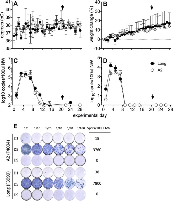
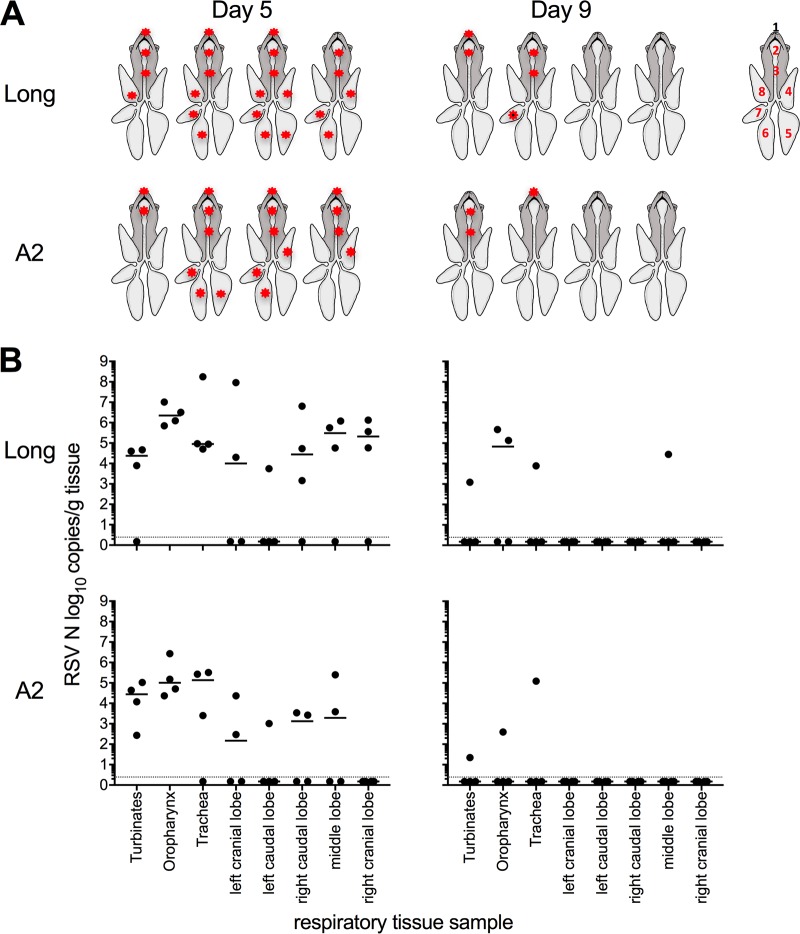
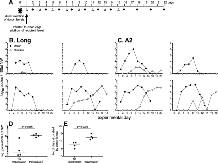
Small-animal models have been used to obtain many insights regarding the pathogenesis and immune responses induced following infection with human respiratory syncytial virus (hRSV). Among those described to date, infections in cotton rats, mice, guinea pigs, chinchillas, and Syrian hamsters with hRSV strains Long and/or A2 have been well characterized, although clinical isolates have also been examined. Ferrets are also susceptible to hRSV infection, but the pathogenesis and immune responses elicited following infection have not been well characterized. Here, we describe the infection of adult ferrets with hRSV Long or A2 via the intranasal route and characterized virus replication, as well as cytokine induction, in the upper and lower airways. Virus replication and cytokine induction during the acute phase of infection (days 0 to 15 postinfection) were similar between the two strains, and both elicited high levels of F glycoprotein-specific binding and neutralizing antibodies following virus clearance (days 16 to 22 postinfection). Importantly, we demonstrate transmission from experimentally infected donor ferrets to cohoused naive recipients and have characterized virus replication and cytokine induction in the upper airways of infected contact animals. Together, these studies provide a direct comparison of the pathogenesis of hRSV Long and A2 in ferrets and highlight the potential of this animal model to study serological responses and examine interventions that limit transmission of hRSV.IMPORTANCE Ferrets have been widely used to study pathogenesis, immunity, and transmission following human influenza virus infections; however, far less is known regarding the utility of the ferret model to study hRSV infections. Following intranasal infection of adult ferrets with the well-characterized Long or A2 strain of hRSV, we report virus replication and cytokine induction in the upper and lower airways, as well as the development of virus-specific humoral responses. Importantly, we demonstrate transmission of hRSV from experimentally infected donor ferrets to cohoused naive recipients. Together, these findings significantly enhance our understanding of the utility of the ferret as a small-animal model to investigate aspects of hRSV pathogenesis and immunity.

Keywords: animal models; ferret; respiratory syncytial virus; respiratory viruses; transmission.


Figures

FIG 1
FIG 1
Viral replication in the upper respiratory tract following experimental i.n. infection with hRSV. Ferrets (n = 4 per group) were infected i.n. with hRSV Long (closed circles) or A2 (open circles) virus, and NW samples were collected every second day. Temperature (A) and weight (B) were measured daily. (A and B) Means ± standard deviations from 4 ferrets per group are shown. (C) Viral shedding was quantified by real-time qPCR detection of the hRSV nucleoprotein (N) gene. (E) A ViroSpot (VS) assay was developed to measure titers of infectious virus in NW. Representative VS images of NW samples from two ferrets are shown at days 1, 5, and 9 p.i. (D) Titers of infectious virus in NW samples as determined by VS assay. Means ± standard deviations from 4 ferrets per group are shown in panels A and C. An arrowhead indicates reinfection with homologous virus strain. Statistical significance for virus shedding between viruses was analyzed by multiple t test with a Bonferroni-Dunn correction (alpha of 0.05), without assuming a consistent standard deviation. No significant differences were observed.
FIG 2
FIG 2
Expression of inflammatory mediator genes in nasal wash samples from ferrets after i.n. infection with hRSV. Ferrets were infected via the i.n. route with hRSV Long or A2 virus. NW samples collected at various times p.i. were assayed for mRNA for the indicated genes using real-time qPCR assays. Reference samples from uninfected ferrets are indicated at day 0, with a fold change of 1. Data are from a single experiment (n = 4 animals/group). Statistical significance for cytokine expression between viruses was analyzed by a multiple t test with a Bonferroni-Dunn correction (alpha of 0.05) without assuming a consistent standard deviation (*, P < 0.05; **, P < 0.01, ***, P < 0.001).
FIG 3
FIG 3
hRSV strains Long and A2 can replicate throughout the respiratory tract of ferrets after i.n. inoculation. Ferrets were infected via the i.n. route with hRSV Long or A2 virus and tissues collected on day 5 or 9 p.i. Levels of vRNA for the viral N gene were determined in tissue homogenates by real-time quantitative RT-PCR assay. (A) Diagrammatic representation of virus spread (indicated by an asterisk) throughout the respiratory tract: 1, turbinates; 2, oropharynx; 3, trachea; 4, left cranial lobe; 5, left caudal lobe; 6, right caudal lobe; 7, middle lobe; 8, right cranial lobe. Figure adapted from reference . (B) Virus load in different tissues from the respiratory tract of ferrets. Results for individual animals are shown (n = 4/group), and the horizontal line represents the median value. The dotted line indicates the limit of detection. Titers for each tissue were compared between virus strains using the Mann-Whitney U test.
FIG 4
FIG 4
Detection of inflammatory mediators in the lung after hRSV infection. Ferrets were infected via the i.n. route with hRSV Long or A2 virus. Lung lobes were collected on day 5 after infection and assayed for mRNA for the indicated genes by real-time qPCR. Note that data from A2/Long were combined and samples were classified as virus positive or virus negative based on vRNA results described in Fig. 3. Circles represent individual lung lobes from up to 4 ferrets, and the horizontal line represents the mean value for each mediator. For each graph, qPCR data first were expressed relative to values for lobes from uninfected animals and then normalized to ATF4, HPRT, and GAPDH housekeeping genes. Values for virus-positive and virus-negative lobes from hRSV-infected animals then were expressed as fold change relative to the corresponding lobes from uninfected animals. For statistical analyses, inflammatory mediators were compared between virus-positive lobes and lobes from uninfected animals (*, P < 0.05; **, P < 0.01, ***, P < 0.001; ****, P < 0.0001), between virus-negative lobes from infected animals and lobes from uninfected animals (÷, P < 0.05; ÷÷, P < 0.01; ÷÷÷, P < 0.001; ÷÷÷÷, P < 0.0001), and between virus-positive and virus-negative lobes from infected animals (±, P < 0.05; ±±, P < 0.01; ±±±, P < 0.001; ±±±, P < 0.0001). Note that the data did not show a normal distribution; therefore, ROUT could not be used to remove outliers. Instead, Kruskal-Wallis one-way analysis of variance (ANOVA) with Dunn's multiple-comparison test was used.
FIG 5
FIG 5
Humoral responses elicited following hRSV infection of ferrets by i.n. inoculation. Sera were collected from animals immediately prior to i.n. infection (day 0) or on day 16 (experiment 1) or 21 (experiment 2) after infection and assayed by ELISA (to detect antibodies capable of binding hRSV F glycoprotein) (A and B) or by ViroSpot microneutralization (VS MN) assay for neutralizing antibodies (C, D, and E). (C) A VS MN assay was developed to measure titers of neutralizing antibodies in ferret serum. Representative images of a VS MN assay using day 0 and day 21 serum samples from a single ferret are shown, including a cell control (CC; 1/4 are shown) and virus control (VC; 1/4 are shown). For panels A, B, D, and E, results from individual animals infected with hRSV Long (closed circles) or A2 (open circles) are shown. For panels A and D, individual titers are shown and the horizontal line represents the GMT. For panels B and E, fold change compared to day 0 are shown and the horizontal line indicates median value.
FIG 6
FIG 6
hRSV can transmit between cohoused ferrets. (A) Experimental plan. Donor ferrets were infected via the i.n. route with hRSV Long (B) or A2 (C) virus. One day later, donor animals were placed in clean cages and a naive recipient ferret was added. NW were collected on alternate days (donor, open triangles; recipients, black diamonds). (B and C) Virus shedding was measured in NW samples by real-time qPCR assay detecting vRNA. (D and E) The peak of virus shedding (D) and the number of days that detectable virus was shed in NW (E) was compared for donor ferrets that did or did not transmit virus to naive contacts. Horizontal bars represent median values, and significance was determined by Mann-Whitney test (P values are shown). Data are pooled from two independent experiments, with 4 donors and 4 recipients for each virus.
FIG 7
FIG 7
Expression of inflammatory mediator genes in nasal wash samples from ferrets after hRSV infection by contact transmission. Naive recipients were cohoused with donor ferrets that had been infected via the i.n. route with hRSV A2 or Long virus. Only recipient animals that shed detectable hRSV were examined, and data have been adjusted such that the first day that vRNA was detected in NW has been designated day 1 p.i. Real-time qPCR assays were used to assess levels of different inflammatory mediators using mRNA derived from NW samples. Data are from two independent experiments (n = 4 animals/group).

Similar articles

Cited by

References

    1. Afonso CL, Amarasinghe GK, Banyai K, Bao Y, Basler CF, Bavari S, Bejerman N, Blasdell KR, Briand FX, Briese T, Bukreyev A, Calisher CH, Chandran K, Cheng J, Clawson AN, Collins PL, Dietzgen RG, Dolnik O, Domier LL, Durrwald R, Dye JM, Easton AJ, Ebihara H, Farkas SL, Freitas-Astua J, Formenty P, Fouchier RA, Fu Y, Ghedin E, Goodin MM, Hewson R, Horie M, Hyndman TH, Jiang D, Kitajima EW, Kobinger GP, Kondo H, Kurath G, Lamb RA, Lenardon S, Leroy EM, Li CX, Lin XD, Liu L, Longdon B, Marton S, Maisner A, Muhlberger E, Netesov SV, Nowotny N, et al. 2016. Taxonomy of the order Mononegavirales: update 2016. Arch Virol 161:2351–2360. doi: 10.1007/s00705-016-2880-1. - DOI - PMC - PubMed
    1. Nair H, Nokes DJ, Gessner BD, Dherani M, Madhi SA, Singleton RJ, O'Brien KL, Roca A, Wright PF, Bruce N, Chandran A, Theodoratou E, Sutanto A, Sedyaningsih ER, Ngama M, Munywoki PK, Kartasasmita C, Simoes EA, Rudan I, Weber MW, Campbell H. 2010. Global burden of acute lower respiratory infections due to respiratory syncytial virus in young children: a systematic review and meta-analysis. Lancet 375:1545–1555. doi: 10.1016/S0140-6736(10)60206-1. - DOI - PMC - PubMed
    1. Scheltema NM, Gentile A, Lucion F, Nokes DJ, Munywoki PK, Madhi SA, Groome MJ, Cohen C, Moyes J, Thorburn K, Thamthitiwat S, Oshitani H, Lupisan SP, Gordon A, Sanchez JF, O'Brien KL, Group PS, Gessner BD, Sutanto A, Mejias A, Ramilo O, Khuri-Bulos N, Halasa N, de-Paris F, Pires MR, Spaeder MC, Paes BA, Simoes EAF, Leung TF, da Costa Oliveira MT, de Freitas Lazaro Emediato CC, Bassat Q, Butt W, Chi H, Aamir UB, Ali A, Lucero MG, Fasce RA, Lopez O, Rath BA, Polack FP, Papenburg J, Roglic S, Ito H, Goka EA, Grobbee DE, Nair H, Bont LJ. 2017. Global respiratory syncytial virus-associated mortality in young children (RSV GOLD): a retrospective case series. Lancet Glob Health 5:e984–e991. doi: 10.1016/S2214-109X(17)30344-3. - DOI - PMC - PubMed
    1. Falsey AR, Hennessey PA, Formica MA, Cox C, Walsh EE. 2005. Respiratory syncytial virus infection in elderly and high-risk adults. N Engl J Med 352:1749–1759. doi: 10.1056/NEJMoa043951. - DOI - PubMed
    1. Cardenas S, Auais A, Piedimonte G. 2005. Palivizumab in the prophylaxis of respiratory syncytial virus infection. Expert Rev Anti Infect Ther 3:719–726. doi: 10.1586/14787210.3.5.719. - DOI - PubMed

Publication types

MeSH terms

LinkOut - more resources