. 2018 Jan 19;293(3):830-846.
doi: 10.1074/jbc.M117.816447. Epub 2017 Nov 29.

Epitope-focused immunogens against the CD4-binding site of HIV-1 envelope protein induce neutralizing antibodies against auto- and heterologous viruses

Affiliations

Epitope-focused immunogens against the CD4-binding site of HIV-1 envelope protein induce neutralizing antibodies against auto- and heterologous viruses

Hua Wang et al. J Biol Chem. .

Abstract

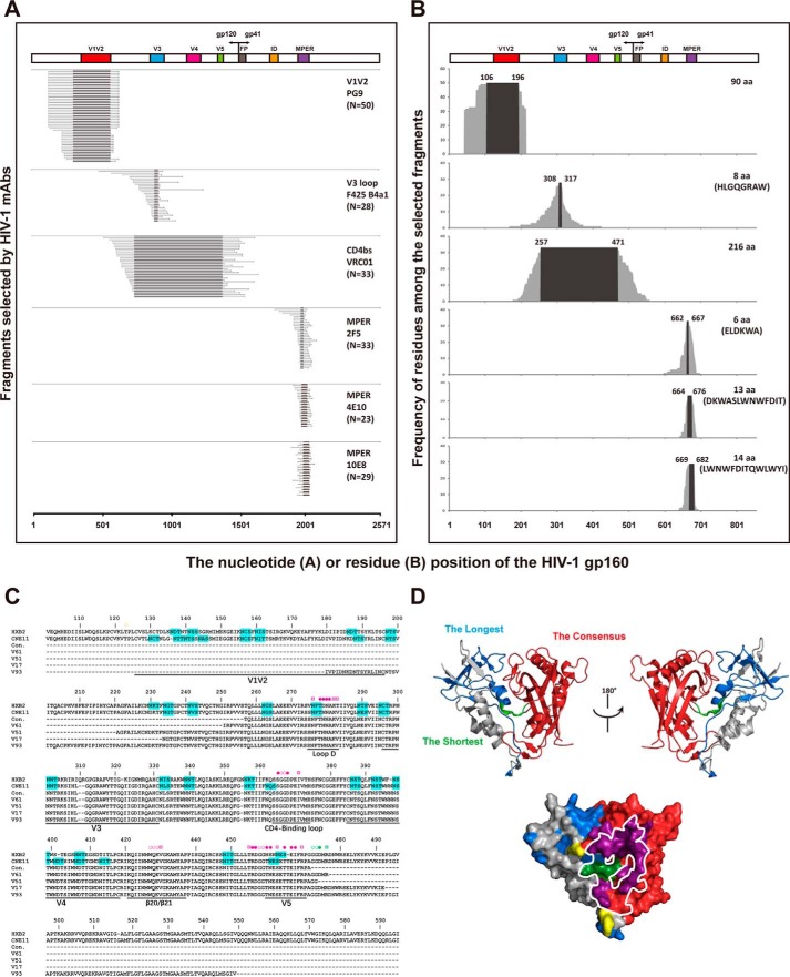
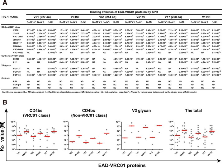
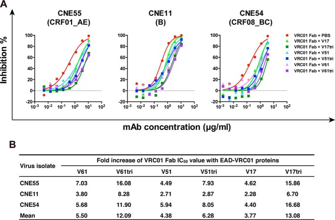
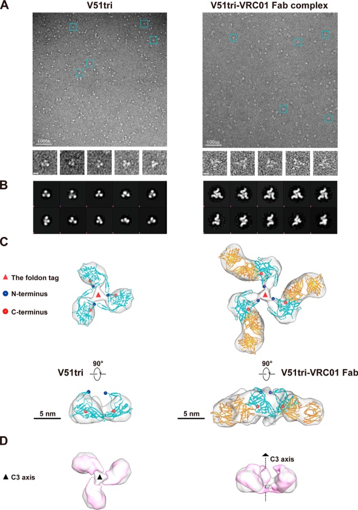
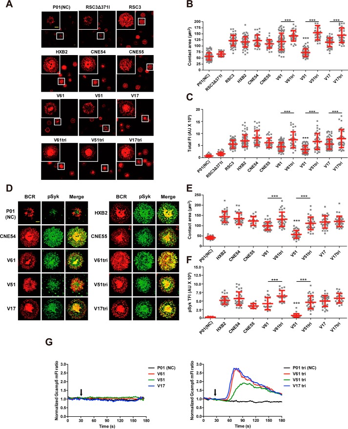
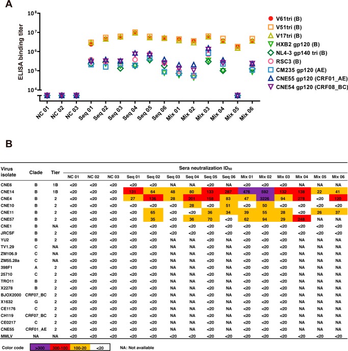
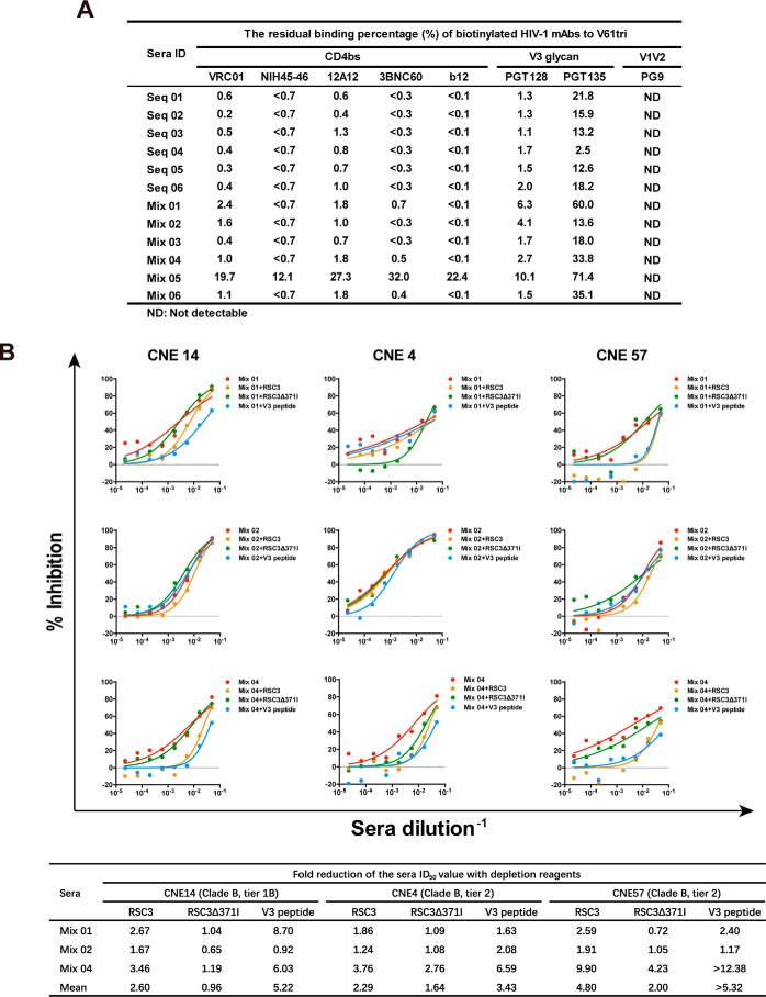
Recent discoveries of broadly neutralizing antibodies (bnAbs) in HIV-1-infected individuals have led to the identification of several major "vulnerable sites" on the HIV-1 envelope (Env) glycoprotein. These sites have provided precise targets for HIV-1 vaccine development, but identifying and utilizing many of these targets remain technically challenging. Using a yeast surface display-based approach, we sought to identify epitope-focused antigenic domains (EADs) containing one of the "vulnerable sites," the CD4-binding site (CD4bs), through screening and selection of a combinatorial antigen library of the HIV-1 envelope glycoprotein with the CD4bs bnAb VRC01. We isolated multiple EADs and found that their trimeric forms have biochemical and structural features that preferentially bind and activate B cells that express VRC01 in vitro More importantly, these EADs could induce detectable levels of neutralizing antibodies against genetically related autologous and heterologous subtype B viruses in guinea pigs. Our results demonstrate that an epitope-focused approach involving a screen of a combinatorial antigen library is feasible. The EADs identified here represent a promising collection of possible targets in the rational design of HIV-1 vaccines and lay the foundation for harnessing the specific antigenicity of CD4bs for protective immunogenicity in vivo.

Keywords: CD4bs; V3; antibody; electron microscopy (EM); epitope; guinea pig; human immunodeficiency virus (HIV); vaccine; yeast.


Conflict of interest statement

The authors declare that they have no conflicts of interest with the contents of this article

Figures

Figure 1.
Figure 1.
Identification and characterization of EAD-VRC01 from the HIV-1 gp160 combinatorial antigen library displayed on the yeast surface. A, EADs selected by the HIV-1 mAbs (PG9, F425 B4a1, VRC01, 2F5, 4E10, and 10E8) aligned to the original full-length CNE11 gp160 sequence used in the construction of the combinatorial yeast library. The target epitopes and the number (N) of sequences isolated by each mAb were indicated on the far right of each panel. The hypervariable regions V1–V5, interspersed conserved regions C1–C5 in gp120, fusion peptide (FP), immunodominant region (ID, and MPER in gp41 are indicated. B, frequency of each amino acid residue among the selected EADs along their corresponding positions in the CNE11 envelope glycoprotein. The consensus of amino acid sequences was numbered on the top of each graph according to HXB2 and highlighted by black background. The length and sequences of each consensus are shown on the far right wherever possible. C, sequence alignment of four EAD-VRC01 together with their consensus (con.) and corresponding sequences from CNE11 and HXB2. The EAD-VRC01 sequences represented the shortest (V61, 227 aa), the longest (V93, 372 aa), and the two intermediate ones (V51, 254 aa and V17, 260 aa). The contact residues with VRC01 were indicated with the symbol ○ for main-chain only contacts; ¤ for side-chain only contacts, and ● for both main- and side-chain contacts. Those colored in yellow were located outside the selected EAD-VRC01 fragments, in purple among the consensus EAD-VRC01 fragment, and in dark green in the shortest EAD-VRC01 V61 fragment. Potential glycosylation sites with the signature sequence NX(T/S) were highlighted in cyan. Dashes represented the gaps introduced to the preserve alignment. The hypervariable regions V1–V5, loop D, CD4-binding loop, and β20/β21 in gp120 were indicated. D, superimposing EAD-VRC01 sequences onto the monomeric gp120 (PDB code 3NGB) in ribbon and surface representations. The longest EAD-VRC01 was highlighted in light blue, the shortest in light green, and the consensus in red. The contact residues with VRC01 were colored as in C, and the contact surface of CD4 on the outer domain of gp120 was outlined by the white line.
Figure 2.
Figure 2.
Binding affinities of monomeric and trimeric EAD-VRC01 to HIV-1 mAbs measured by SPR. A, binding affinity between EAD-VRC01 and HIV-1 mAbs was measured by SPR and presented by Kon (on-rate constant), Koff (off-rate constant), and KD (equilibrium dissociation constant). The tested mAbs were CD4bs-targeted mAbs from multiple donors, including VRC01 class mAbs (VRC01, 12A12, VRC-CH31, 3BNC60, 3BNC117, NIH45–46, VRC-PG04, and VRC-PG20) and non-VRC01 class mAbs (CH103 and b12), V3 glycan-targeted mAbs (PGT121, PGT128, and PGT135), a V1V2-targeted mAb (PG9), CD4-IgG, and an irrelevant influenza hemagglutinin-targeted mAb (3C11) as the negative controls. The sensorgrams were fitted globally in 1:1 Langmuir model, and several KD values (denoted by asterisk symbols) were determined by the steady-state affinity model as the interactions were too fast to provide kinetic information. B, comparison of KD values within and between each mAb or combined group. The median value in each group was represented by the horizontal red line.
Figure 3.
Figure 3.
EAD-VRC01 attenuates the neutralizing activity of VRC01 Fab. A, neutralizing activity of VRC01 Fab in the presence of monomeric and trimeric EAD-VRC01 V61, V51, and V17 (30 μg/ml) against the three representative HIV-1 pseudoviruses bearing the envelope glycoprotein of CNE55 (clade CRF01_AE), CNE11 (clade B), and CNE54 (clade CRF08_BC) from China. B, fold increase in neutralizing IC50 of VRC01 Fab in the presence of EAD-VRC01 as compared with the mock controls in PBS.
Figure 4.
Figure 4.
Negative staining EM and image reconstruction of the purified V51tri and V51tri–VRC01 Fab complex. A, negative staining EM micrographs of the V51tri (left) and V51tri–VRC01 Fab complex (right). Raw micrographs were shown at the top. Representative particles were highlighted by the cyan boxes and shown at the bottom. The scale bars, 100 nm in the raw micrographs and represented 10 nm in the boxed particle images. B, reference-free class averages of the V51tri (left) and V51tri–VRC01 Fab complex (right). C, three-dimensional (3D) reconstructions of the V51tri (left) and V51tri–VRC01 Fab complex (right) were shown in the top (top) and side (bottom) views with the fitted models. The V51tri and V51tri–VRC01 Fab models were generated from the crystal structures of BG505 SOSIP.664 gp140 trimer (PDB code 4NCO) and gp120–VRC01 Fab complex (PDB code 3NGB). The fitted V51 monomers were indicated by the cyan ribbon and the fitted VRC01 Fabs were indicated by the orange ribbon. The N and C termini of the fitted V51 monomers were marked with the blue and red balls, respectively. Positions of the foldon tags were indicated by the red triangles and the scale bars, 5 nm. D, comparison of the V51tri EM map (in gray) and its equivalent map (in magenta) generated from the crystal structure of BG505 SOSIP.664 gp140 (left, top view; right, side view). The position of the 3-fold axis was indicated by a black dotted line.
Figure 5.
Figure 5.
Triggering VRC01-BCR signalings and calcium mobilization by EAD-VRC01. A, synaptic accumulation of BCRs triggered by EAD-VRC01 in the human Ramos B cells expressing VRC01-IgM-BCRs in TIRFM images. The EAD-VRC01 as well as control proteins, such as P01 (a EAD-PG9 as the negative control), RSC3 (the bait protein for isolating VRC01 mAb), RSC3Δ371I (lacked a single amino acid in RSC3 at position 371), HXB2 gp120 (clade B), CNE54 gp120 (clade CRF08_BC), and CNE55 gp120 (clade CRF01_AE), were used to trigger VRC01-BCR clustering. The magnified insets (upper left corner) show the highlighted cells in the original image with the white boxes. The BCR molecules are labeled in red, and the scale bar in the inset window represents 1.5 μm. B and C, statistical analysis for the contact area (B) and total fluorescence intensity (TFI) (C) of accumulated BCRs in the immunological synapses of the Ramos-VRC01 B cells. Each dot in the plot represents the data of one cell. Two-tailed t tests were used for the statistical analysis between the monomer and trimer of the same EAD-VRC01. Bars indicate the mean values and standard deviations. ***, p < 0.001. D, synaptic recruitment of phosphorylated Syk (pSyk) to the contact area of the Ramos B cells after BCR accumulation by EAD-VRC01 in TIRFM images. The BCRs and pSyk molecules are labeled in red and green. The scale bar, 1.5 μm. E and F, statistical analysis for the contact area (E) and TFI (F) of recruited pSyk in the immunological synapses. Each dot in the plot represents the data of one cell. Two-tailed t tests were used for the statistical analysis between the monomer and trimer of the same EAD-VRC01. Bars indicate the mean values and standard deviations. ***, p < 0.001. G, EAD-VRC01 induces the calcium flux in the Ramos B cells. Gcamp5 biosensor reported the Ca2+ flux by flow cytometry in the Ramos B cells after the stimulation of EAD-VRC01 monomers (left) or trimers (right) at the indicated time points (black arrow). P01 and P01tri stated above were negative controls.
Figure 6.
Figure 6.
Immunogenicity of EAD-VRC01 trimers in guinea pigs. A, ELISA binding titers of the immunized guinea pig sera against the autologous EAD-VRC01 trimer immunogens (V61tri, V51tri, V17tri, and clade B), HXB2 gp120 (clade B), NL4-3 SOSIP.664 gp140 trimer (clade B), RSC3 (clade B), CM235 gp120 (clade AE), CNE55 gp120 (clade CRF01_AE), and CNE54 gp120 (clade CRF08_BC). The sera tested here were collected 2 weeks (week = 22) after the fourth inoculation. The ELISAs were performed on two independent experiments. B, neutralizing activity of the immune sera against a panel of pseudoviruses as well as 10 strains from the global panel in the TZM-bl assay. Moloney murine leukemia virus (MMLV) was used as a negative control. The sera tested here were collected 2 weeks (week = 22) after the fourth inoculation. The neutralization assays were conducted on at least two independent occasions, and each was in duplicate. The color code was used to define neutralizing ID50 titers, and those <20 were in black, 20–100 in orange, 100–300 in red, and >300 in purple.
Figure 7.
Figure 7.
Profiling of the guinea pig immune sera by competitive ELISA and neutralization depletion. A, residual binding percentage (%) of biotinylated HIV-1 mAbs to V61tri measured by competitive ELISA. The binding titers of biotinylated mAbs with known epitopes in the presence of the immune guinea pig sera were evaluated and numerically presented. The sera from the NC group and PG9-biotin mAb were used as negative controls. B, reduction of serum-neutralizing activity in the presence of RSC3, RSC3Δ371I, and the autologous V3 peptide. The three relatively potent neutralizing sera (Mix 01, Mix 02, and Mix 04) were studied for their ability to neutralize the three relatively sensitive pseudoviruses (CNE14, CNE4, and CNE57) in the presence of RSC3, RSC3Δ371I, and the autologous V3 peptide in the TZM-bl assay. The actual neutralization curves (top) and fold reduction in neutralization ID50 (bottom) were showed. The > symbol indicates the complete depletion of neutralizing activity.

References

    1. Mascola J. R., and Montefiori D. C. (2010) The role of antibodies in HIV vaccines. Annu. Rev. Immunol. 28, 413–444 10.1146/annurev-immunol-030409-101256 - DOI - PubMed
    1. Fauci A. S., and Marston H. D. (2014) Ending AIDS–is an HIV vaccine necessary? N. Engl. J. Med. 370, 495–498 10.1056/NEJMp1313771 - DOI - PubMed
    1. Haynes B. F., and Bradley T. (2015) Broadly neutralizing antibodies and the development of vaccines. JAMA 313, 2419–2420 10.1001/jama.2015.2427 - DOI - PubMed
    1. Burton D. R., and Mascola J. R. (2015) Antibody responses to envelope glycoproteins in HIV-1 infection. Nat. Immunol. 16, 571–576 10.1038/ni.3158 - DOI - PMC - PubMed
    1. Burton D. R., and Hangartner L. (2016) Broadly neutralizing antibodies to HIV and their role in vaccine design. Annu. Rev. Immunol. 34, 635–659 10.1146/annurev-immunol-041015-055515 - DOI - PMC - PubMed

Publication types