. 2017;24(4):373-414.
doi: 10.1007/s10914-016-9357-6. Epub 2016 Oct 26.

The Skull of Epidolops ameghinoi from the Early Eocene Itaboraí Fauna, Southeastern Brazil, and the Affinities of the Extinct Marsupialiform Order Polydolopimorphia

Affiliations

The Skull of Epidolops ameghinoi from the Early Eocene Itaboraí Fauna, Southeastern Brazil, and the Affinities of the Extinct Marsupialiform Order Polydolopimorphia

Robin M D Beck. J Mamm Evol. 2017.

Abstract

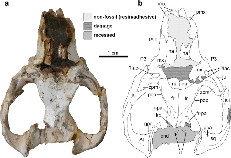
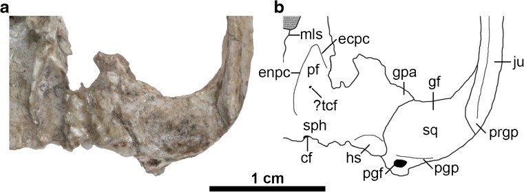
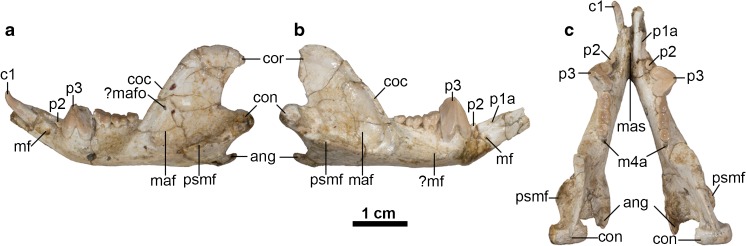
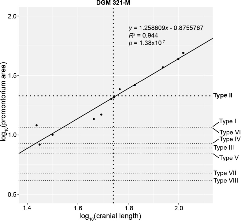
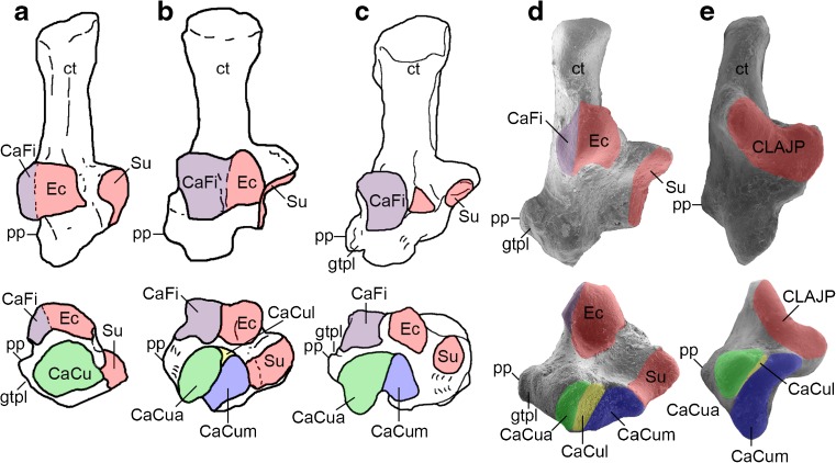
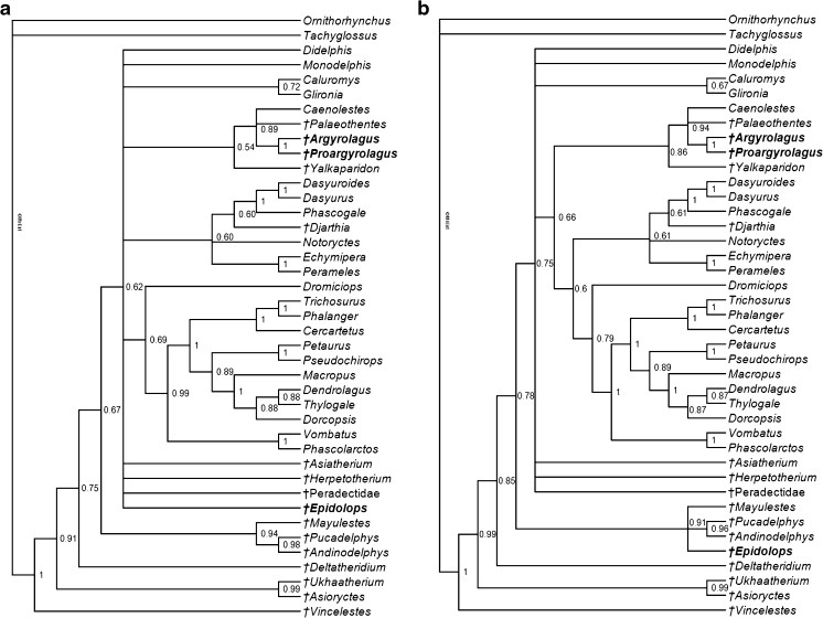
The skull of the polydolopimorphian marsupialiform Epidolops ameghinoi is described in detail for the first time, based on a single well-preserved cranium and associated left and right dentaries plus additional craniodental fragments, all from the early Eocene (53-50 million year old) Itaboraí fauna in southeastern Brazil. Notable craniodental features of E. ameghinoi include absence of a masseteric process, very small maxillopalatine fenestrae, a prominent pterygoid fossa enclosed laterally by a prominent ectopterygoid crest, an absent or tiny transverse canal foramen, a simple, planar glenoid fossa, and a postglenoid foramen that is immediately posterior to the postglenoid process. Most strikingly, the floor of the hypotympanic sinus was apparently unossified, a feature found in several stem marsupials but absent in all known crown marsupials. "Type II" marsupialiform petrosals previously described from Itaboraí plausibly belong to E. ameghinoi; in published phylogenetic analyses, these petrosals fell outside (crown-clade) Marsupialia. "IMG VII" tarsals previously referred to E. ameghinoi do not share obvious synapomorphies with any crown marsupial clade, nor do they resemble those of the only other putative polydolopimorphians represented by tarsal remains, namely the argyrolagids. Most studies have placed Polydolopimorphia within Marsupialia, related to either Paucituberculata, or to Microbiotheria and Diprotodontia. However, diprotodonty almost certainly evolved independently in polydolopimorphians, paucituberculatans and diprotodontians, and Epidolops does not share obvious synapomorphies with any marsupial order. Epidolops is dentally specialized, but several morphological features appear to be more plesiomorphic than any crown marsupial. It seems likely Epidolops that falls outside Marsupialia, as do morphologically similar forms such as Bonapartherium and polydolopids. Argyrolagids differ markedly in their known morphology from Epidolops but share some potential apomorphies with paucituberculatans. It is proposed that Polydolopimorphia as currently recognised is polyphyletic, and that argyrolagids (and possibly other taxa currently included in Argyrolagoidea, such as groeberiids and patagoniids) are members of Paucituberculata. This hypothesis is supported by Bayesian non-clock phylogenetic analyses of a total evidence matrix comprising DNA sequence data from five nuclear protein-coding genes, indels, retroposon insertions, and morphological characters: Epidolops falls outside Marsupialia, whereas argyrolagids form a clade with the paucituberculatans Caenolestes and Palaeothentes, regardless of whether the Type II petrosals and IMG VII tarsals are used to score characters for Epidolops or not. There is no clear evidence for the presence of crown marsupials at Itaboraí, and it is possible that the origin and early evolution of Marsupialia was restricted to the "Austral Kingdom" (southern South America, Antarctica, and Australia).

Keywords: Argyrolagidae; Eocene; Epidolops; Itaboraí; Marsupialia; Marsupialiformes; Polydolopimorphia.


Figures

Fig. 1
Fig. 1
Cranium of Epidolops ameghinoi (DGM 321-M - holotype) in dorsal view. a photograph; b interpretative drawing. Abbreviations: cf = carotid foramen; end = endocranial cavity; fr = frontal; fr-pa = reconstructed path of frontal-parietal suture; fro = foramen rotundum; gpa = glenoid process of the alisphenoid; ju = jugal; ?lac = ?lacrimal; mx = maxilla; na = nasal; pdp = posterodosal process of the premaxilla; pmx = premaxilla = pop = postorbital process; sq = squamosal
Fig. 2
Fig. 2
Cranium of Epidolops ameghinoi (DGM 321-M - holotype) in lateral view. a photograph of left lateral view; b interpretative drawing of left lateral view; c photograph of right lateral view; d interpretative drawing of right lateral view. Abbreviations: C1a = upper canine alveolus; fr = frontal; gf = glenoid fossa; I1a = first upper incisor alveolus; I2a = second upper incisor alveolus; I3a = third upper incisor alveolus; iof = position of infraorbital foramen; ju = jugal; ?lac = ?lacrimal; M1 = first upper molar; M2 = second upper molar; M3a = third upper molar alveoli; M4a = fourth upper molar alveolus; mx = maxilla; na = nasal; pdp = posterodosal process of the premaxilla; P2 = second upper premolar; P3 = third upper premolar; pgp = postglenoid process; pmx = premaxilla; sgf = supraglenoid foramina; sma = sulcus for masseter muscle; sq = squamosal
Fig. 3
Fig. 3
Cranium of Epidolops ameghinoi (DGM 321-M - holotype) in ventral view. a photograph; b interpretative drawing. Abbreviations: ?apf = ?accessory palatal foramen; appf = accessory posterolateral palatal foramen; C1a = upper canine alveolus; cf = carotid foramen; ecpc = ectopterygoid crest; enpc = entopterygoid crest; gf = glenoid fossa; gpa = glenoid process of the alisphenoid; hs = hypotympanic sinus; I1a = first upper incisor alveolus; I2a = second upper incisor alveolus; I3a = third upper incisor alveolus; if = incisive foramen; ju = jugal; M3a = third upper molar alveoli; M4a = fourth upper molar alveolus; ?mpf = ?major palatine foramen; mls = midline suture; mx = maxilla; pal = palatine; ?pc = ?pterygoid canal; pf = pterygoid fossa; pgf = postglenoid foramen; pgp = postglenoid process; plpf = posterolateral palatal foramen; pmx = premaxilla = ppt = postpalatine torus; prgp = preglenoid process; sph = sphenoid complex; sq = squamosal
Fig. 4
Fig. 4
Isolated partial right maxilla of Epidolops ameghinoi (DGM 898-M) in ventral view, with inferred extent of maxillopalatine fenestra indicated. Abbreviations: C1a = upper canine alveolus; if = incisive foramen; M4a = upper fourth molar alveolus; mpf = maxillopalatine fenestra; osm = area of origin of superficial masseter; P1a = upper first premolar alveolus; P3a = upper third premolar alveoli
Fig. 5
Fig. 5
Left basicranial region of Epidolops ameghinoi (DGM 321-M - holotype) in ventral view. a photograph; b interpretative drawing. cf = carotid foramen; ecpc = ectopterygoid crest; enpc = entopterygoid crest; gf = glenoid fossa; gpa = glenoid process of the alisphenoid; hs = hypotympanic sinus; ju = jugal; mls = midline suture; pf = pterygoid fossa; pgf = postglenoid foramen; pgp = postglenoid process; prgp = preglenoid process; sph = sphenoid complex; sq = squamosal
Fig. 6
Fig. 6
Left and right mandibles of Epidolops ameghinoi (DGM 321-M - holotype). a left mandible in lateral view; b right mandible in lateral view; c a left and right mandibles in dorsal view. Abbreviations: ang = angular process; c1 = lower canine; coc = coronoid crest; con = mandibular condyle; cor = coronoid process; m4a = fourth lower molar alveolus; maf = massteric foramen; mas = mandibular symphysis; mf = mental foramen; ?mafo = ?massteric foramen; ?mf = ?mental foramen; p1a = first lower premolar alveolus; p2 = second lower premolar; p3 = third lower premolar; psmf = posterior shelf of the massteric fossa
Fig. 7
Fig. 7
Partial right mandible of Epidolops ameghinoi (MNRJ 2880-V) in anterior view. Abbreviations: c1a = lower canine alveolus; i1a = first lower incisor alveolus; i2a = second lower incisor alveolus; i3a = third lower incisor alveolus; mf = mental foramen; p1a = first lower premolar alveolus
Fig. 8
Fig. 8
Reduced (‘standardized’) major axis regression of log10-transformed measurements of promontorium area against cranial length for 12 extant and fossil marsupialiforms (see Ladevèze and Muizon : table 2 and electronic supplementary material). The solid line represents the line of best fit. The dotted horizontal lines represent the log10(promontorium area) of the eight petrosal morphotypes (Types I-VIII) from Itaboraí described by Ladevèze (2004, 2007) and Ladevèze and Muizon (2010). The dotted vertical line represents log10(estimated cranial length) of Epidolops ameghinoi, based on DGM 321-M. The Type II petrosal morphotype is suitably-sized for referral to Epidolops ameghinoi, whereas the other morphotypes are too small
Fig. 9
Fig. 9
Isolated calcanea of a range of marsupialiforms in dorsal (flexad) and anterior (distal) views. a “Itaboraian Metatherian Group” (IMG) VII tarsal morphotype from Itaborai, which Szalay (1994) tentatively referred to Epidolops ameghinoi (redrawn from Szalay : fig. 6.25); b Unnamed argyrolagid from the Colhuehuapian (early Miocene) Gaiman locality (redrawn from Szalay : fig. 7.28); c Argyrolagus scagliai (redrawn from Szalay : fig. 7.28); d Caenolestes fuliginosus (AMNH M-62915 – see Beck 2012); e Dromiciops gliroides (unregistered UNSW Palaeontology Laboratory specimen– see Beck 2012). Abbreviations: CaCu = calcaneocuboid facet; CaCua = auxiliary calcaneocuboid facet; CaCul = lateral calcaneocuboid facet; CaCum = medial calcaneocuboid facet; CaFi = calcaneofibular facet; CLAJP = continuous lower ankle joint pattern; ct = calcaneal tuber; Ec = ectal facet; gtpl = groove for the tendon of the peroneus longus muscle; pp. = peroneal process; Su = sustentacular facet. Red represents the ectal (Ec) and sustentacular (Su) facets or continuous lower ankle joint pattern (CLAJP – formed by fusion of the ectal and sustentacular facets); green represents the calcaneocuboid (CaCu) facet or auxiliary calcaneocuboid (CaCua) facet (which are probably homologous – see Szalay ; Beck, 2012); yellow represents the lateral calcaneocuboid (CaCul) facet; blue represents the medial calcaneocuboid (CaCum) facet. Specimens are not drawn to scale. Note that the lateral calcaneocuboid (CaCul) facet is perpendicular to the page and so is not visible in Argyrolagus scagliai (c; compare with b, d and e)
Fig. 10
Fig. 10
Dentaries of Epidolops ameghinoi and putative relatives. a Epidolops ameghinoi (DGM 321-M; modified from Paula Couto 1952: fig. 6A); b the polydolopid Kramadolops abanicoi (SGOPY 2941 [reversed]; modified from Flynn and Wyss : fig. 1); c the argyrolagid Anargyrolagus primus (MPEF-PV 5299 [reversed]; modified from Goin and Abello : fig. 4.18); d the caenolestid Lestoros inca (USNM 194383; modified from Martin : fig. 4A). Abbreviations: c1 = lower canine; i1 = first lower incisor; i2a = second lower incisor alveolus; i3 = third lower incisor; p1 = first lower premolar; p1a = first lower premolar alveolus; p2 = second lower premolar; p3 = third lower premolar
Fig. 11
Fig. 11
Upper premolars and first upper molar of Epidolops ameghinoi and putative relatives. a Epidolops ameghinoi (DGM 800-M; modified from Marshall 1982: fig. 67b); b the bonapartheriid Bonapartherium hinakusijum (MMP 1416); c the gashterniid Gashternia carioca (MCN-PV 1801 [reversed]; modified from Goin and Oliveira : fig. 1); d the polydolopid Polydolops thomasi (MACN 10338; modified from Marshall 1982: fig. 32);. e the argyrolagid Anargyrolagus primus (MACN-ch-1305; modified from Carlini et al. : fig. 2A). Abbreviations: M1 = first upper molar; P2 = second upper premolar; P3 = third upper premolar
Fig. 12
Fig. 12
Basicranial region of Epidolops ameghinoi and other marsupialiforms. a Epidolops ameghinoi (DGM 321-M); b the mayulestid Mayulestes ferox (MHNC 1249; modified from Muizon : fig. 8A); c the caenolestid Caenolestes convelatus (modified from Animal Diversity Web); d the argyrolagid Argyrolagus scagliai (MMP 5538 – specimen is damaged and is missing the occipital region). Abbreviations: atp = alisphenoid tympanic process; cf = carotid foramen; ect = ectotympanic; fo = foramen ovale; gf = glenoid fossa; hs = hypotympanic sinus; pgf = postglenoid foramen; pgp = postglenoid process; pr = promontorium of the petrosal; rtpp = rostral tympanic process of the petrosal; tcf = transverse canal foramen. Note that a-c are in ventral view, whereas d is in ventromedial view
Fig. 13
Fig. 13
Comparison of the morphology of the retromolar space of the argyrolagid Argyrolagus and the caenolestid Caenolestes. a Partial left mandible of Argyrolagus sp. (MACN 17590) in medial view, with arrows indicating the path of the retrodental canal (= maxillary canal sensu Babot and García-López ; modified from Babot and García-López : fig. 9.1); b Left mandible of Caenolestes caniventer (BMNH 1954.302) in medial view, with vertical red line corresponding to the plane of the coronal section shown in c; c coronal section of left mandible of Caenolestes fuliginosus (KU 124015) in anterior view, based on CT scan data (modified from Digimorph). Abbreviations: can = canal; cor = coronoid process; m4 = fourth lower molar; manc = mandibular canal; manf = mandibular foramen; p3 = third lower premolar
Fig. 14
Fig. 14
Phylogenetic relationships of Epidolops, argyrolagids, and other metatherians, based on Bayesian undated analyses of a total evidence matrix modified from Beck et al. (2014); both analyses comprised four independent runs of 50 × 106 generations each, sampling trees every 2000 generations, and discarding the first 25 % (i.e., 12.5 × 106 generations) as burn-in. a 50 % majority rule consensus of post-burn-in trees from analysis of matrix in which the Type II petrosals and IMG VII tarsals from Itaboraí were not used to score characters for Epidolops (“Matrix A”); the harmonic mean of lnL across all four runs was −62,311.01; b 50 % majority rule consensus of post-burn-in trees from analysis of matrix in which the Type II petrosals and IMG VII tarsals from Itaboraí were used to score characters for Epidolops (“Matrix B″); the harmonic mean of lnL across all four runs was −62,336.95. Values at nodes are Bayesian posterior probabilities. Extinct taxa are identified by daggers, and Epidolops and the argyrolagids are highlighted in bold

Similar articles

Cited by

References

    1. Abbie AA. A masticatory adaptation peculiar to some diprotodont marsupials. Proc Zool Soc Lond. 1939;B109(2):261–279.
    1. Abello MA. Analysis of dental homologies and phylogeny of Paucituberculata (Mammalia: Marsupialia) Biol J Linn Soc. 2013;109(2):441–465.
    1. Abello MA, Candela AM. Postcranial skeleton of the Miocene marsupial Palaeothentes (Paucituberculata, Palaeothentidae): paleobiology and phylogeny. J Vertebr Paleontol. 2010;30(5):1515–1527.
    1. Abello MA, Rubilar-Rogers D. Revisión del género Abderites Ameghino, 1887 (Marsupialia, Paucituberculata) Ameghiniana. 2012;48(3):605–620.
    1. Ameghino F. Notices préliminaires sur les mammifères nouveaux des terrains crétacés de Patagonie. Boletín de la Academia Nacional de Ciencias de Córdoba. 1902;17:5–70.

LinkOut - more resources