Review
. 2017 Nov 15:8:2252.
doi: 10.3389/fmicb.2017.02252. eCollection 2017.

A Multilayered Control of the Human Survival Motor Neuron Gene Expression by Alu Elements

Affiliations
Review

A Multilayered Control of the Human Survival Motor Neuron Gene Expression by Alu Elements

Eric W Ottesen et al. Front Microbiol. .

Abstract

Humans carry two nearly identical copies of Survival Motor Neuron gene: SMN1 and SMN2. Mutations or deletions of SMN1, which codes for SMN, cause spinal muscular atrophy (SMA), a leading genetic disease associated with infant mortality. Aberrant expression or localization of SMN has been also implicated in other pathological conditions, including male infertility, inclusion body myositis, amyotrophic lateral sclerosis and osteoarthritis. SMN2 fails to compensate for the loss of SMN1 due to skipping of exon 7, leading to the production of SMNΔ7, an unstable protein. In addition, SMNΔ7 is less functional due to the lack of a critical C-terminus of the full-length SMN, a multifunctional protein. Alu elements are specific to primates and are generally found within protein coding genes. About 41% of the human SMN gene including promoter region is occupied by more than 60 Alu-like sequences. Here we discuss how such an abundance of Alu-like sequences may contribute toward SMA pathogenesis. We describe the likely impact of Alu elements on expression of SMN. We have recently identified a novel exon 6B, created by exonization of an Alu-element located within SMN intron 6. Irrespective of the exon 7 inclusion or skipping, transcripts harboring exon 6B code for the same SMN6B protein that has altered C-terminus compared to the full-length SMN. We have demonstrated that SMN6B is more stable than SMNΔ7 and likely functions similarly to the full-length SMN. We discuss the possible mechanism(s) of regulation of SMN exon 6B splicing and potential consequences of the generation of exon 6B-containing transcripts.

Keywords: Alu; SMA; SMN; SMN6B; exonization; spinal muscular atrophy; survival motor neuron; transposable elements.


Figures
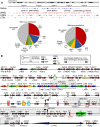
FIGURE 1
FIGURE 1
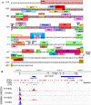
High prevalence of Alu-derived repeats in SMN locus. (A) Genomic overview of the duplicated genomic region in chromosome 5 encompassing the SMN genes. Upper panel indicates the location of genes and the three most prevalent types of repeats: SINEs, LINEs, and long terminal repeats (LTRs). Lower panel pie charts indicate the total percentage of sequence occupied by different types of repeats in either the whole SMN locus including other duplicated genes (left) or in the SMN gene itself (right). (B) Detailed view of the SMN gene and nearby surrounding sequences. SMN exons are indicated with colored boxes. Repeat sequences are indicated as colored arrows, with the direction of the arrow indicating the orientation of the repeat-derived sequence. An Alu-mediated recombination event which resulted in a deletion in an SMA patient (Wirth et al., 1999) is indicated. Two boxed regions indicate the location of known antisense transcripts derived from the SMN locus (d’Ydewalle et al., 2017; Woo et al., 2017).
FIGURE 2
FIGURE 2
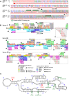
Alu repeats reshape transcriptional regulation of SMN. (A) Overview of the promoter region of SMN. Lowercase letters indicate untranscribed or untranslated regions, uppercase letters indicate coding sequences. A partial AluJb element is inserted from 289 to 54 bases upstream of the canonical TSS (TSS1a) and is indicated with a dark pink colored box. Other colored boxes indicate the locations of putative promoter elements and/or transcription factor binding sites. TSSs are indicated with black arrows. The start codon is written in blue and is indicated with a red arrow and circle. Promoter elements were either described previously (Singh et al., 2016) or were computationally predicted by ConSite (consite.genereg.net). (B) Locations of Polycomb-associated antisense transcripts derived from SMN. A genomic view of the SMN gene is shown. Location of SMN exons (E1–E8) and antisense transcripts are indicated with blue boxes, introns are indicated by lines with arrows indicating the direction of transcription. The locations of SINEs, LINEs, and LTRs are indicated. Four ChIP-Seq outputs are given below. The red peaks indicate the read depth from ChIP-Seq of human embryonic stem cells using antibodies against acetylated H3K27 (upper) or tri-methylated H3K4 (lower). The blue peaks indicate the same readout from umbilical cord-derived HUVEC cells. ChIP-Seq data was obtained from ENCODE and previously described by d’Ydewalle et al. (2017).
FIGURE 3
FIGURE 3
Consequences of Alu–Alu base pairing within SMN pre-mRNA. Diagrammatic representation of the partial SMN pre-mRNA (not to the scale) showing inclusion, skipping and circularization of exon 5. Exons are shown in colored boxes, whereas, red arrows indicated Alu elements. For simplicity, only two Alu elements per intron are shown. Base pairing between complementary Alu sequences are shown by stacked lines. Various hypothetical scenarios of splicing reactions involving different combinations of splice site pairings are shown by broken lines. Base pairing between Alu sequences within intron 5 promotes inclusion of exon 5, whereas, base pairing between intronic Alu sequences flanking exon 5 promotes exon 5 skipping. Base pairing between Alu elements within intron 5 combined with the base pairing between intronic Alu sequences upstream of exon 5 and downstream of exon 6 promotes circularization event.
FIGURE 4
FIGURE 4
Exon 6B is derived from an intronic Alu element. (A) Alignment of SMN intron 6 region spanning exon 6B. Numbering starts from the beginning of intron 6. Stars signify sequence identity. Hyphens designate the positions where gaps were introduced to maximize sequence identity. The gray box indicates exon 6B sequences and the green boxes indicate putative binding sites for hnRNP C. The black arrows indicate splice site (ss) positions of exon 6B. The red arrow indicates position and direction of AluY insertion (reverse and complement) which are obtained from Dfam (Accession: DF0000002). (B) Predicted splicing cis-elements. The exon 6B and 109 nt of upstream and downstream intronic sequences are shown. Colored boxes indicate potential regulatory elements identified by Human Splicing Finder (Desmet et al., 2009). Potential splicing enhancers are indicated above the SMN sequence and splicing silencers are below. Color code is explained in the bottom panel, where numbers indicate the software tool used for identification or publications in which motifs were originally described. Exonic splicing enhancer (ESE) finder is described in (Cartegni et al., 2003). RESCUE refers to an algorithm that predicts ESEs (Fairbrother et al., 2002). Octamer motifs are described in (Zhang and Chasin, 2004). Motifs 1-3 are described in (Sironi et al., 2004). Silencer motifs highlighted in pink are described in (Wang et al., 2004). (C) Secondary structure of SMN exon 6B. Numbering starts from the beginning of exon 6B. Exon 6B sequences are shown in capital letters, while adjacent intronic sequences are shown in lower-case letters. The red arrows indicate ss positions of exon 6B. The green arrows indicate sequence differences of exon 6B between human and primates. The secondary structure was predicted using mfold algorithm (Zuker, 2003).
FIGURE 5
FIGURE 5
A model of exon 6B action. (Left) Describes the transcription of the SMN gene and pre-mRNA splicing producing either the 6B-skipped (upper) or 6B-included (lower mRNA). Exons are indicated as colored boxes, the Alu element from which exon 6B is derived is indicated as a red arrow, introns are shown as lines. Potential splicing events are shown as red (exon 6B-skipped) or blue (exon 6B-included) dotted lines. Locations of stop codons generated by each potential transcript are indicated. (Right) Shows the computationally predicted glycine zipper dimers formed by the YG boxes at the C termini of each of the SMN protein isoforms. Both SMNΔ7 and SMN6B have altered YG boxes resulting in an increase in the inter-helical distances of the coiled-coil interaction, potentially reducing oligomerization. In SMNΔ7 this results in an unstable degron (Cho and Dreyfuss, 2010), whereas in SMN6B the destabilization is less pronounced (Seo et al., 2016a).

Similar articles

Cited by

References

    1. Ahmad S., Bhatia K., Kannan A., Gangwani L. (2016). Molecular mechanisms of neurodegeneration in spinal muscular atrophy. J. Exp. Neurosci. 10 39–49. 10.4137/jen.s33122 - DOI - PMC - PubMed
    1. Aktaş T., Ilik I. A., Maticzka D., Bhardwaj V., Rodrigues C. P., Mittler G., et al. (2017). DHX9 suppresses RNA processing defects originating from the Alu invasion of the human genome. Nature 544 115–119. 10.1038/nature21715 - DOI - PubMed
    1. Antonaki A., Demetriades C., Polyzos A., Banos A., Vatsellas G., Lavigne M. D., et al. (2011). Genomic analysis reveals a novel nuclear factor-kappa B (NF-kappa B)-binding Site in Alu-repetitive elements. J. Biol. Chem. 286 38768–38782. 10.1074/jbc.M111.234161 - DOI - PMC - PubMed
    1. Attig J., Mozos I., Haberman N., Wang Z., Emmett W., Zarnack K., et al. (2016). Splicing repression allows the gradual emergence of new Alu-exons in primate evolution. Elife. 5:e19545. 10.7554/eLife.19545 - DOI - PMC - PubMed
    1. Bouttier M., Laperriere D., Memari B., Mangiapane J., Fiore A., Mitchell E., et al. (2016). Alu repeats as transcriptional regulatory platforms in macrophage responses to M-tuberculosis infection. Nucleic Acids Res. 44 10571–10587. 10.1093/nar/gkw782 - DOI - PMC - PubMed

LinkOut - more resources