

Kaempferol Inhibits the Primary Attachment Phase of Biofilm Formation in Staphylococcus aureus
- PMID: 29187848
- PMCID: PMC5694784
- DOI: 10.3389/fmicb.2017.02263
Kaempferol Inhibits the Primary Attachment Phase of Biofilm Formation in Staphylococcus aureus
Abstract
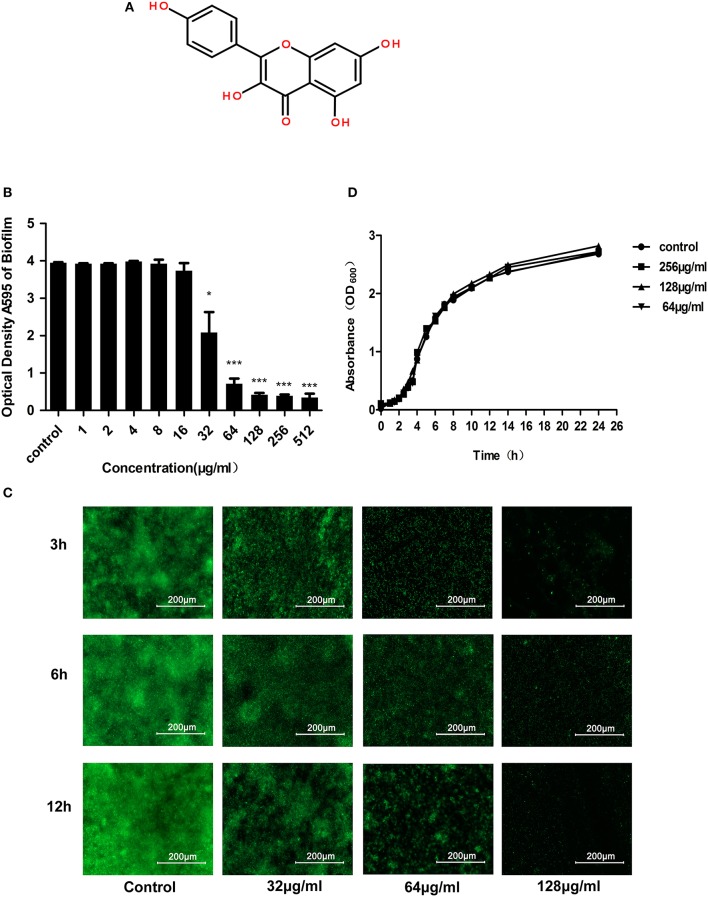
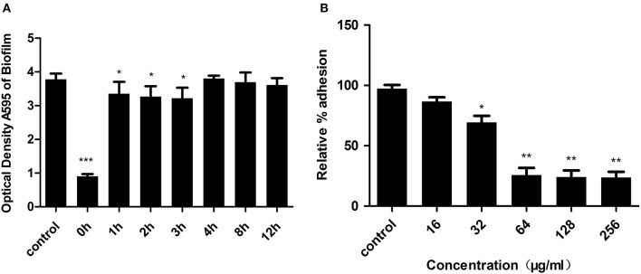
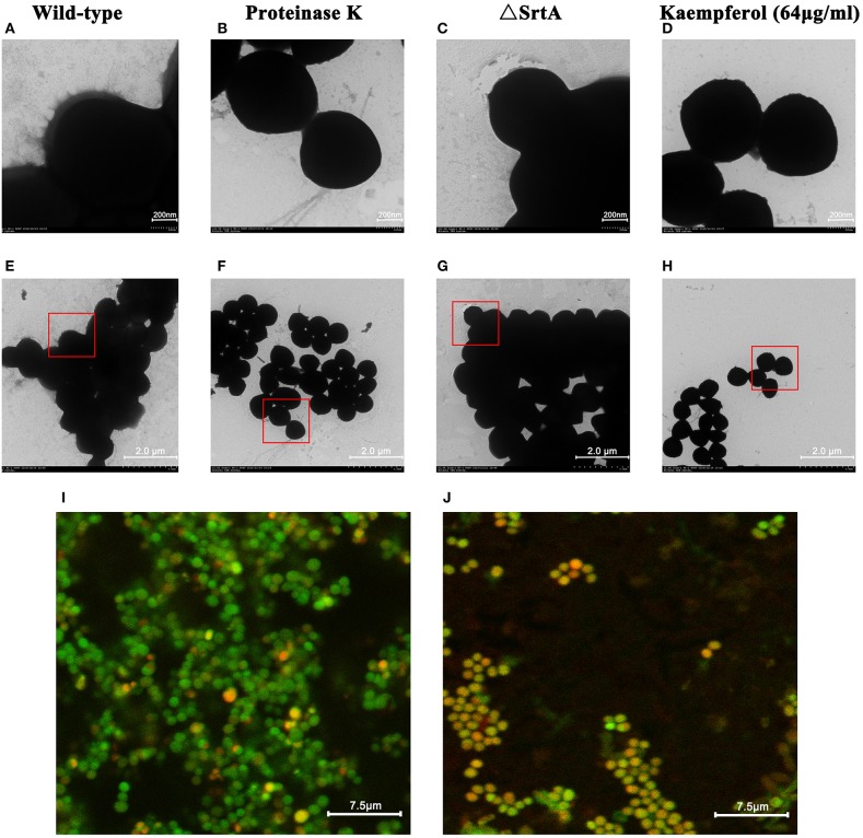
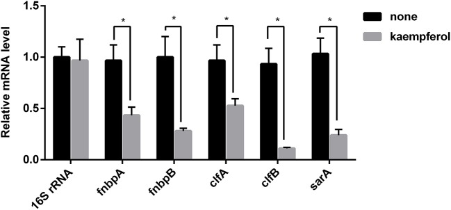
The ability to form biofilms on surfaces makes Staphylococcus aureus the main pathogenic factor in implanted medical device infections. The aim of this study was to discover a biofilm inhibitor distinct from the antibiotics used to prevent infections resulting from S. aureus biofilms. Here, we describe kaempferol, a small molecule with anti-biofilm activity that specifically inhibited the formation of S. aureus biofilms. Crystal violet (CV) staining and fluorescence microscopy clearly showed that 64 μg/ml kaempferol inhibited biofilm formation by 80%. Meanwhile, the minimum inhibitory concentration (MIC) and growth curve results indicated that kaempferol had no antibacterial activity against the tested bacterial strain. Kaempferol inhibited the primary attachment phase of biofilm formation, as determined by a fibrinogen-binding assay. Moreover, a fluorescence resonance energy transfer (FRET) assay and quantitative real-time reverse transcription polymerase chain reaction (qRT-PCR) analyses revealed that kaempferol reduced the activity of S. aureus sortaseA (SrtA) and the expression of adhesion-related genes. Based on these results, kaempferol provides a starting point for the development of novel anti-biofilm drugs, which may decrease the risk of bacterial drug resistance, to prevent S. aureus biofilm-related infections.
Keywords: Staphylococcus aureus; adhesion; biofilm; inhibitor; kaempferol; primary.
Figures

Similar articles
-
Small-Molecule Compound CY-158-11 Inhibits Staphylococcus aureus Biofilm Formation.Microbiol Spectr. 2023 Jun 15;11(3):e0004523. doi: 10.1128/spectrum.00045-23. Epub 2023 May 11. Microbiol Spectr. 2023. PMID: 37166296 Free PMC article.
-
Baicalein Inhibits Staphylococcus aureus Biofilm Formation and the Quorum Sensing System In Vitro.PLoS One. 2016 Apr 29;11(4):e0153468. doi: 10.1371/journal.pone.0153468. eCollection 2016. PLoS One. 2016. PMID: 27128436 Free PMC article.
-
Inhibitory effect of a natural phenolic compound, 3-p-trans-coumaroyl-2-hydroxyquinic acid against the attachment phase of biofilm formation of Staphylococcus aureus through targeting sortase A.RSC Adv. 2019 Oct 11;9(56):32453-32461. doi: 10.1039/c9ra05883d. eCollection 2019 Oct 10. RSC Adv. 2019. PMID: 35529766 Free PMC article.
-
Effect of paeonol against bacterial growth, biofilm formation and dispersal of Staphylococcus aureus and Listeria monocytogenes in vitro.Biofouling. 2022 Feb;38(2):173-185. doi: 10.1080/08927014.2022.2045014. Epub 2022 Feb 23. Biofouling. 2022. PMID: 35196921
-
20S-ginsenoside Rg3 inhibits the biofilm formation and haemolytic activity of Staphylococcus aureus by inhibiting the SaeR/SaeS two-component system.J Med Microbiol. 2022 Oct;71(10). doi: 10.1099/jmm.0.001587. J Med Microbiol. 2022. PMID: 36288093
Cited by
-
Comparative metabolite profiling of four polyphenol rich Morus leaves extracts in relation to their antibiofilm activity against Enterococcus faecalis.Sci Rep. 2022 Nov 23;12(1):20168. doi: 10.1038/s41598-022-24382-4. Sci Rep. 2022. PMID: 36424446 Free PMC article.
-
Comparative evaluation of small molecules reported to be inhibitors of Staphylococcus aureus biofilm formation.Microbiol Spectr. 2024 Jan 11;12(1):e0314723. doi: 10.1128/spectrum.03147-23. Epub 2023 Dec 7. Microbiol Spectr. 2024. PMID: 38059629 Free PMC article.
-
Insight into the effect of quinic acid on biofilm formed by Staphylococcus aureus.RSC Adv. 2019 Jan 29;9(7):3938-3945. doi: 10.1039/c8ra09136f. eCollection 2019 Jan 25. RSC Adv. 2019. PMID: 35518066 Free PMC article.
-
Unveiling the anti-inflammatory potential of Acalypha indica L. and analyzing its research trend: digging deep to learn deep.Naunyn Schmiedebergs Arch Pharmacol. 2024 Apr;397(4):1935-1956. doi: 10.1007/s00210-023-02734-9. Epub 2023 Oct 5. Naunyn Schmiedebergs Arch Pharmacol. 2024. PMID: 37796311 Review.
-
Co-regulation of Thermosensor Pathogenic Factors by C-di-GMP-Related Two-Component Systems and a cAMP Receptor-like Protein (Clp) in Stenotrophomonas maltophilia.Foods. 2024 Apr 15;13(8):1201. doi: 10.3390/foods13081201. Foods. 2024. PMID: 38672874 Free PMC article.
References
-
- Aaron S. D., Ferris W., Ramotar K., Vandemheen K., Chan F., Saginur R. (2002). Single and combination antibiotic susceptibilities of planktonic, adherent, and biofilm-grown Pseudomonas aeruginosa isolates cultured from sputa of adults with cystic fibrosis. J. Clin. Microbiol. 40, 4172–4179. 10.1128/JCM.40.11.4172-4179.2002 - DOI - PMC - PubMed
-
- Abouelhassan Y., Garrison A. T., Burch G. M., Wong W., Norwood V. M., IV., Huigens R. W., III. (2014). Discovery of quinoline small molecules with potent dispersal activity against methicillin-resistant Staphylococcus aureus and Staphylococcus epidermidis biofilms using a scaffold hopping strategy. Bioorg. Med. Chem. Lett. 24, 5076–5080. 10.1016/j.bmcl.2014.09.009 - DOI - PubMed
-
- Aendekerk S., Diggle S. P., Song Z., Hoiby N., Cornelis P., Williams P., et al. . (2005). The MexGHI-OpmD multidrug efflux pump controls growth, antibiotic susceptibility and virulence in Pseudomonas aeruginosa via 4-quinolone-dependent cell-to-cell communication. Microbiology 151, 1113–1125. 10.1099/mic.0.27631-0 - DOI - PubMed
Grants and funding
LinkOut - more resources
Full Text Sources
Other Literature Sources