. 2017 Nov 15:8:1529.
doi: 10.3389/fimmu.2017.01529. eCollection 2017.

Modulation of P2X7 Receptor during Inflammation in Multiple Sclerosis

Affiliations

Modulation of P2X7 Receptor during Inflammation in Multiple Sclerosis

Susanna Amadio et al. Front Immunol. .

Abstract

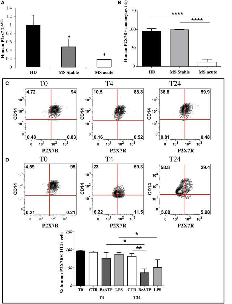
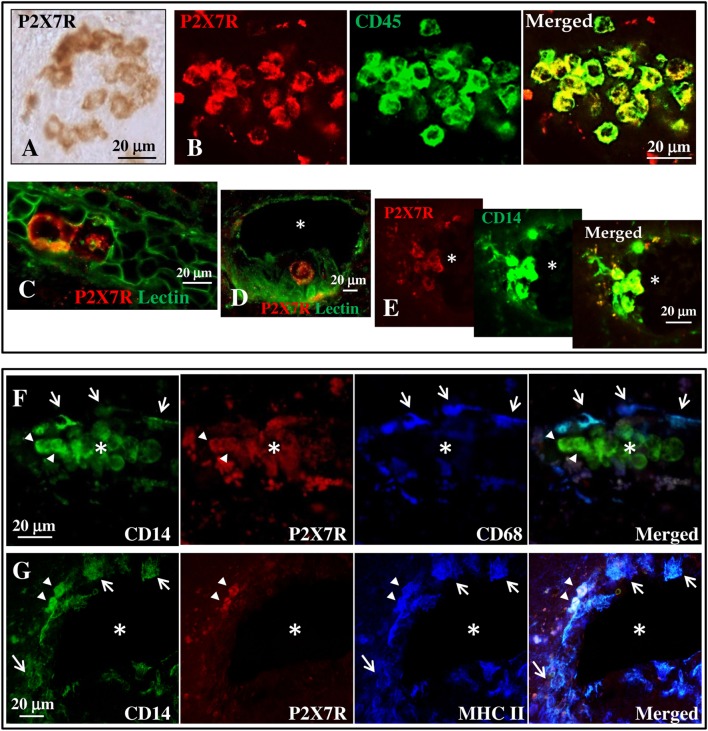
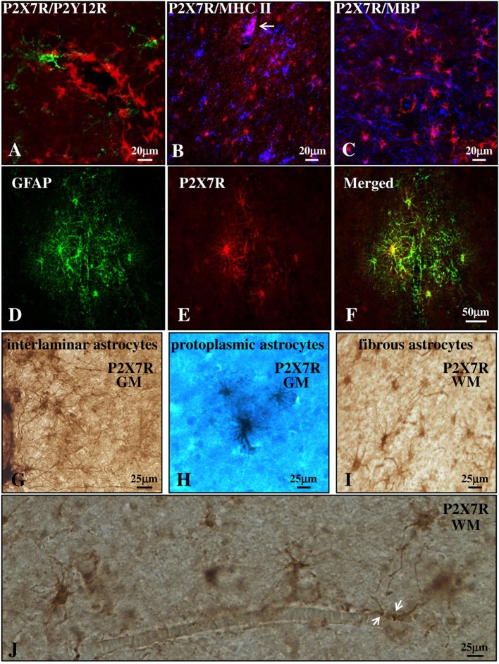
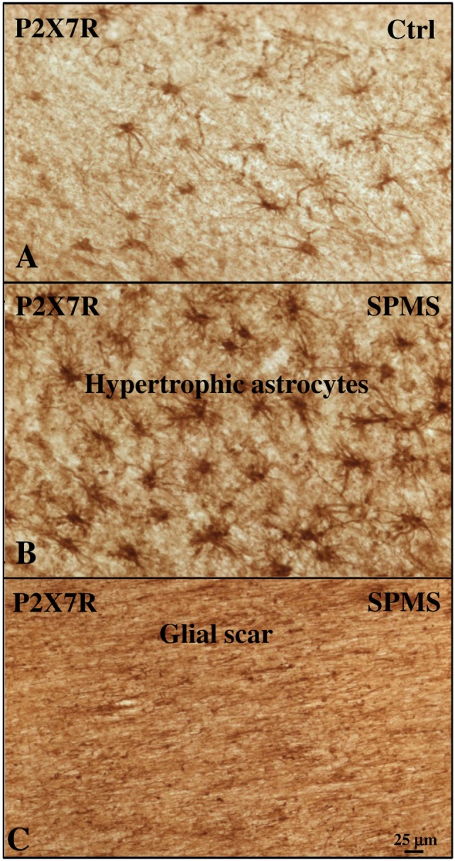
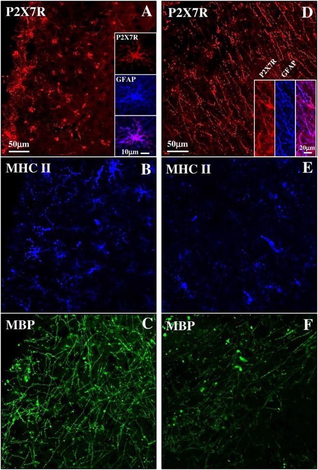
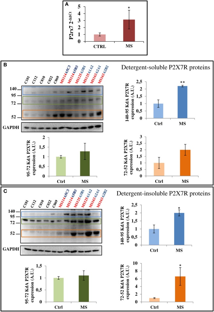
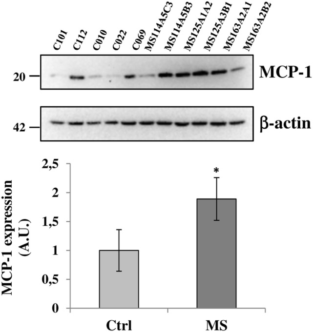
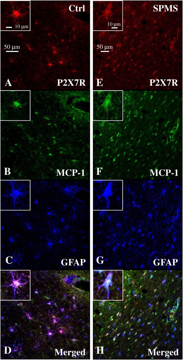
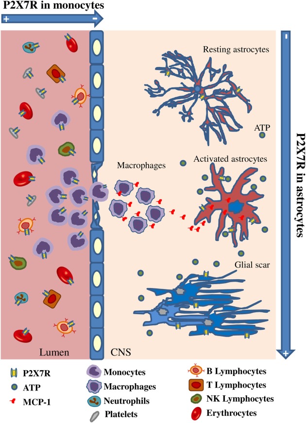
Multiple sclerosis (MS) is characterized by macrophage accumulation and inflammatory infiltrates into the CNS contributing to demyelination. Because purinergic P2X7 receptor (P2X7R) is known to be abundantly expressed on cells of the hematopoietic lineage and of the nervous system, we further investigated its phenotypic expression in MS and experimental autoimmune encephalomyelitis conditions. By quantitative reverse transcription polymerase chain reaction and flow cytometry, we analyzed the P2X7R expression in human mononuclear cells of peripheral blood from stable and acute relapsing-remitting MS phases. Human monocytes were also challenged in vitro with pro-inflammatory stimuli such as the lipopolysaccharide, or the P2X7R preferential agonist 2'(3')-O-(4 Benzoylbenzoyl)adenosine 5'-triphosphate, before evaluating P2X7R protein expression. Finally, by immunohistochemistry and immunofluorescence confocal analysis, we investigated the P2X7R expression in frontal cortex from secondary progressive MS cases. We demonstrated that P2X7R is present and inhibited on peripheral monocytes isolated from MS donors during the acute phase of the disease, moreover it is down-regulated in human monocytes after pro-inflammatory stimulation in vitro. P2X7R is instead up-regulated on astrocytes in the parenchyma of frontal cortex from secondary progressive MS patients, concomitantly with monocyte chemoattractant protein-1 chemokine, while totally absent from microglia/macrophages or oligodendrocytes, despite the occurrence of inflammatory conditions. Our results suggest that inhibition of P2X7R on monocytes and up-regulation in astrocytes might contribute to sustain inflammatory mechanisms in MS. By acquiring further knowledge about P2X7R dynamics and identifying P2X7R as a potential marker for the disease, we expect to gain insights into the molecular pathways of MS.

Keywords: P2X7 receptor; astrocytes; monocyte chemoattractant protein-1; monocytes; multiple sclerosis; neuroinflammation.


Figures

Figure 1
Figure 1
P2X7 receptor (P2X7R) is down-regulated on circulating multiple sclerosis (MS) monocytes and on healthy donors (HD) monocytes after in vitro induced inflammation. (A) RT-qPCR analysis of P2X7R was performed with freshly isolated monocytes from MS stable (n = 8), acute patients (n = 8), and HD (n = 8). GAPDH was used for normalization. (B) Flow cytometry analysis was used to isolate cluster of differentiation 14 (CD14)-positive monocytes and P2X7R-CD14 double-positive cells within freshly isolated peripheral blood mononuclear cells from acute, stable MS patients, and HD. Cumulative data of P2X7R-positive cells within monocytes are reported as % mean ± SEM (n = 5). Statistical significance was calculated by ANOVA-Student’s t-test, ****p < 0.0001 and *p < 0.05. Circulating monocytes purified from HD were incubated with inflammatory lipopolysaccharide (250 ng/ml) (C) or 2′3′-O-(4-benzoyl-benzoyl)adenosine5′-triphosphate (250 µM) (D) for 4 and 24 h. Flow cytometry analysis and representative plots of P2X7R expression are shown (C,D), together with cumulative data from three independent experiments are presented (E). Statistical significance was calculated by ANOVA-Student’s t-test *p < 0.05, **p < 0.01.
Figure 2
Figure 2
P2X7 receptor (P2X7R) is present on monocytes in blood vessels of secondary progressive multiple sclerosis (SPMS) frontal cortex. (A) Immunohistochemistry on sections from human frontal cortex shows roundish P2X7R-positive cells distributed within distinct clusters throughout the entire SPMS tissue. Confocal double immunofluorescence analysis of these clusters exhibits colocalization of P2X7R protein (red) with CD45 leukocyte marker [(B), green]. Staining with Lectin from Lycopersicon esculentum for vascular endothelial cells [(C,D), green] clearly shows the presence of P2X7R-positive roundish cells (red) within the lumen of blood vessels (asterisk). Double immunofluorescence of P2X7R-positive clusters shows colocalization of P2X7R (red) with cluster of differentiation 14 (CD14) monocyte/macrophage marker [(E), green]. Confocal triple immunofluorescence analysis is then performed with antibodies for CD14 [(F,G), green], P2X7R [(F,G), red], and microglia/macrophages markers CD68 (F) or major histocompatibility complex II [(G), blue]. The asterisk always indicates the lumen of blood vessels, arrows indicate P2X7R-negative cells, and arrowheads P2X7R-positive cells.
Figure 3
Figure 3
P2X7 receptor (P2X7R) is present on astrocytes in the parenchyma of secondary progressive multiple sclerosis (SPMS) frontal cortex. Confocal analysis of SPMS frontal cortex parenchyma shows absence of colocalization of P2X7R (red) with P2Y12R [(A), green], major histocompatibility complex II (MHC II) [(B), blue], and myelin basic protein [(C), blue], but the presence of merged P2X7R/glial fibrillary acidic protein signals (D–F). P2X7R/MHC II-positive signal is also visible but confined in the lumen of a blood vessel [(B), arrow, pink]. Immunohistochemistry analysis of cortical parenchyma reveals the abundant presence of P2X7R in GM on interlaminar (G) and protoplasmic astrocytes (E), and in white matter on fibrous astrocytes (I,J). In (J), astrocytic “vascular feet” are visible adjacent to the blood vessel walls (arrows).
Figure 4
Figure 4
P2X7 receptor (P2X7R) immunoreactivity is increased with astrogliosis in white matter (WM) of secondary progressive multiple sclerosis (SPMS) frontal cortex parenchyma. Immunohistochemistry analysis of two adjacent WM fields from SPMS frontal cortex characterized, respectively, by hypertrophic astrocytes (B) and glial scar (C) reveals a noteworthy increase in P2X7R-positive astrocytes (B,C), compared with control case (A).
Figure 5
Figure 5
P2X7 receptor (P2X7R) expression in both active and inactive subpial lesions of secondary progressive multiple sclerosis (SPMS) frontal cortex. Confocal triple immunofluorescence analysis performed with antibodies for P2X7R [(A,D), red], major histocompatibility complex II (MHC II) [(B,E), blue], and myelin basic protein (MBP) [(C,F), green] on SPMS frontal cortex sections, shows a chronic active subpial lesion (A–C) with abundant glial fibrillary acidic protein (GFAP)/P2X7R-positive signal (A, inset), with reactive MHC II-positive monocyets/macrophages/microglia (blue) and with MBP-positive myelin fibers (green). In a chronic inactive lesion (D–F), an intense GFAP/P2X7R glial scar is shown [(D), inset], with decreased MHC II-positive [(E), blue] and MBP-positive [(F), green] immunoreactivities.
Figure 6
Figure 6
P2X7 receptor (P2X7R) mRNA and protein are augmented in secondary progressive multiple sclerosis. (A) Total RNA was extracted from three different snap-frozen blocks from MS patients (cases MS114, MS125, and MS163) and six controls (cases C12–101, C12–112, C13–010, C13–022, C14–069, and C14–053) and the expression of P2X7R mRNA was examined by RT-qPCR. Cortical protein extracts (15 μg/well) from different tissue blocks (A5C3, A5B3, A3B1, A1A2, A2A1, and A2B2) of MS cases MS114, MS125, and MS163 were analyzed by western blotting for the modulation of P2X7R, with respect to control cases (C12–101, C12–112, C13–010, C13–022, and C14–069), in both detergent-soluble (B) and -insoluble fractions (C). GAPDH was used for protein normalization. Statistical significance was calculated by Student’s t-test, *p < 0.05, **p < 0.01. Results are shown as mean ± SEM.
Figure 7
Figure 7
Monocyte chemoattractant protein-1 (MCP-1) chemokine is up-regulated in secondary progressive multiple sclerosis frontal cortex. Equal amount of total protein extracts of cortical tissue (15 μg/well) from different tissue blocks (A5C3, A5B3, A3B1, A1A2, A2A1, and A2B2) of multiple sclerosis (MS) cases MS114, MS125, and MS163 was analyzed by western blotting for the expression of MCP-1 chemokine with respect to control cases (C12–101, C12–112, C13–010, C13–022, and C14–069). β-actin was used for protein normalization. Statistical significance was calculated by Student’s t-test, *p < 0.05. Results are shown as mean ± SEM.
Figure 8
Figure 8
Monocyte chemoattractant protein-1 (MCP-1) chemokine colocalizes with P2X7 receptor (P2X7R) on astrocytes in secondary progressive multiple sclerosis (SPMS) frontal cortex. Triple immunofluorescence confocal analysis performed on sections of frontal cortex from control (A–D) and SPMS (E–H) with antibodies for P2X7R [(A,E), red], MCP-1 [(B,F), green], and glial fibrillary acidic protein [(C,G), blue] shows colocalization [(D,H), white signal] and strong up-regulation of signals in white matter of SPMS (E–H).
Figure 9
Figure 9
Spatiotemporal profile of P2X7 receptor (P2X7R) expression in multiple sclerosis (MS). The cartoon describes that P2X7R is down-regulated in monocytes during their activation and extravasation from blood vessel to MS cerebral cortex, while the receptor and the monocyte chemoattractant protein-1 chemokine are up-regulated on MS astrocytes concurrently with increased glial fibrillary acidic protein signal and glial scar formation.

Similar articles

Cited by

References

    1. Yamasaki R, Lu H, Butovsky O, Ohno N, Rietsch AM, Cialic R, et al. Differential roles of microglia and monocytes in the inflamed central nervous system. J Exp Med (2014) 211:1533–49.10.1084/jem.20132477 - DOI - PMC - PubMed
    1. Williams A, Piaton G, Lubetzki C. Astrocytes—friends or foes in multiple sclerosis? Glia (2007) 55:1300–12.10.1002/glia.20546 - DOI - PubMed
    1. Nair A, Frederick TJ, Miller SD. Astrocytes in multiple sclerosis: a product of their environment. Cell Mol Life Sci (2008) 65:2702–20.10.1007/s00018-008-8059-5 - DOI - PMC - PubMed
    1. Brosnan CF, Raine CS. The astrocyte in multiple sclerosis revisited. Glia (2013) 61:453–65.10.1002/glia.22443 - DOI - PubMed
    1. Correale J, Farez MF. The role of astrocytes in multiple sclerosis progression. Front Neurol (2015) 6:180.10.3389/fneur.2015.00180 - DOI - PMC - PubMed