doi: 10.14791/btrt.2017.5.2.105.
Epub 2017 Oct 31.
Supratentorial Pilocytic Astrocytoma Mimicking Convexity Meningioma with Early Anaplastic Transformation: A Case Report
Affiliations
- PMID: 29188212
- PMCID: PMC5700022
- DOI: 10.14791/btrt.2017.5.2.105
Item in Clipboard
Supratentorial Pilocytic Astrocytoma Mimicking Convexity Meningioma with Early Anaplastic Transformation: A Case Report
Brain Tumor Res Treat.
2017 Oct.
Abstract
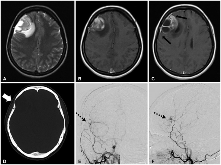
Meningiomas and pilocytic astrocytomas are benign intracranial tumors. Pilocytic astrocytomas arises frequently at the posterior fossa in childhood. Meningiomas have several image findings, such as a dural tail sign, bony erosion, and sunburst appearance on angiography. However, pilocytic astrocytomas with these findings have been rarely reported. In this report, we describe a mass with typical image findings of a meningioma, but diagnosed as a supratentorial pilocytic astrocytoma with early anaplastic transformation.
Keywords: Anaplasia; Astrocytoma; Meningioma; Radiography.
Conflict of interest statement
Conflicts of Interest: The authors have no financial conflicts of interest.
Figures
Similar articles
-
A pilocytic astrocytoma mimicking a clinoidal meningioma.Case Rep Radiol. 2014;2014:524574. doi: 10.1155/2014/524574. Epub 2014 Mar 13. Case Rep Radiol. 2014. PMID: 24744944 Free PMC article.
-
Primary anaplastic pilocytic astrocytoma.J Clin Neurosci. 2009 Dec;16(12):1704-6. doi: 10.1016/j.jocn.2009.04.012. Epub 2009 Oct 7. J Clin Neurosci. 2009. PMID: 19815416
-
Anaplastic astrocytoma with piloid features, a novel molecular class of IDH wildtype glioma with recurrent MAPK pathway, CDKN2A/B and ATRX alterations.Acta Neuropathol. 2018 Aug;136(2):273-291. doi: 10.1007/s00401-018-1837-8. Epub 2018 Mar 21. Acta Neuropathol. 2018. PMID: 29564591
-
Juvenile pilocytic astrocytomas do not undergo spontaneous malignant transformation: grounds for designation as hamartomas.Br J Ophthalmol. 2008 Jan;92(1):40-6. doi: 10.1136/bjo.2007.125567. Epub 2007 Oct 25. Br J Ophthalmol. 2008. PMID: 17962395 Review.
-
Recurrence patterns and anaplastic change in a long-term study of pilocytic astrocytomas.Pediatr Neurosurg. 1997 Jul;27(1):1-11. doi: 10.1159/000121218. Pediatr Neurosurg. 1997. PMID: 9486830 Review.
Cited by
-
Involvement of the Olfactory Apparatus by Gliomas.AJNR Am J Neuroradiol. 2020 Apr;41(4):712-717. doi: 10.3174/ajnr.A6471. Epub 2020 Mar 12. AJNR Am J Neuroradiol. 2020. PMID: 32165363 Free PMC article.
-
Low-Grade Astrocytoma Causing Dural and Calvarial Destruction.Asian J Neurosurg. 2023 Mar 31;18(1):223-227. doi: 10.1055/s-0043-1764325. eCollection 2023 Mar. Asian J Neurosurg. 2023. PMID: 37056894 Free PMC article.
References
-
- Shibahara I, Kawaguchi T, Kanamori M, et al. Pilocytic astrocytoma with histological malignant features without previous radiation therapy--case report. Neurol Med Chir (Tokyo) 2011;51:144–147. - PubMed
-
- Qi ST, Liu Y, Pan J, Chotai S, Fang LX. A radiopathological classification of dural tail sign of meningiomas. J Neurosurg. 2012;117:645–653. - PubMed
LinkOut - more resources
Full Text Sources
Other Literature Sources