. 2017 Dec;12(12):2531-2553.
doi: 10.1038/nprot.2017.120. Epub 2017 Nov 16.

Multimodal profiling of single-cell morphology, electrophysiology, and gene expression using Patch-seq

Affiliations

Multimodal profiling of single-cell morphology, electrophysiology, and gene expression using Patch-seq

Cathryn R Cadwell et al. Nat Protoc. 2017 Dec.

Abstract

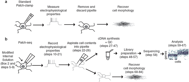
Neurons exhibit a rich diversity of morphological phenotypes, electrophysiological properties, and gene-expression patterns. Understanding how these different characteristics are interrelated at the single-cell level has been difficult because of the lack of techniques for multimodal profiling of individual cells. We recently developed Patch-seq, a technique that combines whole-cell patch-clamp recording, immunohistochemistry, and single-cell RNA-sequencing (scRNA-seq) to comprehensively profile single neurons from mouse brain slices. Here, we present a detailed step-by-step protocol, including modifications to the patching mechanics and recording procedure, reagents and recipes, procedures for immunohistochemistry, and other tips to assist researchers in obtaining high-quality morphological, electrophysiological, and transcriptomic data from single neurons. Successful implementation of Patch-seq allows researchers to explore the multidimensional phenotypic variability among neurons and to correlate gene expression with phenotype at the level of single cells. The entire procedure can be completed in ∼2 weeks through the combined efforts of a skilled electrophysiologist, molecular biologist, and biostatistician.


Conflict of interest statement

COMPETING FINANCIAL INTERESTS

The authors declare no competing financial interests.

Figures

Figure 1.
Figure 1.. Schematic of Patch-seq technique.
a) Standard whole-cell patch clamp recording technique is used to record the electrophysiological properties of a neuron and dialyze the cell with a dye (e.g. biocytin) for later histological staining and recovery of cell morphology. b) For Patch-seq, a modified internal solution and patching approach is used. Cell contents are aspirated into the pipette after recording the cell’s electrophysiological properties and used for cDNA synthesis, amplification, and sequencing. The collapsed soma and axodendritic processes remain embedded in the tissue slice and can still be stained for morphological recovery. Adapted from (24).
Figure 2.
Figure 2.. Ideal patching approach for aspiration of cell contents.
a) The target cell (Step 14) is approached from the side (Step 17–18) rather than the top to provide the most direct path for cell contents to enter the pipette. After obtaining a gigaseal, the cell membrane is quickly ruptured so that the patch pipette contents can freely diffuse into the cell (Step 19). If using a high osmolarity internal solution, the cell will gradually swell over the next 5–15 min and can be helpful to move the pipette back a few microns (Step 21) to release the tension on the membrane and prevent current leakage. At the end of the recording session (~30 min if aiming for morphological reconstruction), the content of the cell are aspirated into the pipette using gentle, negative pressure (Step 22). The cell is continuously monitored, both visually and electrophysiologically, to ensure that no extracellular contents are entering the pipette and the seal between the pipette and the membrane is intact (no large current leakage). Once aspiration is complete, the negative pressure is released and the pipette is slowly pulled away from the cell to separate the cell membrane from the pipette, leaving the cell body intact in the slice (Step 23). Once the membrane has released, the pipette can be quickly removed from the tissue and the single-cell contents collected in a 0.2 mL PCR tube. b) Examples of collapse of the cell body under two-photon imaging guidance (2PI) following aspiration of cell contents into the pipette. For these experiments, Alexa-488 was added to the internal solution to enable visualization of the pipette and patched neuron. Scale bars, 10 μm. Adapted from (24) c) Examples of collapse of the cell body after aspiration of cell contents, visualized using differential interference contrast (DIC) imaging. See Supplementary Movie S1 for video of sample collection from DIC Cell 2. Scale bar, 10 μm. All animal procedures were approved by the Institutional Animal Care and Use Committee (IACUC) at Baylor College of Medicine.
Figure 3.
Figure 3.. Custom equipment.
a) 1 mL syringe with 0.2 μm syringe filter and tapered pipette tip, used for filtering internal solution and backfilling glass pipettes (Steps 8–9, 15). b) Patch pipette marked at approximately 0.3 μL volume, used as a reference while loading other pipettes (Step 15). c) Positive pressure device used to eject pipette contents into PCR tube after aspirating cell contents (Step 24). d) Staining chamber used for immunohistochemistry (Step 68).
Figure 4.
Figure 4.. cDNA yield using physiological and high osmolarity internal solution.
Amplified cDNA yield (a) and mean size (b) as a function of recording time for both types of internal solution used. The physiological internal results in slightly lower yield of cDNA compared to the high osmolarity internal solution (likely due to a reduction in the amount of RRI), however no further decrease in yield is noted when recording time is extended up to 45 min. The grey and black “X”‘s indicate negative controls (no cell patched) for the high osmolarity and physiologic osmolarity internal solutions, respectively. All animal procedures were approved by the Institutional Animal Care and Use Committee (IACUC) at Baylor College of Medicine.
Figure 5.
Figure 5.. Quality check of full-length single-cell cDNA libraries.
Bioanalyzer profiles of single-cell amplified cDNA from several cells from a typical experiment (a) showing variable cDNA yield, as well as a negative control from the same experiment that consisted of ERCC spike-in RNA only (diluted 1:4×10⁶ in the RT reaction). Profiles from degraded (b) or contaminated (c) samples are also shown. The low molecular weight fragments in (c) were traced back to bacterial contamination of a particular reagent. All animal procedures were approved by the Institutional Animal Care and Use Committee (IACUC) at Baylor College of Medicine.
Figure 6.
Figure 6.. Bioanalyzer profile of pooled cDNA library.
Example of bioanalyzer profile showing the size distribution of the final pooled sequencing library for 224 Patch-seq neurons. The mean size of the cDNA fragments for this library (integrated from ~100 bp to 8 kb) is 336 bp and the concentration is ~3 ng/μL. All animal procedures were approved by the Institutional Animal Care and Use Committee (IACUC) at Baylor College of Medicine.
Figure 7.
Figure 7.. Initial Patch-seq data processing steps.
Quality of the samples is assessed by analyzing the sequencing depth (a), number of detected genes (b), maximum spearman correlation between each cell and all other cells (c), and fraction of reads mapped to exons, introns and intergenic segments (d). This example dataset originally contained of 224 neurons which were primarily excitatory (n = 218/224, ~97%). Twenty-eight cells were discarded based on QC criteria (dashed lines in a and b represent the QC cutoffs), leaving 196 for subsequent analyses. e) Histogram of mean normalized expression across cells for all genes in the example dataset. Genes with a mean expression less than one (dotted line) may be excluded to minimize the impact of gene dropout on further analyses. Panels a, b, and e were generated by following the tutorial described in (30). All animal procedures were approved by the Institutional Animal Care and Use Committee (IACUC) at Baylor College of Medicine.
Figure 8.
Figure 8.. Direct morphological recovery of Patch-seq cells.
a) cDNA yield as a function of recording time. Points are color coded according to the quality of the morphological recovery. b) Venn diagram summarizing success rate for obtaining electrophysiological, morphological and cDNA information from single cells. Examples of two excitatory (c,e) and two inhibitory (d,f) neurons demonstrating successful characterization of all three data modalities: morphology (left), electrophysiology (right, upper panel) and full-length cDNA profile assessed using an Agilent Bioanalyzer (right, lower panel). Scale bars for immunohistochemical staining, 50 μm. Scale bars for electrophysiological traces, 100 ms and 50 mV/500pA. All animal procedures were approved by the Institutional Animal Care and Use Committee (IACUC) at Baylor College of Medicine.

References

Key References

    1. Cadwell CR, Palasantza A, Jiang X, Berens P, Deng Q, Yilmaz M, Reimer J, Shen S, Bethge M, Tolias KF, Sandberg R & Tolias AS (2016). Electrophysiological, transcriptomic and morphologic profiling of single neurons using Patch-seq. Nat Biotechnol, 34 (2), 199–203. - PMC - PubMed
    1. Cadwell CR & Tolias AS (2016) Correlating cellular morphology, physiology, and gene expression using Patch-Seq. In: Using single-cell genomics to analyze neurons, glia, and circuits. (McCarroll S, ed) pp. 41–51. San Diego, CA: Society for Neuroscience.
    1. Cadwell CR, Sandberg R, Jiang X & Tolias AS (2017). Q&A: using Patch-seq to profile single cells. BMC Biology, 15 (1), 58. - PMC - PubMed

References

    1. Tang F et al., mRNA-Seq whole-transcriptome analysis of a single cell. Nature methods 6, 377–382 (2009). - PubMed
    1. Picelli S et al., Smart-seq2 for sensitive full-length transcriptome profiling in single cells. Nature methods 10, 1096–1098 (2013). - PubMed
    1. Macosko EZ et al., Highly Parallel Genome-wide Expression Profiling of Individual Cells Using Nanoliter Droplets. Cell 161, 1202–1214 (2015). - PMC - PubMed
    1. Hashimshony T, Wagner F, Sher N, Yanai I, CEL-Seq: single-cell RNA-Seq by multiplexed linear amplification. Cell Rep 2, 666–673 (2012). - PubMed
    1. Jaitin DA et al., Massively parallel single-cell RNA-seq for marker-free decomposition of tissues into cell types. Science 343, 776–779 (2014). - PMC - PubMed