. 2017 Nov 29;551(7682):644-647.
doi: 10.1038/nature24659.

A semi-synthetic organism that stores and retrieves increased genetic information

Affiliations

A semi-synthetic organism that stores and retrieves increased genetic information

Yorke Zhang et al. Nature. .

Abstract

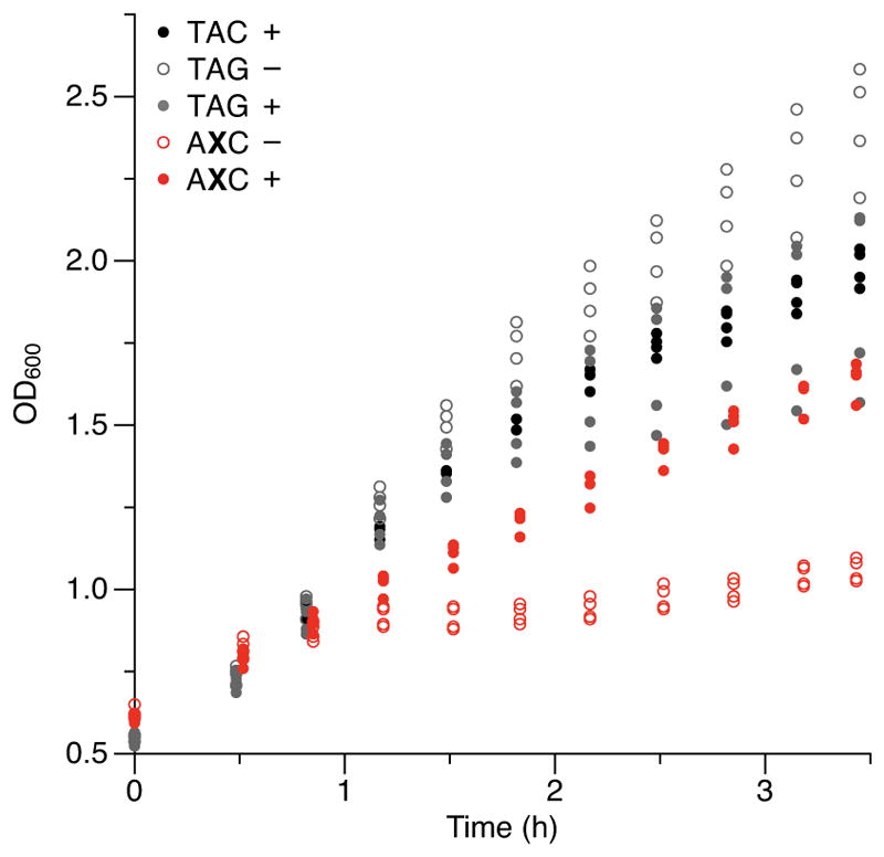
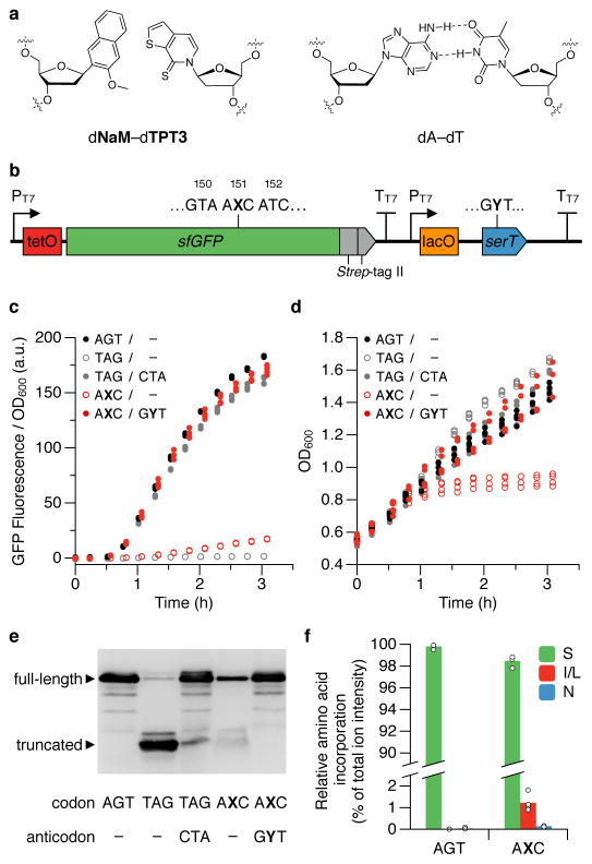
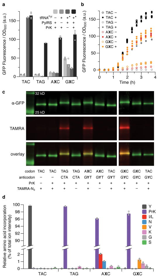
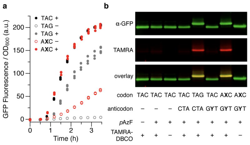
Since at least the last common ancestor of all life on Earth, genetic information has been stored in a four-letter alphabet that is propagated and retrieved by the formation of two base pairs. The central goal of synthetic biology is to create new life forms and functions, and the most general route to this goal is the creation of semi-synthetic organisms whose DNA harbours two additional letters that form a third, unnatural base pair. Previous efforts to generate such semi-synthetic organisms culminated in the creation of a strain of Escherichia coli that, by virtue of a nucleoside triphosphate transporter from Phaeodactylum tricornutum, imports the requisite unnatural triphosphates from its medium and then uses them to replicate a plasmid containing the unnatural base pair dNaM-dTPT3. Although the semi-synthetic organism stores increased information when compared to natural organisms, retrieval of the information requires in vivo transcription of the unnatural base pair into mRNA and tRNA, aminoacylation of the tRNA with a non-canonical amino acid, and efficient participation of the unnatural base pair in decoding at the ribosome. Here we report the in vivo transcription of DNA containing dNaM and dTPT3 into mRNAs with two different unnatural codons and tRNAs with cognate unnatural anticodons, and their efficient decoding at the ribosome to direct the site-specific incorporation of natural or non-canonical amino acids into superfolder green fluorescent protein. The results demonstrate that interactions other than hydrogen bonding can contribute to every step of information storage and retrieval. The resulting semi-synthetic organism both encodes and retrieves increased information and should serve as a platform for the creation of new life forms and functions.


Conflict of interest statement

The authors declare the following competing financial interests: A provisional patent application has been filed by Synthorx and The Scripps Research Institute (application #62/531,325; inventors J.L.P, C.E.C., H.R.A, Y.Z., E.C.F., A.W.F., V.T. Dien, and F.E.R.) covering the use of UBPs in tRNAs and mRNAs to produce proteins containing ncAAs. J.L.P, C.E.C., H.R.A., K.S.J., and F.E.R. have a financial interest (shares) in Synthorx, Inc., a company that has commercial interests in the UBP.

Figures

Extended data Figure 1
Extended data Figure 1. Fluorescence of cells expressing sfGFP with various codons at position 151
Strain YZ3 cells carrying a plasmid expressing TetR and an sfGFP plasmid with the indicated position-151 codon were grown to an OD600 ~0.5 and induced with IPTG and aTc. Fluorescence measurements were taken after 3 h of induction. Data shown as mean with individual values, n = 3 cultures originating from the same colony and grown in parallel (technical replicates).
Extended data Figure 2
Extended data Figure 2. Decoding of the AXC codon with natural near-cognate anticodons
a, b, Fluorescence (a) and growth (b) of cells expressing sfGFP(AXC)151 with or without tRNASer with the indicated anticodon. Cells were induced as described in Fig. 1c,d and fluorescence measurements correspond to the last time point shown in Fig. 1c. Values for the GYT anticodon and in the absence of tRNASer (−tRNA) correspond to the same values in Fig. 1c,d. Individual data points (with means in a) shown from n = 4 cultures, each propagated from an individual colony (biological replicates).
Extended data Figure 3
Extended data Figure 3. Western blots and growth of cells decoding AXC and GXC codons with tRNAPyl
a, Western blot of lysates (normalized by OD600) from cells expressing sfGFP with the indicated position 151-codon, in the presence (+) or absence (−) of a tRNAPyl with a cognate anticodon, PylRS, or 20 mM PrK in the media. Blots were probed with an α-GFP antibody (N-terminal epitope). Cells were induced and collected at an equivalent time point as described in Fig. 2b. For blot source data, see Supplemental Figure 1. b, Growth of cultures analyzed in Fig. 2a. The fold change in OD600 between induction of sfGFP (t = 1 h) and the final time point is greatest when all components necessary for aminoacylating tRNAPyl are present. Variations in the absolute value of OD600 are due to small variations in cell density at the start of T7 RNAP (and if present tRNAPyl) induction (t = 0). Individual data points shown from n = 4 cultures, each propagated from an individual colony (biological replicates).
Extended data Figure 4
Extended data Figure 4. Decoding of AXC and GXC codons with tRNAPyl and cell growth as a function of added unnatural ribotriphosphates
a, Fluorescence (upper panel) and western blots of purified sfGFP (lower panel) from cells expressing sfGFP and tRNAPyl with the position 151-codon/anticodon indicated, in the presence (+) or absence (−) of each unnatural ribotriphosphate in the media, and with or without 20 mM PrK. Cells were induced as described in Fig. 2b and fluorescence measurements were taken at the end of induction (~3.5 h), prior to collecting the cells and purifying the sfGFP protein for click conjugation of TAMRA and western blotting. Western blots were probed with an α-GFP antibody and imaged to detect both sfGFP and the conjugated TAMRA; all lanes correspond to sfGFP purified from cells grown with added PrK. Data shown as mean with individual values, n = 3 cultures, each propagated from an individual colony (biological replicates); n.d., not determined. For blot source data, see Supplemental Figure 1. b, Fluorescence and growth of cells expressing sfGFP(TAC)151 in the presence (+) or absence (−) of both unnatural deoxyribotriphosphates and each unnatural ribotriphosphate. t = 0 corresponds to the addition of IPTG to induce expression of T7 RNAP; aTc was added at t = 1 h to induce expression of sfGFP. Individual data points shown from n = 3 cultures, each propagated from an individual colony (biological replicates). At the concentrations used (see Methods), dNaMTP and dTPT3TP do not inhibit cell growth, whereas both unnatural ribotriphosphates, particularly TPT3TP, show some inhibition of growth. c, Cell growth corresponding to the cultures with added PrK (20 mM) whose fluorescence is shown in Fig. 2b. Cells expressing sfGFP with natural codons were grown without any unnatural triphosphates, whereas cells expressing sfGFP with unnatural codons were grown with both unnatural deoxy- and ribotriphosphates. Individual data points shown from n = 4 cultures, each propagated from an individual colony (biological replicates).
Extended data Figure 5
Extended data Figure 5. Decoding of AXC and GXC codons with tRNAPyl as a function of PrK concentration in the media
a, Western blots of sfGFP purified from cells expressing sfGFP and tRNAPyl with the indicated position-151 codon/anticodon, with conjugation of TAMRA and the addition of PrK to the media at the indicated concentrations. sfGFP was induced and purified from cells collected as described in Fig. 2b. Western blots were probed with an α-GFP antibody and imaged to detect both sfGFP and the conjugated TAMRA. For blot source data, see Supplemental Figure 1. b, Fluorescence of cells (measured at the last time point shown in c) expressing sfGFP and tRNAPyl with the indicated position-151 codon and anticodon, respectively, as a function of PrK concentration in the media. Fluorescence values for the 0 and 20 mM PrK conditions are the same as the (−) and (+) PrK conditions, respectively, shown in Fig. 2b. Individual data points shown from n = 4 cultures, each propagated from an individual colony (biological replicates). c, Timecourse analysis of fluorescence and cell growth in b. For clarity, only one representative culture (of four) is shown for each codon/anticodon pair and PrK concentration. We attribute the low level of sfGFP produced in the absence of PrK to decoding by endogenous tRNAs and loss of UBP retention in sfGFP (Extended data Table 2). However, the relative amount of sfGFP that contains PrK (a) and absolute amount of sfGFP expressed (b,c) increased in a dose-dependent manner with increasing PrK in the media, ultimately resulting in nearly full incorporation of PrK, suggesting that endogenous read-through of the AXC and GXC codons can be efficiently suppressed with sufficient concentrations of charged PrK-tRNAPyl(GYT) or PrK-tRNAPyl(GYC).
Extended data Figure 6
Extended data Figure 6. Cell growth of the cultures whose fluorescence is shown in Fig. 3a
Individual data points shown from n = 4 cultures, each propagated from an individual colony (biological replicates).
Figure 1
Figure 1. UBP and decoding with tRNASer
a, Chemical structure of the dNaM-dTPT3 UBP and a natural dA-dT base pair. b, Schematic illustration of the gene cassette used to express sfGFP(AXC)151 and tRNASer(GYT). PT7 and TT7 denote the T7 RNAP promoter and terminator, respectively. In controls where sfGFP is expressed in the absence of serT, the sequence following the sfGFP T7 terminator is absent. c, d, Fluorescence (c) and growth (d) of cells expressing sfGFP and tRNASer with the indicated position 151-codon and anticodon, respectively. Minus sign denotes the absence of serT in the expression cassette. t = 0 corresponds to the addition of IPTG to induce expression of T7 RNAP and tRNASer (if present); aTc was added at t = 0.5 h to induce expression of sfGFP. Individual data points shown from n = 4 cultures, each propagated from an individual colony (biological replicates). e, Western blot of lysates (normalized by OD600) from cells collected at the last time point shown in c and d, probed with an α-GFP antibody (N-terminal epitope). For blot source data, see Supplemental Figure 1. f, Relative abundance of amino acids at position 151 of sfGFP purified from cells expressing sfGFP(AGT)151 or sfGFP(AXC)151 and tRNASer(GYT), as determined by LC-MS/MS and precursor ion intensity based quantitation; amino acids detected at <0.1% (on average, for both codons) are not shown. Data shown as mean with individual values, n = 4 purified sfGFP samples, each from a culture propagated from an individual colony and collected at the last time point shown in c and d.
Figure 2
Figure 2. Incorporation of PrK using PylRS/tRNAPyl
a, Fluorescence of cells expressing sfGFP with the indicated position 151-codon, in the presence (+) or absence (−) of a tRNAPyl with a cognate anticodon, PylRS, or 20 mM PrK in the media, determined at the last time point in b. Asterisk denotes the absence of tRNAPyl in cells expressing sfGFP(TAC)151; n.d., not determined. Data shown as mean with individual values, n = 4 cultures, each propagated from an individual colony (biological replicates). b, Fluorescence timecourse of a subset of conditions in a (presence (+) or absence (−) of PrK in the media). t = 0 corresponds to the addition of IPTG to induce expression of PylRS, T7 RNAP, and tRNAPyl; aTc was added at t = 1 h to induce expression of sfGFP. Individual data points shown from n = 4 cultures, each propagated from an individual colony (biological replicates). c, Western blots and fluorescence scans of sfGFP purified from cells expressing sfGFP and tRNAPyl (if present) with the indicated position-151 codon and anticodon, respectively, with or without conjugation of TAMRA or addition of PrK to the media. sfGFP was purified from cultures collected at the last time point shown in b. For blot source data, see Supplemental Figure 1. d, Relative abundance of amino acids at position 151 of sfGFP purified from cells expressing sfGFP(TAC)151 or sfGFP and tRNAPyl with the indicated position-151 codon and a cognate anticodon, respectively, as determined by LC-MS/MS and precursor ion intensity based quantitation; amino acids detected at <0.1% (on average, for all codons) are not shown. Data shown as mean with individual values, n = 4 purified sfGFP samples, each from a culture propagated from an individual colony, grown in the presence of PrK, and collected at the last time point shown in b.
Figure 3
Figure 3. Incorporation of pAzF using pAzFRS/tRNApAzF
a, Fluorescence of cells expressing sfGFP(TAC)151 or sfGFP and tRNApAzF with the indicated position-151 codon and a cognate anticodon, respectively, in the presence (+) or absence (−) of 5 mM pAzF in the media. t = 0 corresponds to the addition of IPTG to induce expression of pAzFRS, T7 RNAP, and tRNApAzF; aTc was added at t = 0.5 h to induce expression of sfGFP. Individual data points shown from n = 4 cultures, each propagated from an individual colony (biological replicates). The fluorescence observed with sfGFP(AXC)151 in the absence of pAzF is attributed to charging of tRNApAzF(GYT) with a natural amino acid. b, Western blot and fluorescence scan of sfGFP purified from cells expressing sfGFP and tRNApAzF (if present) with the indicated position-151 codon and anticodon, respectively, with or without conjugation of TAMRA or addition of pAzF to the media. sfGFP was purified from cultures collected at the last time point shown in a. For blot source data, see Supplemental Figure 1.

Comment in

Similar articles

Cited by

References

    1. Leduc S. The Mechanisms of Life. Rebman Company; 1911.
    1. Zhang Y, et al. A semisynthetic organism engineered for the stable expansion of the genetic alphabet. Proc Natl Acad Sci USA. 2017;114:1317–1322. - PMC - PubMed
    1. Pedelacq JD, Cabantous S, Tran T, Terwilliger TC, Waldo GS. Engineering and characterization of a superfolder green fluorescent protein. Nat Biotechnol. 2006;24:79–88. - PubMed
    1. Mukai T, Lajoie MJ, Englert M, Soll D. Rewriting the genetic code. Annu Rev Microbiol. 2017;71 - PMC - PubMed
    1. Liu CC, Schultz PG. Adding new chemistries to the genetic code. Annu Rev Biochem. 2010;79:413–444. - PubMed

Publication types

MeSH terms