

Diffusion tractography reveals pervasive asymmetry of cerebral white matter tracts in the bottlenose dolphin (Tursiops truncatus)
- PMID: 29189908
- PMCID: PMC5884918
- DOI: 10.1007/s00429-017-1525-9
Diffusion tractography reveals pervasive asymmetry of cerebral white matter tracts in the bottlenose dolphin (Tursiops truncatus)
Abstract
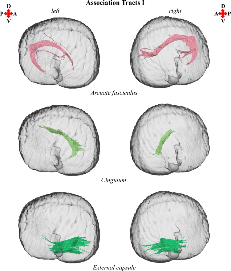
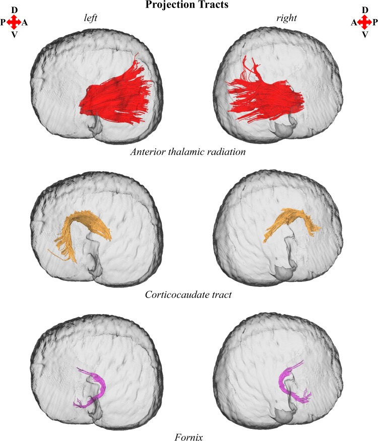
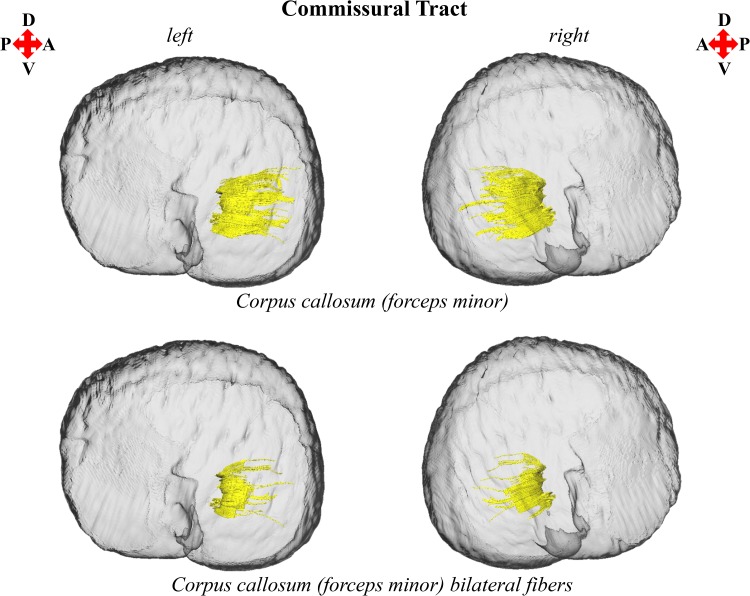
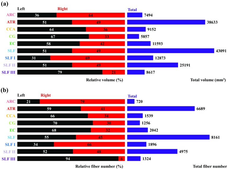
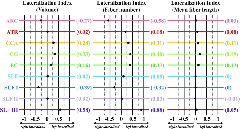
Brain enlargement is associated with concomitant growth of interneuronal distance, increased conduction time, and reduced neuronal interconnectivity. Recognition of these functional constraints led to the hypothesis that large-brained mammals should exhibit greater structural and functional brain lateralization. As a taxon with the largest brains in the animal kingdom, Cetacea provides a unique opportunity to examine asymmetries of brain structure and function. In the present study, diffusion tensor imaging and tractography were used to investigate cerebral white matter asymmetry in the bottlenose dolphin (Tursiops truncatus). Widespread white matter asymmetries were observed with the preponderance of tracts exhibiting leftward structural asymmetries. Leftward lateralization may reflect differential processing and execution of behaviorally variant sensory and motor functions by the cerebral hemispheres. The arcuate fasciculus, an association tract linked to human language evolution, was isolated and exhibited rightward asymmetry suggesting a right hemisphere bias for conspecific communication unlike that of most mammals. This study represents the first examination of cetacean white matter asymmetry and constitutes an important step toward understanding potential drivers of structural asymmetry and its role in underpinning functional and behavioral lateralization in cetaceans.
Keywords: Arcuate fasciculus; Asymmetry; Bottlenose dolphin (Tursiops truncatus); Diffusion tensor imaging (DTI); Tractography; White matter.
Conflict of interest statement
Conflict of interest
AKW, RJT, SHR, and MS declare no conflict of interest.
Funding
AKW was supported by the National Science Foundation Graduate Research Fellowship Program, Scripps Institution of Oceanography Graduate Department, and University of California-San Diego Graduate Division. SHR was partially supported by the Office of Naval Research (Project N0001417WX01558). The funders had no role in the study design, data collection, analysis, or interpretation, preparation of the manuscript, or decision to publish.
Ethics statement
No live animals were used for this study.
Figures

Similar articles
-
Hemispheric asymmetries in dorsal language pathway white-matter tracts: A magnetic resonance imaging tractography and functional magnetic resonance imaging study.Neuroradiol J. 2017 Oct;30(5):470-476. doi: 10.1177/1971400917720829. Epub 2017 Jul 12. Neuroradiol J. 2017. PMID: 28699372 Free PMC article.
-
Gray and white matter asymmetries in healthy individuals aged 21-29 years: a voxel-based morphometry and diffusion tensor imaging study.Hum Brain Mapp. 2011 Oct;32(10):1762-73. doi: 10.1002/hbm.21145. Epub 2010 Sep 30. Hum Brain Mapp. 2011. PMID: 20886579 Free PMC article.
-
From structure to function in the lateralized brain: how structural properties of the arcuate and uncinate fasciculus are associated with dichotic listening performance.Neurosci Lett. 2014 Sep 19;580:32-6. doi: 10.1016/j.neulet.2014.07.044. Epub 2014 Aug 2. Neurosci Lett. 2014. PMID: 25093701
-
Handedness and White Matter Networks.Neuroscientist. 2021 Feb;27(1):88-103. doi: 10.1177/1073858420937657. Epub 2020 Jul 29. Neuroscientist. 2021. PMID: 32723129 Review.
-
Imaging white-matter pathways of the auditory system with diffusion imaging tractography.Handb Clin Neurol. 2015;129:277-88. doi: 10.1016/B978-0-444-62630-1.00016-0. Handb Clin Neurol. 2015. PMID: 25726275 Review.
Cited by
-
In vivo Diffusion Tensor Magnetic Resonance Tractography of the Sheep Brain: An Atlas of the Ovine White Matter Fiber Bundles.Front Vet Sci. 2019 Oct 16;6:345. doi: 10.3389/fvets.2019.00345. eCollection 2019. Front Vet Sci. 2019. PMID: 31681805 Free PMC article.
-
Behavioural laterality in foraging bottlenose dolphins (Tursiops truncatus).R Soc Open Sci. 2019 Nov 27;6(11):190929. doi: 10.1098/rsos.190929. eCollection 2019 Nov. R Soc Open Sci. 2019. PMID: 31827837 Free PMC article.
-
Morphology of the Amazonian Teleost Genus Arapaima Using Advanced 3D Imaging.Front Physiol. 2020 Apr 27;11:260. doi: 10.3389/fphys.2020.00260. eCollection 2020. Front Physiol. 2020. PMID: 32395105 Free PMC article.
-
Neuroanatomy of the Cetacean Sensory Systems.Animals (Basel). 2023 Dec 23;14(1):66. doi: 10.3390/ani14010066. Animals (Basel). 2023. PMID: 38200796 Free PMC article. Review.
-
Postnatal growth and spatial conformity of the cranium, brain, eyeballs and masseter muscles in the macaque (Macaca mulatta).J Anat. 2023 Oct;243(4):590-604. doi: 10.1111/joa.13911. Epub 2023 Jun 9. J Anat. 2023. PMID: 37300248 Free PMC article.
References
-
- Beaulieu C. The biological basis of diffusion anisotropy. In: Johansen-Berg H, Behrens TEJ, editors. Diffusion MRI: from quantitative measurement to in vivo neuroanatomy. 2. Amsterdam: Elsevier Academic Press; 2014. pp. 155–183.
MeSH terms
Grants and funding
LinkOut - more resources
Full Text Sources
Other Literature Sources
Research Materials
