

Cell wall biosynthesis impairment affects the budding lifespan of the Saccharomyces cerevisiae yeast
- PMID: 29189912
- PMCID: PMC5765204
- DOI: 10.1007/s10522-017-9740-6
Cell wall biosynthesis impairment affects the budding lifespan of the Saccharomyces cerevisiae yeast
Abstract
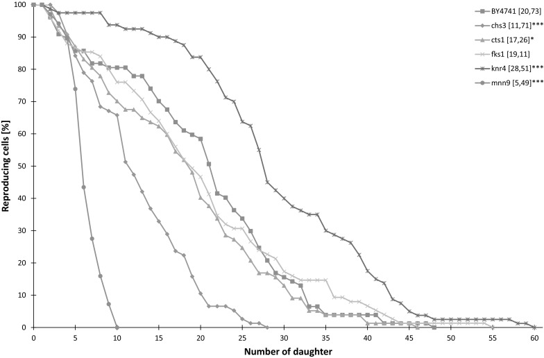
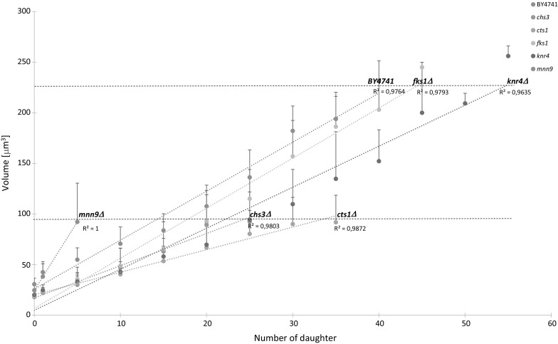
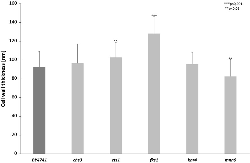
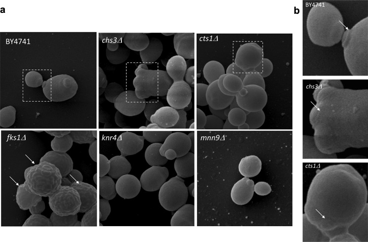
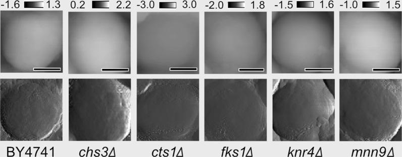
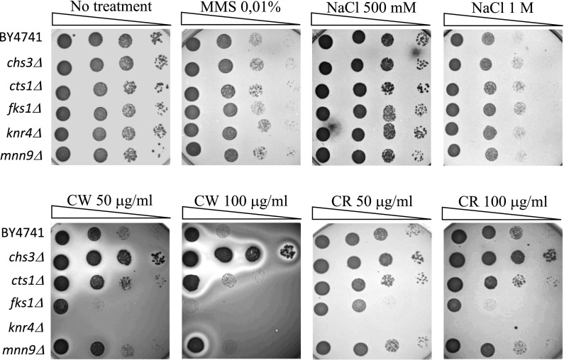
The Saccharomyces cerevisiae yeast is one of the most widely used model in studies of cellular and organismal biology, including as aging and proliferation. Although several constraints of aging and budding lifespan have been identified, these processes have not yet been fully understood. Previous studies of aging in yeast have focused mostly on the molecular basics of the underlying mechanisms, while physical aspects, particularly those related to the cell wall, were rather neglected. In this paper, we examine for the first time, to our knowledge, the impact of cell wall biosynthesis disturbances on the lifespan in the budding yeast. We have used a set of cell wall mutants, including knr4Δ, cts1Δ, chs3Δ, fks1Δ and mnn9Δ, which affect biosynthesis of all major cell wall compounds. Our results indicated that impairment of chitin biosynthesis and cell wall protein mannosylation reduced the budding lifespan, while disruption in the 1,3-β-glucan synthase activity had no adverse effect on that parameter. The impact varied in the severity and the most notable effect was observed for the mnn9Δ mutant. What was interesting, in the case of the dysfunction of the Knr4 protein playing the role of the transcriptional regulator of cell wall chitin and glucan synthesis, the lifespan increased significantly. We also report the phenotypic characteristics of cell wall-associated mutants as revealed by imaging of the cell wall using transmission electron microscopy, scanning electron microscopy and atomic force microscopy. In addition, our findings support the conviction that achievement of the state of hypertrophy may not be the only factor that determines the budding lifespan.
Keywords: AFM; Aging; Budding lifespan; Cell wall; SEM; TEM; Yeast.
Conflict of interest statement
The authors declare that they have no conflict of interest.
Figures
Similar articles
-
An atomic force microscopy analysis of yeast mutants defective in cell wall architecture.Yeast. 2010 Aug;27(8):673-84. doi: 10.1002/yea.1801. Yeast. 2010. PMID: 20602335
-
HKR1 encodes a cell surface protein that regulates both cell wall beta-glucan synthesis and budding pattern in the yeast Saccharomyces cerevisiae.J Bacteriol. 1996 Jan;178(2):477-83. doi: 10.1128/jb.178.2.477-483.1996. J Bacteriol. 1996. PMID: 8550469 Free PMC article.
-
Crh1p and Crh2p are required for the cross-linking of chitin to beta(1-6)glucan in the Saccharomyces cerevisiae cell wall.Mol Microbiol. 2007 Feb;63(3):921-35. doi: 10.1111/j.1365-2958.2006.05565.x. Mol Microbiol. 2007. PMID: 17302808
-
Biosynthesis of cell wall and septum during yeast growth.Arch Med Res. 1993 Autumn;24(3):301-3. Arch Med Res. 1993. PMID: 8298281 Review.
-
Signalling towards cell wall synthesis in budding yeast.Acta Univ Palacki Olomuc Fac Med. 1998;141:7-16. Acta Univ Palacki Olomuc Fac Med. 1998. PMID: 9684473 Review.
Cited by
-
Loss of Smi1, a protein involved in cell wall synthesis, extends replicative life span by enhancing rDNA stability in Saccharomyces cerevisiae.J Biol Chem. 2021 Jan-Jun;296:100258. doi: 10.1016/j.jbc.2021.100258. Epub 2021 Jan 7. J Biol Chem. 2021. PMID: 33837734 Free PMC article.
-
Disturbances in cell wall biogenesis as a key factor in the replicative aging of budding yeast.Biogerontology. 2025 Feb 5;26(2):54. doi: 10.1007/s10522-025-10196-0. Biogerontology. 2025. PMID: 39907841
-
Deletion of Atg22 gene contributes to reduce programmed cell death induced by acetic acid stress in Saccharomyces cerevisiae.Biotechnol Biofuels. 2019 Dec 27;12:298. doi: 10.1186/s13068-019-1638-x. eCollection 2019. Biotechnol Biofuels. 2019. PMID: 31890026 Free PMC article.
-
Tri-methylation of histone H3 lysine 4 facilitates gene expression in ageing cells.Elife. 2018 Oct 2;7:e34081. doi: 10.7554/eLife.34081. Elife. 2018. PMID: 30274593 Free PMC article.
-
Hydrogen sulfide treatment at the late growth stage of Saccharomyces cerevisiae extends chronological lifespan.Aging (Albany NY). 2021 Mar 19;13(7):9859-9873. doi: 10.18632/aging.202738. Epub 2021 Mar 19. Aging (Albany NY). 2021. PMID: 33744847 Free PMC article.
References
-
- Bilinski T, Bartosz G. Hypothesis: cell volume limits cell divisions. Acta Biochim Pol. 2006;53:833–835. - PubMed
Publication types
MeSH terms
Substances
LinkOut - more resources
Full Text Sources
Other Literature Sources
Medical
Molecular Biology Databases
Miscellaneous
