. 2018 Jan;20(1):32-43.
doi: 10.1016/j.neo.2017.11.002. Epub 2017 Nov 27.

BIGH3 Promotes Osteolytic Lesions in Renal Cell Carcinoma Bone Metastasis by Inhibiting Osteoblast Differentiation

Affiliations

BIGH3 Promotes Osteolytic Lesions in Renal Cell Carcinoma Bone Metastasis by Inhibiting Osteoblast Differentiation

Tianhong Pan et al. Neoplasia. 2018 Jan.

Abstract

Background: Bone metastasis is common in renal cell carcinoma (RCC), and the lesions are mainly osteolytic. The mechanism of bone destruction in RCC bone metastasis is unknown.

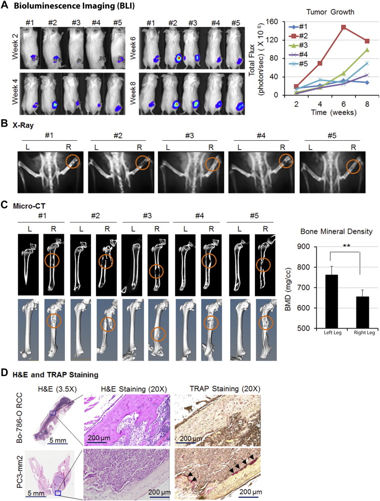
Methods: We used a direct intrafemur injection of mice with bone-derived 786-O RCC cells (Bo-786) as an in vivo model to study if inhibition of osteoblast differentiation is involved in osteolytic bone lesions in RCC bone metastasis.

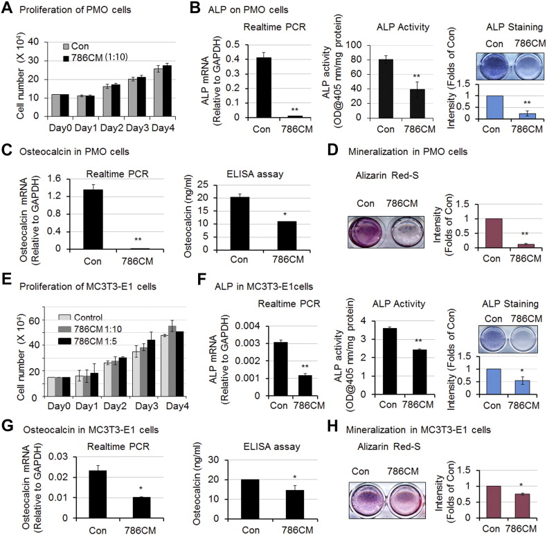
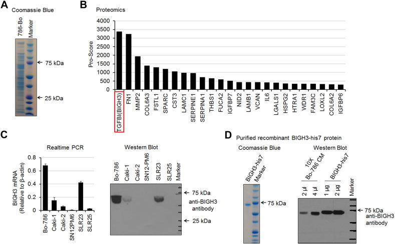
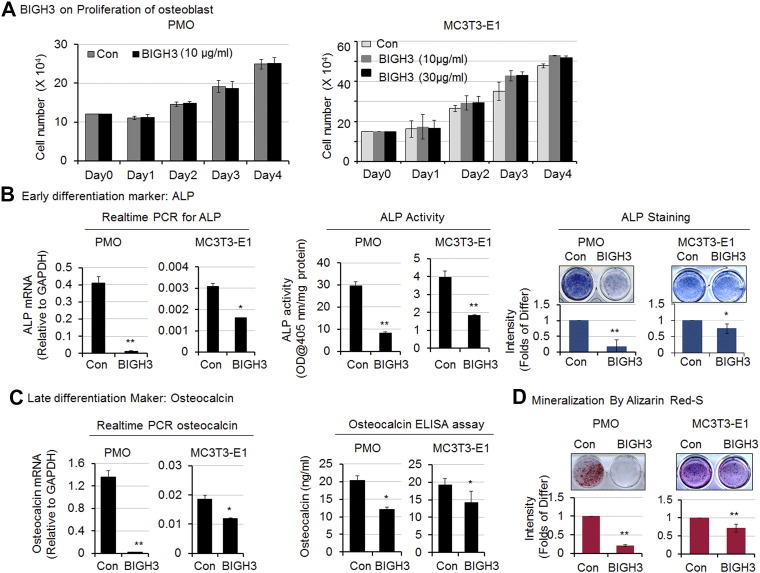
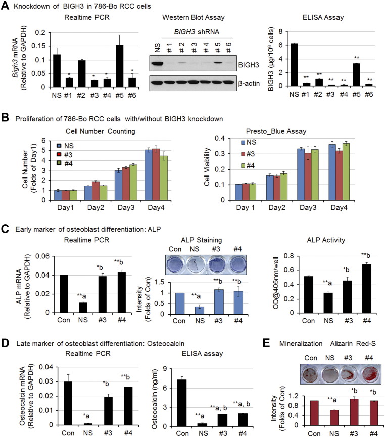
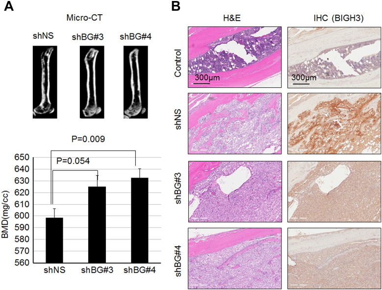
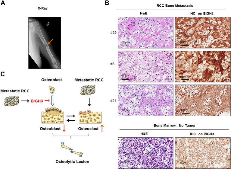
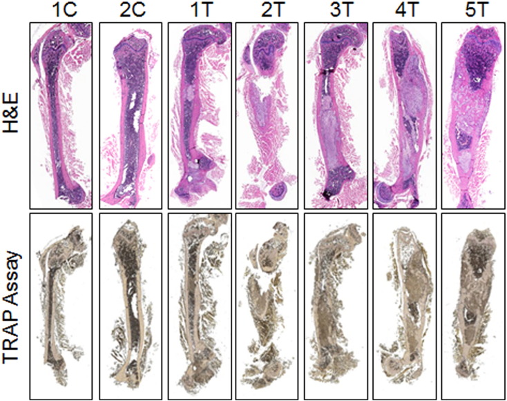
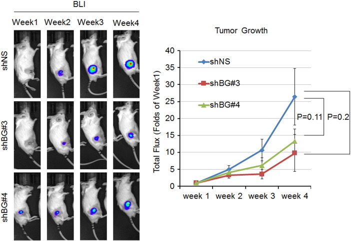
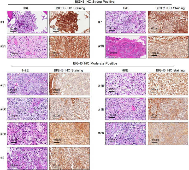
Results: We showed that bone-derived Bo-786 cells induced osteolytic bone lesions in the femur of mice. We examined the effect of conditioned medium of Bo-786 cells (Bo-786 CM) on both primary mouse osteoblasts and MC3T3-E1 preosteoblasts and found that Bo-786 CM inhibited osteoblast differentiation. Secretome analysis of Bo-786 CM revealed that BIGH3 (Beta ig h3 protein), also known as TGFBI (transforming growth factor beta-induced protein), is highly expressed. We generated recombinant BIGH3 and found that BIGH3 inhibited osteoblast differentiation in vitro. In addition, CM from Bo-786 BIGH3 knockdown cells (786-BIGH3 KD) reduced the inhibition of osteoblast differentiation compared to CM from vector control. Intrafemural injection of mice with 786-BIGH3 KD cells showed a reduction in osteolytic bone lesions compared to vector control. Immunohistochemical staining of 18 bone metastasis specimens from human RCC showed strong BIGH3 expression in 11/18 (61%) and moderate BIGH3 expression in 7/18 (39%) of the specimens.

Conclusions: These results suggest that suppression of osteoblast differentiation by BIGH3 is one of the mechanisms that enhance osteolytic lesions in RCC bone metastasis, and raise the possibilty that treatments that increase bone formation may improve therapy outcomes.


Figures

Figure 1
Figure 1
Bo-786 cells generated osteolytic lesions in mouse femur. Bo-786 cells were injected into the femur of mouse right leg. (A) The growth of cancer cells in the right legs was monitored by BLI over 8 weeks. (B) Osteolytic bone lesions were detected by X-ray. (C) Micro-CT of the femurs with or without tumors. Quantification of BMD was done on whole femurs using Microview software. (D) A representative image of H&E-stained bone section of the leg shows the presence of tumor cells. A representative image of TRAP staining shows strong staining in PC3-mm2–injected femur but only weakly in Bo-786–injected femur. “L”: normal left femur; “R”: tumor cell injected right femur. **P < .01.
Figure 2
Figure 2
Bo-786 culture medium inhibited osteoblast differentiation. (A) PMO cell proliferation with or without treatment with Bo-786 CM (786CM). (B) ALP gene expression (left), ALP activity (middle), and ALP staining (right) in Bo-786 CM-treated PMO cells. (C) Osteoclacin mRNA (left) and osteocalcin protein in 786-O CM-treated PMO cells (right). (D) Effect of Bo-786 CM on mineralization of PMO cells as determined by Alizarin Red S staining. (E) Proliferation of MC3T3-E1 cells with or without Bo-786 CM treatment. (F) ALP gene expression (left), ALP activity (middle), and ALP staining (right) in Bo-786 CM-treated MC3T3-E1cells. (G) Osteoclacin mRNA (left) and osteocalcin protein in 786-O CM-treated MC3T3-E1 cells (right). (H) MC3T3-E1 mineralization was determined by Alzarin Red S staining. “Con”: Control. *P < .05; **P < .01.
Figure 3
Figure 3
Mass spectrometry analysis of Bo-786 CM. (A) SDS-PAGE analysis of Bo-786 CM. The proteins were stained with Coomassie blue. (B) Lists of 25 secretory proteins with highest protein score from tandem mass spectrometry analysis. (C) Expression of BIGH3 in kidney cell lines as determined by real-time PCR and Western blot. (D) SDS-PAGE analysis of BIGH3-his7 protein expressed and purified from HEK293 cells (left). Western blot of purified BIGH3-his7 protein and Bo-786 CM with anti-BIGH3 antibody (right).
Figure 4
Figure 4
Recombinant BIGH3-his7 protein inhibited osteoblast differentiation. (A) Cell proliferation was determined on PMO (left) and MC3T3-E1 cells (right) after cells were treated with recombinant BIGH3-his7 protein. (B) Effects of BIGH3 on ALP gene expression (left), ALP activity (middle), and ALP staining (right) in PMO and MC3T3-E1 cells. (C) Effects of BIGH3 on osteoclacin mRNA (left) and osteocalcin protein (right) in PMO and MC3T3-E1 cells. (D) Effects of BIGH3 on PMO and MC3T3-E1 cell mineralization as determined by Alizarin Red S staining. “Con”: [200 mM] imidazole vehicle control; “BIGH3”: recombinant purified BIGH3-his7 protein. *P < .05; **P < .01.
Figure 5
Figure 5
Inhibition of osteoblast differentiation by Bo-786 CM was partially mediated through BIGH3. (A) Generation of Bo-786 RCC cells with knockdown of BIGH3. Knockdown of BIGH3 was confirmed by real-time PCR, Western blot assay, and ELISA. (B) Cell proliferation and cell viability of BIGH3 knockdown cell lines. (C) Effects of CM from BIGH3 knockdown cell lines on PMO differentiation. ALP mRNA (left), ALP staining (middle), and ALP activity (right). (D) Osteocalcin mRNA and protein levels. (E) Mineralization. “NS”: 786-shNS nonsilencing control cells; “#3”: 786-shBG#3 cells; “#4”: 786-shBG#4 cells. *P < .05; **P < .01; “a”: compared to “Con”; “b”: compared to “NS”.
Figure 6
Figure 6
Knockdown of BIGH3 gene in Bo-786 cells reduced Bo-786-induced osteolytic lesions. (A) Representative micro-CT images of mouse femur 4 weeks after being injected with 786-shNS, 786-shBG#3, or 786-shBG#4 cells (upper). BMD of whole femur was quantified using Microview software (lower). (B) H&E-stained bone section of an injected femur showed the presence of tumor cells and IHC of BIGH3 on the adjacent sections of H&E. “shNS”: 786-shNS cells (nonsilencing control cells); “shBG#3”: 786-shBG#3 cells; “shBG#4”: 786-shBG#4 cells.
Figure 7
Figure 7
BIGH3 expression in RCC bone metastasis. (A) A representative X-ray from a patient showing a pathologic fracture due to bone metastasis from RCC. (B) BIGH3 expression in three specimens of human RCC bone metastasis samples (upper) and normal bone marrow (lower) was determined by IHC staining. Both H&E staining and IHC staining for BIGH3 are shown at the magnification of 40×. (C) The proposed role of BIGH3 in enhancing osteolytic bone lesions from RCC bone metastasis. RCC cells in bone secrete BIGH3 that inhibits osteoblast differentiation that would normally counterbalance osteoclast activity, thus enhancing RCC-mediated osteolytic bone destruction.
Figure S1
Figure S1
H&E and TRAP staining on femurs injected with Bo-786-O cells. “C”: control non–tumor-bearing femur; “T”: tumor-bearing femur.
Figure S2
Figure S2
Growth of 786-shNS and BIGH3 knockdown cells in mouse femurs as monitored by BLI. Mice were injected with 786-shNS or BIGH3 knockdown 786-shBG#3 and 786-shBG#4 cells at right femurs. 786-shNS cells were used as control. The tumor growth was monitored by BLI over a 4-week time period, and the total flux was expressed as folds of week 1.
Figure S3
Figure S3
H&E and IHC of BIGH3 in human RCC bone metastasis tissue samples.

References

    1. Chen SC, Kuo PL. Bone metastasis from renal cell carcinoma. Int J Mol Sci. 2016;17 pii: E987. - PMC - PubMed
    1. Guise TA. The vicious cycle of bone metastases. J Musculoskelet Neuronal Interact. 2002;2:570–572. - PubMed
    1. Mikami S, Katsube K, Oya M, Ishida M, Kosaka T, Mizuno R, Mochizuki S, Ikeda T, Mukai M, Okada Y. Increased RANKL expression is related to tumour migration and metastasis of renal cell carcinomas. J Pathol. 2009;218:530–539. - PubMed
    1. Takahashi N, Udagawa N, Suda T. A new member of tumor necrosis factor ligand family, ODF/OPGL/TRANCE/RANKL, regulates osteoclast differentiation and function. Biochem Biophys Res Commun. 1999;256:449–455. - PubMed
    1. Raggatt LJ, Partridge NC. Cellular and molecular mechanisms of bone remodeling. J Biol Chem. 2010;285:25103–25108. - PMC - PubMed

Publication types

MeSH terms

Substances