. 2017 Nov 30;12(11):e0188292.
doi: 10.1371/journal.pone.0188292. eCollection 2017.

Contrast-enhanced magnetic resonance imaging for the detection of ruptured coronary plaques in patients with acute myocardial infarction

Affiliations

Contrast-enhanced magnetic resonance imaging for the detection of ruptured coronary plaques in patients with acute myocardial infarction

Christian H P Jansen et al. PLoS One. .

Abstract

Purpose: X-ray coronary angiography (XCA) is the current gold standard for the assessment of lumen encroaching coronary stenosis but XCA does not allow for early detection of rupture-prone vulnerable plaques, which are thought to be the precursor lesions of most acute myocardial infarctions (AMI) and sudden death. The aim of this study was to investigate the potential of delayed contrast-enhanced magnetic resonance coronary vessel wall imaging (CE-MRCVI) for the detection of culprit lesions in the coronary arteries.

Methods: 16 patients (13 male, age 61.9±8.6 years) presenting with sub-acute MI underwent CE-MRCVI within 24-72h prior to invasive XCA. CE-MRCVI was performed using a T1-weighted 3D gradient echo inversion recovery sequence (3D IR TFE) 40±4 minutes following the administration of 0.2 mmol/kg gadolinium-diethylenetriamine-pentaacetic acid (DTPA) on a 3T MRI scanner equipped with a 32-channel cardiac coil.

Results: 14 patients were found to have culprit lesions (7x LAD, 1xLCX, 6xRCA) as identified by XCA. Quantitative CE-MRCVI correctly identified the culprit lesion location with a sensitivity of 79% and excluded culprit lesion formation with a specificity of 99%. The contrast to noise ratio (CNR) of culprit lesions (9.7±4.1) significantly exceeded CNR values of segments without culprit lesions (2.9±1.9, p<0.001).

Conclusion: CE-MRCVI allows the selective visualization of culprit lesions in patients immediately after myocardial infarction (MI). The pronounced contrast uptake in ruptured plaques may represent a surrogate biomarker of plaque activity and/or vulnerability.


Conflict of interest statement

Competing Interests: The authors acknowledge financial support from the British Heart Foundation (https://www.bhf.org.uk/) (PG/10/044/28343) and the BHF Centre of Excellence (https://www.bhf.org.uk/research/where-we-fund-research/centres-of-research-excellence) (RE/08/03). The Division of Imaging Sciences receives also support from the Centre of Excellence in Medical Engineering (funded by the Welcome Trust and EPSRC; grant number WT 088641/Z/09/Z) (https://www.epsrc.ac.uk/) and the Department of Health through the National Institute for Health Research (NIHR) Biomedical Research Centre award to Guy’s and St Thomas’ NHS Foundation Trust in partnership with King’s College London, and by the NIHR Healthcare Technology Co- operative for Cardiovascular Disease at Guy’s and St Thomas’ NHS Foundation Trust. The views expressed are those of the author(s) and not necessarily those of the NHS, the NIHR or the Department of Health. The author MRM is grateful for the financial support from the Deutsche Forschungsgemeinschaft (http://www.dfg.de/) (DFG, 594331/41/91). The author AW is affiliated to Philips Healthcare. This does not alter our adherence to PLOS ONE policies on sharing data and materials.

Figures

Fig 1
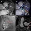
Fig 1. CE-MRCVI findings in a patient with troponin positive ACS in the RCA.
Coronary MRA (A) of a 48-year-old male with troponin positive ACS showed decreased vessel lumen size of the mid RCA (red arrows). To highlight the relationship between CE-MRCVI (B) and morphology (A), images were fused in a way similar to PET/CT (C). CE-MRCVI displays high signal intensity (red arrows) within the mid RCA. Analysis of signal enhancement on CE-MRCVI (E, red) suggestive for culprit lesion yielded a contrast-to-noise ratio (CNR) of 12.7. Corresponding XCA (F) confirmed CE-MRCVI findings with mid RCA de-novo lesion (pre-treatment stenosis of 75–94%). MRA: magnetic resonance angiography, CE-MRCVI: contrast enhanced magnetic resonance coronary vessel wall imaging, PET/CT: positron emission tomography/computed tomography. CE-MRCVI:contrast enhanced magnetic resoance coronary vessel wall imaging, ACS: acute coronary syndrome, RCA: right coronary artery, MRA: magnetic resonance angiography, XCA: x-ray coronary angiography, PET/CT: positron emission tomography/computed tomography.
Fig 2
Fig 2. CE-MRCVI findings with troponin positive ACS in the LCX.
Coronary MRA (A) with troponin positive ACS showed decreased vessel lumen size of the proximal LCX and first marginal branch (grey arrows). To highlight the relationship between and morphology (A) and CE-MRCVI, images were fused in a way similar to PET/CT (B). CE-MRCVI displays high signal intensity (red arrows) within the first marginal branch. Corresponding XCA (C) confirmed CE-MRCVI findings with mid LCX lesion. Short axis delayed enhancement scan shows predominantly transmural myocardial infarction of the mid-ventricular infero-lateral segment. MRA: magnetic resonance angiography, CE-MRCVI: contrast enhanced magnetic resonance coronary vessel wall imaging, PET/CT: positron emission tomography/computed tomography.
Fig 3
Fig 3. Contrast-to-noise ratio of culprit lesion and coronary vessel wall.
Contrast to noise ratio (CNR) of segmented culprit lesion in comparison to segments with and without culprit lesion formation. Absolute CNR values of segmented culprit lesion area (Mean: 9.7, 95% CI: 7.6–11.9), segments with (Mean: 4.7, 95% CI: 3.4–6.0) and “normal” segments without visual apparent culprit lesion formation (Mean: 2.9, 95% CI: 2.5–3.3) were found to differ significantly (p<0.05).
Fig 4
Fig 4. Relative contrast-to-noise ratio of culprit lesion and coronary vessel wall.
Relative CNR values were calculated as ratio between visually apparent culprit lesion area, the affected coronary segment (including culprit lesion formation) or “normal” segments without apparent culprit lesion formation and the averaged CNR of the corresponding entire left or right coronary system. Relative CNR values of segmented culprit lesion area (Mean: 329%, 95% CI: 249–409%), affected coronary segments with culprit lesion (Mean: 130%, 95% CI: 116–144%), and “normal” segments without visual apparent culprit lesion formation (Mean: 96%, 95% CI: 88.54–103.46) were found to differ significantly (p<0.05). A relative CNR threshold of 230 (red line) resulted in a true positive detection and localization in 11 out of 14 patients and true exclusion in 104 out of 105 coronary segments. The false positive classification of the left main stem (red dot) was observed in a patient without culprit lesion in XCA (inferior STEMI).

References

    1. Clinical-pathological correlations of coronary disease progression and regression. 1992;86: III1–11. Available: http://eutils.ncbi.nlm.nih.gov/entrez/eutils/elink.fcgi?dbfrom=pubmed&id... - PubMed
    1. Human monocyte-derived macrophages induce collagen breakdown in fibrous caps of atherosclerotic plaques. Potential role of matrix-degrading metalloproteinases and implications for plaque rupture. 1995;92: 1565–1569. Available: http://eutils.ncbi.nlm.nih.gov/entrez/eutils/elink.fcgi?dbfrom=pubmed&id... - PubMed
    1. Coronary plaque disruption. 1995;92: 657–671. Available: http://circ.ahajournals.org/cgi/content/full/92/3/657 - PubMed
    1. Virmani R, Burke A, Farb A, Kolodgie F. Pathology of the Vulnerable Plaque. J Am Coll Cardiol. 2006;47: C13–C18. doi: 10.1016/j.jacc.2005.10.065 - DOI - PubMed
    1. Mechanisms of plaque formation and rupture. Lippincott Williams & Wilkins; 2014;114: 1852–1866. doi: 10.1161/CIRCRESAHA.114.302721 - DOI - PubMed