. 2017 Aug 14;8(54):91928-91937.
doi: 10.18632/oncotarget.20248. eCollection 2017 Nov 3.

Metformin activates type I interferon signaling against HCV via activation of adenosine monophosphate-activated protein kinase

Affiliations

Metformin activates type I interferon signaling against HCV via activation of adenosine monophosphate-activated protein kinase

Wei-Lun Tsai et al. Oncotarget. .

Abstract

Activation of the type I interferon (IFN) signaling pathway is essential for the eradication of hepatitis C virus (HCV). Metformin can activate adenosine monophosphate-activated protein kinase (AMPK) to reduce insulin resistance. Cross talks between AMPK and IFN signaling remain unclear. To understand the influence of metformin on the type I IFN signaling pathway and HCV infection, the full-length HCV replicon OR6 cells and the infectious HCV clones JFH1 were used to assess the anti-HCV effect of the insulin sensitizers, metformin and pioglitazone. Immunofluorescence staining and the immunoblotting of HCV viral protein demonstrated that metformin, but not pioglitazone, inhibited HCV replication in OR-6 and JFH-1-infected Huh 7.5.1 cells. Immunoblotting data showed that metformin activated the phosphorylation of STAT-1 and STAT-2 in OR-6 and JFH-1 infected Huh 7.5.1 cells. Metformin enhanced the phosphorylation of AMPK, and the metformin-activated IFN signaling was down-regulated by AMPK inhibitor. After treatment of AMPK inhibitor, the level of HCV core protein decreased by metformin can be rescued. In conclusion, metformin activates type I interferon signaling and inhibits the replication of HCV via activation of AMPK.

Keywords: AMPK; hepatits C virus; interferon; metformin.


Conflict of interest statement

CONFLICTS OF INTEREST The authors declare no competing financial interests.

Figures

Figure 1
Figure 1. Effects of metformin on HCV replication in OR-6 cells HCV core protein expression was determined in OR-6 cells treated with different doses of metformin for 48 h
(A) Immunofluorescence assay (IFA) with anti-HCV core antibody (upper panels) and DAPI staining of nuclei (lower panels) were shown. (B) Quantification of IFA was performed. *P < 0.05, **P < 0.01, ***P < 0.001 in metformin treated versus untreated control group. (C) The cell lysates from OR-6 cells treated with different doses of metformin for 48 h, were analyzed by immunoblotting with anti-HCV core protein antibody, and the β-actin was shown as the loading control.
Figure 2
Figure 2. Effects of metformin on HCV replication in JFH-1 infected Huh 7.5.1 cells
The Huh7.5.1 cells were infected by JFH1 for 72 h and then treated with different doses of metformin for 48 h. (A) Immunofluorescence assay using anti-HCV core antibody was performed. IFA of anti-HCV core antibody (upper panels) and DAPI staining of nuclei (lower panels) were shown. (B) Quantification of IFA was performed. *P < 0.05, **P < 0.01, ***P < 0.001 in metformin treated versus untreated control group. (C) JFH-1 infected Huh 7.5.1 cells were treated with different doses of metformin for 48 h and the cell lysates were analyzed by immunoblotting with anti-HCV core protein antibody.
Figure 3
Figure 3. Effects of pioglitazone on HCV replication in OR-6 cells
HCV core protein expression was determined in OR-6 cells treated with different doses of pioglitazone for 48 h. (A) Immunofluorescence assay (IFA) of anti-HCV core antibody (upper panels) and DAPI staining of nuclei (lower panels) were shown. (B) Quantification of IFA was performed and no significant difference between pioglitazone treated and untreated control group was found. (C) The cell lysates harvested from OR-6 cells treated with different doses of pioglitazone and were analyzed by immunoblotting with anti-HCV core protein antibody.
Figure 4
Figure 4. Effects of pioglitazone on HCV replication in JFH-1 infected Huh 7.5.1 cells
The Huh7.5.1 cells were infected by JFH1 for 72 h and then treated with different doses of pioglitazone for 48 h. (A) Immunofluorescence assay using anti-HCV core antibody was performed. IFA of anti-HCV core antibody (upper panels) and DAPI staining of nuclei (lower panels) were shown. (B) Quantification of IFA was performed and no significant difference between pioglitazone treated and untreated control group was found. (C) JFH-1 infected Huh 7.5.1 cells were treated with different doses of pioglitazone for 48 h, then and cell lysates were analyzed by immunoblotting with anti-HCV core protein antibody.
Figure 5
Figure 5. Effects of metformin on type I IFN signaling pathway
The cell extracts were harvested from OR6 cells (A) or JFH1-infected Huh7.5.1 cells (B) with or without metformin (10∼320 μM) treatment for 48 h, the expression of IFN signaling proteins were analyzed by immunoblotting with the specific antibodies.
Figure 6
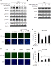
Figure 6. Metformin activates IFN signaling pathway in an AMPK dependent manner
(A) OR6 cells were treated with AMPK inhibitor (0.1∼10 μM) for 30 min then stimulated with metformin (160 μM) for 48 hrs. The whole cell extracts were analyzed by immunoblotting with indicated antibodies. (B) OR6 cells were treated with AMPK inhibitor (1 and 10 μM) for 30 min then stimulated with metformin (320 μM) for 6 hrs. The whole cell extracts were analyzed by Immunoblotting with indicated antibodies. (C) Immunofluorescence assay using anti-HCV core antibody was performed in OR6 cells treated with AMPK inhibitor (1 and 10 μM) for 30 min then stimulated with metformin (320 μM) for 6 hrs. IFA of anti-HCV core antibody (upper panels) and DAPI staining of nuclei (lower panels) were shown. (D) Quantification of IFA was performed. *P < 0.05, **P < 0.01 in AMPK inhibitor and metformin treated group versus metformin alone treated group. (E) Immunofluorescence assay of HCV core protein was conducted in OR6 cells with AMPK siRNA knockdown, DAPI staining indicates cell nuclei. (F) The infectivity of HCV in AMPK knockdown cells was quantified. Student’s t test between groups, **P < 0.01, ***P < 0.001

References

    1. Kao JH, Chen DS. Changing disease burden of hepatocellular carcinoma in the Far East and Southeast Asia. Liver Int. 2005;25:696–703. - PubMed
    1. Santantonio T, Wiegand J, Gerlach JT. Acute hepatitis C: current status and remaining challenges. J Hepatol. 2008;49:625–633. - PubMed
    1. Fattovich G, Stroffolini T, Zagni I, Donato F. Hepatocellular carcinoma in cirrhosis: incidence and risk factors. Gastroenterology. 2004;127:S35–S50. - PubMed
    1. Shintani Y, Fujie H, Miyoshi H, Tsutsumi T, Tsukamoto K, Kimura S, Moriya K, Koike K. Hepatitis C virus infection and diabetes: direct involvement of the virus in the development of insulin resistance. Gastroenterology. 2004;126:840–848. - PubMed
    1. Sheikh MY, Choi J, Qadri I, Friedman JE, Sanyal AJ. Hepatitis C virus infection: molecular pathways to metabolic syndrome. Hepatology. 2008;47:2127–2133. - PubMed