. 2017 Sep 21;8(54):92227-92239.
doi: 10.18632/oncotarget.21152. eCollection 2017 Nov 3.

The tissue distribution and significance of B7-H4 in laryngeal carcinoma

Affiliations

The tissue distribution and significance of B7-H4 in laryngeal carcinoma

Lili Chen et al. Oncotarget. .

Abstract

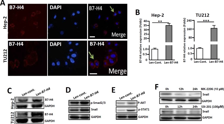
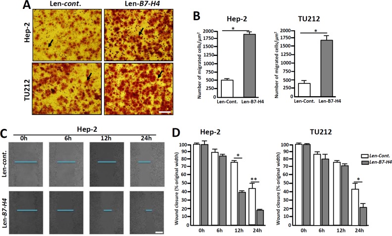
The costimulatory signals CD28 and B7 have been shown to control tumor invasion and metastasis by regulating T cell activation, whereas the distribution characteristics of B7-associated proteins in laryngeal carcinoma (LC) tissue are still unclear. Here, the expression of members of the B7 superfamily, including B7-H1 (PD-L1), B7-DC (PD-L2) and B7-H4, in fifty-two LC samples was determined by immunohistochemistry, and the relationship between B7-H4 and epithelial-mesenchymal transition (EMT)-associated markers was further assessed by immunofluorescence double staining. Furthermore, the human LC cell lines, Hep-2 and TU212 cells, were further transfected to overexpress B7-H4, and cell invasion and metastasis were analyzed. The results showed that B7-H1, B7-DC and B7-H4 were expressed in the tumor cells, and their expression was restricted to the cell membrane and the cytoplasm. The positive rates of these molecules in the tumor tissues were 57.7% (30/52), 32.7% (17/52) and 34.6% (18/52), respectively. Interestingly, double immunofluorescence staining showed that B7-H4 is coexpression with EMT-related markers, including p-Smad2/3, Snail and Vimentin, in carcinoma cells. Moreover, overexpression of B7-H4 in Hep-2 cells promotes the expression of pSmad2/3 and Snail by activating AKT-STAT3 signaling. Transwell and wound-healing assays demonstrated that B7-H4 enhanced both Hep-2 and TU212 cell invasion and metastasis. Our results suggest that B7-H4 transmits feedback signaling to tumor cells and promotes invasion and metastasis by promoting EMT progression. Therefore, blocking B7-H4 signaling might be a novel treatment strategy for LC.

Keywords: B7-H4; EMT; STAT3; invasion and metastasis; laryngeal carcinoma.


Conflict of interest statement

CONFLICTS OF INTEREST The authors declare no financial or commercial conflicts of interest.

Figures

Figure 1
Figure 1. Expression of B7-H1 in LC tissues detected by immunohistochemistry
(A) Sections were stained with isotype-specific mouse IgG1 control antibodies. (B) Low levels of B7-H1 were seen in the infiltrated T cells and epithelial cells of peritumoral normal tissues. (C) B7-H1 was expressed on the surface and in the cytoplasm of the tumor cells, (D) tissue-infiltrating lymphocytes and (E) the blood capillaries of tumor tissues. The arrows indicate positive cells. Scale bar = 20 μm, ▲ indicated blood capillaries.
Figure 2
Figure 2. Expression of B7-DC in LC tissues detected by immunohistochemistry
(A) Sections were stained with isotype-specific mouse IgG1 control antibodies. (B) B7-DC was absent in peritumoral normal tissues. B7-DC was expressed on the surface and in the cytoplasm of tumor cells (C), tissue-infiltrating lymphocytes (D), blood capillaries and epithelial cells (E). Arrows indicate positive cells, ▲ indicates blood capillaries and ● indicates epithelial cells. Scale bar = 20 μm.
Figure 3
Figure 3. Expression of B7-H4 in LC tissues detected by immunohistochemistry
(A) Sections were stained with isotype-specific mouse IgG1 control antibodies. (B) B7-H4 was absent in peritumoral normal tissues. In tumor tissues, B7-H4 was expressed on the surface and in the cytoplasm of tumor cells (C) and in tissue-infiltrating macrophages (D), and tissue-infiltrating lymphocytes (E), whereas it was absent in the blood capillaries (F). ▲ indicates blood capillaries and ● indicates epithelial cells. Arrows indicate positive cells. Scale bar = 20 μm.
Figure 4
Figure 4. Expression of the cell proliferation marker PCNA and EMT-related markers in human LC samples detected by immunohistochemistry
(A) The expression of PCNA, CK18, p-Smad2/3, p-Smad3, Vimentin and Snail in laryngeal carcinoma cells was detected by immunohistochemistry. Arrows indicate positive cells. Scale bar = 20 μm. (B) The co-expression of the EMT-related markers and B7-H4 in LC cells was detected by immunofluorescence double staining. The arrows indicate positive cells and DAPI indicates nuclear staining. Scale bar = 20 μm. (C) Statistical analyzed the relationship of %B7-H4positive and % Snail positive cells in total tumor cells of these fifty-two LC samples, r=0.247.
Figure 5
Figure 5. Overexpression of B7-H4 in both Hep-2 and TU212 cells promotes the expression of EMT-associated markers
(A) Immunofluorescence shows endogenous expression of B7-H4 in Hep-2/TU212 cells. The arrow indicates positive cells. Blue, DAPI; Red, B7-H4. Scale bar =20 μm. (B) qRT-PCR was used to detect B7-H4 in Hep-2/TU212 cells by lentiviral infection (Len-B7-H4) and control (Len-Cont.). (C) B7-H4 protein expression in Hep-2/TU212 cells was measured by western blot. (D) The expression of EMT-associated markers, including p-Smad2/3 and Snail, in Hep-2 cells in which B7-H4 was overexpressed and the control counterparts was compared by western blot. (E) The expression of p-AKT and p-STAT-1 in Hep-2 cells in which B7-H4 was overexpressed and the control counterparts was compared by western blot. (F) Hep-2 cells in which B7-H4 was overexpressed were treated with the Akt inhibitor MK-2206 or the STAT-1 inhibitor S3I-201 for 0 h, 12 h and 24 h, and the expression of Snail was detected by western blot. One of three experiments that had comparable results is shown.
Figure 6
Figure 6. Overexpression of B7-H4 in Hep-2/TU212 cells promotes cell invasion and metastasis
(A) Representative images of cell invasion by transwell chamber assay. (B) Cell invasion assays of Hep-2/TU212 cells with or without B7-H4 overexpression. The results are the mean ± SEM (standard error of the mean) from three independent experiments. * p<0.05. (C) and (D) Cell migration by wound healing analysis. The confluent cells were wounded by sterile pipettes and the status of wound closure were observed and photographed after 6 h, 12 h and 24 h of culture. All the experiments were repeated for three times. Scale bar=20μm. * p<0.05 and ** p<0.01.

References

    1. Mannelli G, Cecconi L, Gallo O. Laryngeal preneoplastic lesions and cancer: challenging diagnosis. Qualitative literature review and meta-analysis. Crit Rev Oncol Hematol. 2016;106:64–90. - PubMed
    1. Curado MP, Hashibe M. Recent changes in the epidemiology of head and neck cancer. Curr Opin Oncol. 2009;21:194–200. - PubMed
    1. Herbella FA, Neto SP, Santoro IL, Figueiredo LC. Gastroesophageal reflux disease and non-esophageal cancer. World J Gastroenterol. 2015;21:815–9. - PMC - PubMed
    1. Dedivitis RA, Aires FT, Cernea CR, Brandão LG. Pharyngocutaneous fistula after total laryngectomy: systematic review of risk factors. Head Neck. 2015;37:1691–7. - PubMed
    1. Bellmunt J, Powles T, Vogelzang NJ. A review on the evolution of PD-1/PD-L1 immunotherapy for bladder cancer: the future is now. Cancer Treat Rev. 2017;54:58–67. - PubMed