Review
. 2018 Oct:71-72:174-187.
doi: 10.1016/j.matbio.2017.11.009. Epub 2017 Nov 27.

Laminin-deficient muscular dystrophy: Molecular pathogenesis and structural repair strategies

Affiliations
Review

Laminin-deficient muscular dystrophy: Molecular pathogenesis and structural repair strategies

Peter D Yurchenco et al. Matrix Biol. 2018 Oct.

Abstract

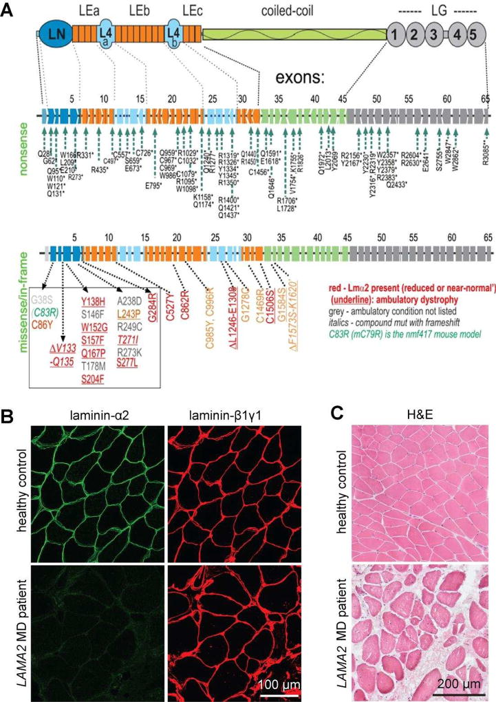
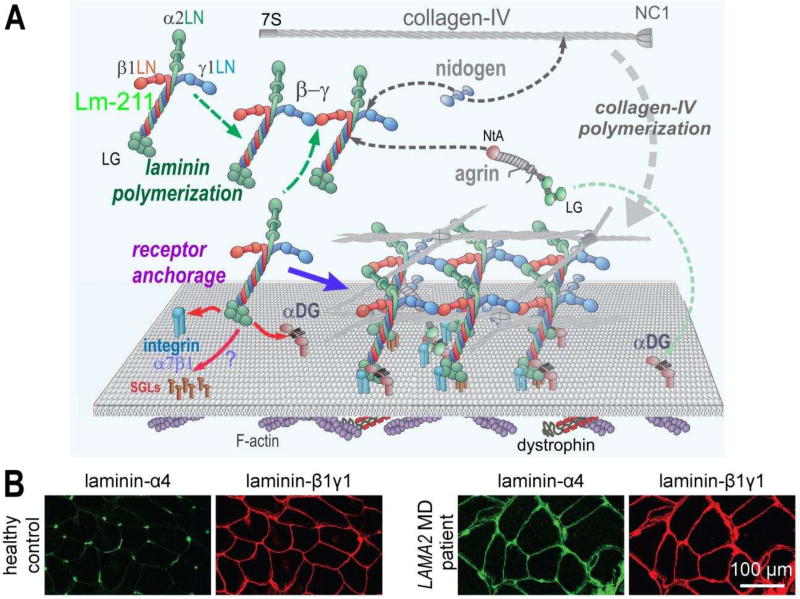
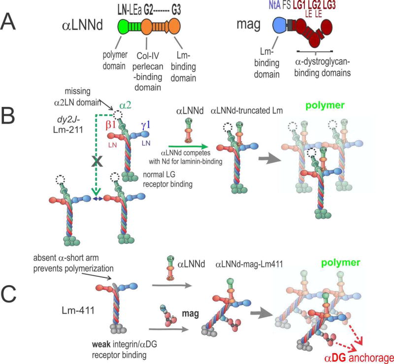
Laminins are large heterotrimers composed of the α, β and γ subunits with distinct tissue-specific and developmentally regulated expression patterns. The laminin-α2 subunit, encoded by the LAMA2 gene, is expressed in skeletal muscle, Schwann cells of the peripheral nerve and astrocytes and pericytes of the capillaries in the brain. Mutations in LAMA2 cause the most common type of congenital muscular dystrophies, called LAMA2 MD or MDC1A. The disorder manifests mostly as a muscular dystrophy but slowing of nerve conduction contributes to the disease. There are severe, non-ambulatory or milder, ambulatory variants, the latter resulting from reduced laminin-α2 expression and/or deficient laminin-α2 function. Lm-211 (α2β1γ1) is responsible for initiating basement membrane assembly. This is primarily accomplished by anchorage of Lm-211 to dystroglycan and α7β1 integrin receptors, polymerization, and binding to nidogen and other structural components. In LAMA2 MD, Lm-411 replaces Lm-211; however, Lm-411 lacks the ability to polymerize and bind to receptors. This results in a weakened basement membrane leading to the disease. The possibility of introducing structural repair proteins that correct the underlying abnormality is an attractive therapeutic goal. Recent studies in mouse models for LAMA2 MD reveal that introduction of laminin-binding linker proteins that restore lost functional activities can substantially ameliorate the disease. This review discusses the underlying mechanism of this repair and compares this approach to other developing therapies employing pharmacological treatments.


Figures

Figure 1
Figure 1. Mutations and muscle phenotype in LAMA2 MD
A. Domain structure of laminin-α2 (top), location of mutations that cause the severe, non-ambulatory form (middle) or the ambulatory form of LAMA2 MD (bottom). The mutations of the severe form span the entire length of the gene and its 65 exons and intervening introns. They completely lack expression of the laminin-α2 resulting from premature chain-termination mutations. The ambulatory dystrophy is typically seen with missense mutations causing reduced and sometimes even normal expression of laminin-α2, resulting from amino acid substitutions or small deletions. Note that 17 of these mutations are clustered in the N-terminal LN domain that enables the laminin to polymerize. B. Cross-sections of muscle biopsies from normal and LAMA2 MD patients stained with antibodies directed to laminin-α2 or laminin-β1 and –γ1. Note that LAMA2 patient biopsies are negative for laminin-α2 but express similar amount of the laminin-β1 and -γ1. C. Haematoxylineosin stained muscle cross-sections from healthy controls and LAMA2 MD patients. Note the loss of muscle fibers and replacement with non-muscle tissue and the heterogeneous size of muscle fibers in LAMA2 MD patients compared to healthy controls.
Figure 2
Figure 2. Normal and dystrophic basement membrane assembly and composition
A. In skeletal muscle, Lm-211 forms the initial polymer nascent scaffolding by binding to integrin α7β1, αDG, and possibly also to sulfated glycolipids (SGL) via the LG domains. The α7β1 integrin and αDG form linkages through cellular adaptor proteins to actin and other cytoskeletal cables. Lm-21 1 also polymerizes through the three different LN domains that interact to form a ternary node. Nidogen-1 (and −2) binds to the laminin-γ1subunit (domain LEb3) and to collagen-IV, acting as a bridge, with the collagen polymerizing to form a second network. Agrin binds to the coiled-coil of the Lm-211 via its NtA domain and to αDG with the C-terminal LG domains, possibly acting as a collateral linker. All structural components become directly or indirectly tethered to cell receptors through the polymerizing laminin. Lm-411 is a component of the peripheral nerve Schwann cell basement membrane but absent in normal sarcolemmal basement membrane. B. Immuno-stained cross-section of skeletal muscle from a healthy control and a LAMA2 MD patient. In healthy controls, laminin-α4 is largely restricted to blood vessel and the neuromuscular junction. In LAMA2 MD patients, laminin-α4 is found in muscle basement membrane hereby substituting for laminin-α2.
Figure 3
Figure 3. αLNNd and mag repair of laminin function
A. Domain structure and functional activities of αLNNd and mag. Regions derived from laminin-α1 are in green; regions derived from nidogen-1 are in orange. Mag is a miniaturized version of agrin with N-terminal regions (blue) and C-terminal parts (red). B. In the ambulatory form of LAMA2 MD and its dy2J/dy2J mouse model, a truncated version of Lm-211 (“dy2J–Lm-211”) is expressed. αLNNd binds to the nidogen-binding site and creates an artificial short arm with a functional LN domain. This enables polymerization and promotes assembly of a stable basement membrane. C. In the absence of laminin-α2, which causes the severe non-ambulatory form of LAMA2 MD, the amount of Lm-41 1 is increased. Lm-411 is unable to polymerize and binds poorly to integrin α7β1 and to αDG. Co-expression of αLNNd and mag provide the necessary domains for polymerization and αDG anchorage. Together the two linker proteins restore laminin assembly and muscle binding, which results in strong amelioration of the severe muscular dystrophy in dyW/dyW mice.

References

    1. Yurchenco PD, Patton BL. Developmental and pathogenic mechanisms of basement membrane assembly. Current Pharmaceutical Design. 2009;15:1277–1294. - PMC - PubMed
    1. Yurchenco PD. Basement membranes: cell scaffoldings and signaling platforms. Cold Spring Harb Perspect Biol. 2011;3(2):a004911. - PMC - PubMed
    1. Pozzi A, Yurchenco PD, Iozzo RV. The nature and biology of basement membranes. Matrix Biol. 2017;57–58:1–11. - PMC - PubMed
    1. Macdonald PR, Lustig A, Steinmetz MO, Kammerer RA. Laminin chain assembly is regulated by specific coiled-coil interactions. J Struct Biol. 2010;170(2):398–405. - PMC - PubMed
    1. Aumailley M, Bruckner-Tuderman L, Carter WG, Deutzmann R, Edgar D, Ekblom P, Engel J, Engvall E, Hohenester E, Jones JC, Kleinman HK, Marinkovich MP, Martin GR, Mayer U, Meneguzzi G, Miner JH, Miyazaki K, Patarroyo M, Paulsson M, Quaranta V, Sanes JR, Sasaki T, Sekiguchi K, Sorokin LM, Talts JF, Tryggvason K, Uitto J, Virtanen I, von der Mark K, Wewer UM, Yamada Y, Yurchenco PD. A simplified laminin nomenclature. Matrix Biol. 2005;24(5):326–32. - PubMed

Publication types