Review
. 2018 Mar:43:1-10.
doi: 10.1016/j.cellsig.2017.11.005. Epub 2017 Nov 28.

TGF-β1/p53 signaling in renal fibrogenesis

Affiliations
Review

TGF-β1/p53 signaling in renal fibrogenesis

Stephen P Higgins et al. Cell Signal. 2018 Mar.

Abstract

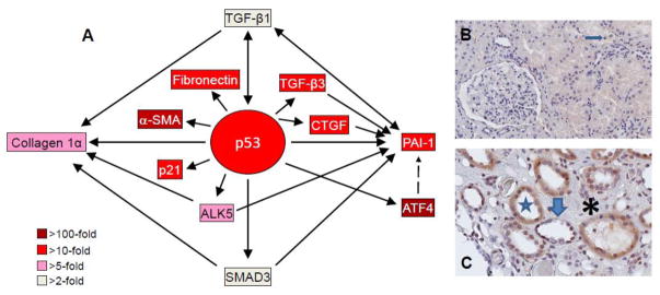
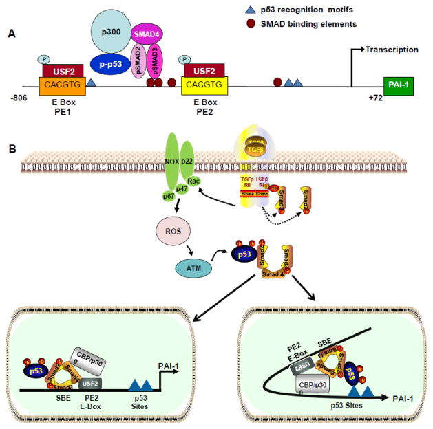
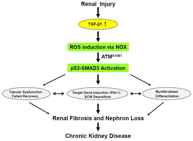
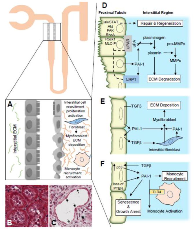
Fibrotic disorders of the renal, pulmonary, cardiac, and hepatic systems are associated with significant morbidity and mortality. Effective therapies to prevent or curtail the advancement to organ failure, however, remain a major clinical challenge. Chronic kidney disease, in particular, constitutes an increasing medical burden affecting >15% of the US population. Regardless of etiology (diabetes, hypertension, ischemia, acute injury, urologic obstruction), persistently elevated TGF-β1 levels are causatively linked to the activation of profibrotic signaling networks and disease progression. TGF-β1 is the principal driver of renal fibrogenesis, a dynamic pathophysiologic process that involves tubular cell injury/apoptosis, infiltration of inflammatory cells, interstitial fibroblast activation and excess extracellular matrix synthesis/deposition leading to impaired kidney function and, eventually, to chronic and end-stage disease. TGF-β1 activates the ALK5 type I receptor (which phosphorylates SMAD2/3) as well as non-canonical (e.g., src kinase, EGFR, JAK/STAT, p53) pathways that collectively drive the fibrotic genomic program. Such multiplexed signal integration has pathophysiological consequences. Indeed, TGF-β1 stimulates the activation and assembly of p53-SMAD3 complexes required for transcription of the renal fibrotic genes plasminogen activator inhibitor-1, connective tissue growth factor and TGF-β1. Tubular-specific ablation of p53 in mice or pifithrin-α-mediated inactivation of p53 prevents epithelial G2/M arrest, reduces the secretion of fibrotic effectors and attenuates the transition from acute to chronic renal injury, further supporting the involvement of p53 in disease progression. This review focuses on the pathophysiology of TGF-β1-initiated renal fibrogenesis and the role of p53 as a regulator of profibrotic gene expression.

Keywords: Fibrosis; Kidney; Plasminogen activator Inhibitor-1; TGF-β1; p53.


Conflict of interest statement

Conflicts of Interest: The authors declare no conflict of interest.

Figures

Figure 1
Figure 1. p53 induction and target gene expression in the UUO-injured kidney and in renal allografts
Microarray analyses of TGF-β1-stumulated proximal tubular epithelial cells illustrating the relative expression levels of target genes within the p53 transcriptome node (A). p53-dependent up-regulation of ALK5, SMAD3, TGF-β1, TGF-β3, CTGF, α-smooth muscle actin (α-SMA) and PAI-1 may constitute a complex feed-forward loop that maintains a profibrotic renal microenvironment. Compared to a normal appearing patient allograft (B) in which only infrequent p-p53S15 tubular epithelial cells were evident (arrow), a renal transplant exhibiting dysmorphic tubules (star) with a flattened and occasionally denuded epithelium (thick arrow) and a markedly expanded interstitial region (asterisk) has abundant nuclear and cytoplasmic p-p53S15 immunoreactivity (C).
Figure 2
Figure 2. Topography of transcriptional motifs in the PAI-1 promoter and pathways involved in TGF-β1-induced expression
Downstream p53 binding sites (AcACATGCCT, cAGCAAGTCC) map to nucleotides -224 to -204 relative to the transcription start site and the upstream 4G/5G polymorphic sequence (blue triangles) in the PAI-1 promoter. pSMAD2/3/p-p53 interactions, at the PE2 USF2-binding E Box site located immediately 3′ of three clustered SMAD-binding elements (SBEs), are critical for PAI-1 transcription (A). Ligand-dependent TGF-β1 receptor activation initiates SMAD2/3 phosphorylation (by the ALK5/TGF-β1 type I receptor) (B). Rapid TGF-β1-induced generation of ROS stimulates non-SMAD-mediated signaling (e.g., upon p53 phosphorylation/acetylation). The SMAD and non-SMAD pathways collectively regulate target gene expression. In one model (B, left panel), p53 interacts directly with SMAD2; such SMAD2-p53 interactions may occur independently of p53 occupancy of its consensus motif. Alternatively, certain bHLH-LZ factors (including USF) bend DNA toward the minor groove potentially promoting interactions between p53, bound to its two downstream half-site motifs, with SMAD2 tethered to SBE sites immediately upstream of the CACGTG E-Box [72] (B, right panel).
Figure 3
Figure 3. Events downstream of renal injury-induced TGF-β1 expression that contribute to fibrotic disease
Activated ATM (pATMS1981) increased significantly in the tubulointerstitial region of the UUO-injured kidney, likely in response to elevated TGF-β1 levels and expression of the p22phox subunit of the NADP(H) oxidases, correlating with SMAD3 and p53S15 phosphorylation and induction of the fibrotic markers PAI-1 and fibronectin [71]. Stable silencing or pharmacological inhibition of ATM attenuated TGF-β1-induced p53 activation and expression of the downstream targets PAI-1, fibronectin, CTGF and p21. Silencing of the NADPH oxidase (NOX) subunits, p22phox and p47phox in HK-2 cells blocked TGF-β1-stimulated phosphorylation of ATM (pATMS1981) and target gene induction via p53- dependent mechanisms. Thus, TGF-β1 promotes NOX-dependent ATM activation leading to p53-mediated fibrotic gene reprogramming and growth arrest in HK-2 cells. Depletion of ATM or p53 in HK-2 cells, moreover, resulted in a bypass of TGF-β1-mediated cytostasis [71]. Furthermore, TGF-β1/ATM-initiated paracrine factor secretion by the dysfunctional renal epithelium promotes interstitial fibroblast growth, suggesting a role for tubular ATM in mediating epithelial-mesenchymal cross-talk highlighting the translational benefit of targeting the NOX/ATM/p53 axis in renal disease.
Figure 4
Figure 4. Multifunctional roles of PAI-1 in renal fibrosis
Development of renal disease upon proximal tubule injury is characterized by major functional and morphological changes including epithelial dedifferentiation, ECM accumulation, cell cycle arrest and tubular dysmorphism (A left, B = normal kidney, A right, C = fibrosis). Thick and thin arrows denote flattened epithelium and denuded regions, respectively. Following tissue injury, an inter-dependent plasmin-generating/matrix metalloproteinase (MMP) pericellular proteolytic cascade is finely titered, both temporally and spatially, by local PAI-1 levels (D). Collectively, these highly integrated systems cooperate to regulate ECM degradation and stromal remodeling. Elevated PAI-1 abundance in the wounded kidney compromises ECM degradation and fibrin clearance promoting increased matrix accumulation that contributes to the initiation of the fibrotic process and eventual development of CKD. Increased TGF-β levels or activation in response to trauma facilitate the transition of pericytes and resident interstitial fibroblasts (with perhaps minor contributions from other cell types) toward a myofibroblastic phenotype (E). An increased persistence and/or density of myofibroblasts further accelerates ECM deposition and eventual loss of tissue function. In conjunction with high p53 expression and loss of PTEN, TGF-β expression in the injury microenvironment mediates epithelial growth arrest with loss of regenerative repair, exacerbating ECM accumulation, likely through elevation of local levels of profibrotic factors including PAI-1 (F). Microenvironmental cues in the injured epithelial and interstitial compartments initiate TGF-β1-dependent monocyte recruitment/activation (perhaps via PAI-1 modulation of TLR4 signaling) (E,F). Additional mechanistic details are provided in the text. Collectively, these events (D–F) illustrate the clinical significance of the collaborative effects of TGF-β1 and TGF-β1 target genes (e.g., PAI-1) on the development of renal fibrosis.

References

    1. Zeisberg M, Neilson EG. Mechanisms of tubulointestinal fibrosis. J Am Soc Neprhol. 2010;21:181901834. - PubMed
    1. Venkatachalam MA, Weinberg JM, Kriz W, Bidani AK. Failed tubule recovery, AKI-CKD transition, and kidney disease progress. J Am Soc Neprhol. 2015;26:1765–1776. - PMC - PubMed
    1. Schnaper HW. The tubulointerstitial pathophysiology of progressive kidney disease. Adv Chronic Kidney Dis. 2017;24:107–116. - PMC - PubMed
    1. Webster AC, Nagler EV, Morton RL, Masson P. Chronic kidney disease. Lancet. 2017;389:1238–1252. - PubMed
    1. Mei C, Zheng F. Chronic inflammation potentiates kidney aging. Semin Nephrol. 2009;29:555–568. - PubMed

Publication types

Substances