. 2018 Jan 26;293(4):1138-1148.
doi: 10.1074/jbc.M117.817197. Epub 2017 Nov 30.

Hydrogen-deuterium exchange reveals long-range dynamical allostery in soybean lipoxygenase

Affiliations

Hydrogen-deuterium exchange reveals long-range dynamical allostery in soybean lipoxygenase

Adam R Offenbacher et al. J Biol Chem. .

Abstract

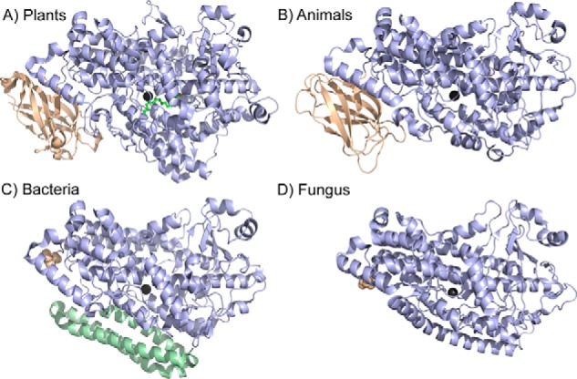
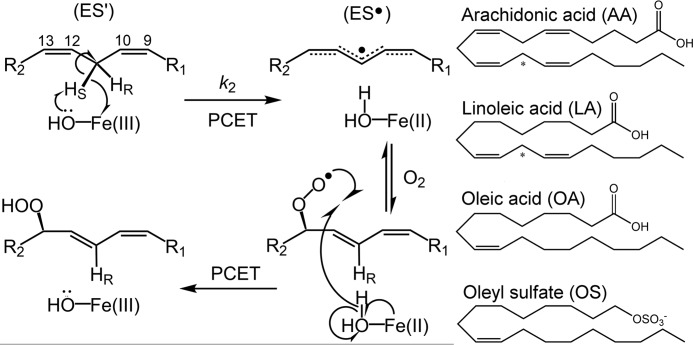
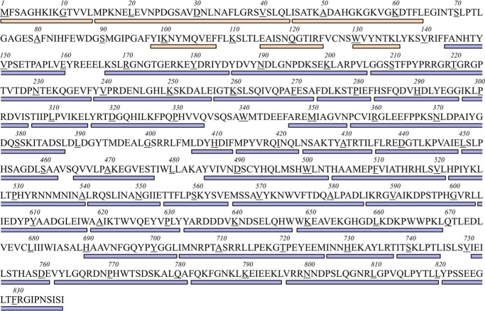
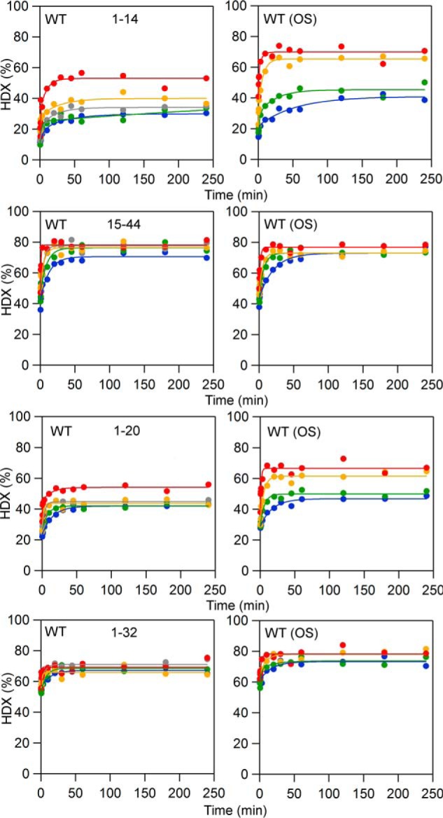
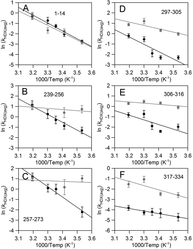
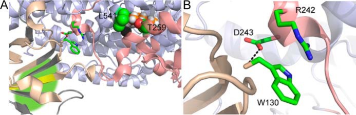
In lipoxygenases, the topologically conserved C-terminal domain catalyzes the oxidation of polyunsaturated fatty acids, generating an assortment of biologically relevant signaling mediators. Plant and animal lipoxygenases also contain a 100-150-amino acid N-terminal C2-like domain that has been implicated in interactions with isolated fatty acids and at the phospholipid bilayer. These interactions may lead to increased substrate availability and contribute to the regulation of active-site catalysis. Because of a lack of structural information, a molecular understanding of this lipid-protein interaction remains unresolved. Herein, we employed hydrogen-deuterium exchange MS (HDXMS) to spatially resolve changes in protein conformation upon interaction of soybean lipoxygenase with a fatty acid surrogate, oleyl sulfate (OS), previously shown to act at a site separate from the substrate-binding site. Specific, OS-induced conformational changes are detected both at the N-terminal domain and within the substrate portal nearly 30 Å away. Combining previously measured kinetic properties in the presence of OS with its impact on the Kd for linoleic acid substrate binding, we conclude that OS binding brings about an increase in rate constants for both the ingress and egress of substrate. We discuss the role of OS-induced changes in protein flexibility in the context of changes in the mechanism of substrate acquisition.

Keywords: SLO; allosteric regulation; allostery; enzyme inhibitor; fatty acid; hydrogen exchange mass spectrometry; hydrogen-deuterium exchange (HDX); inhibition mechanism; lipoxygenase.


Conflict of interest statement

The authors declare that they have no conflicts of interest with the contents of this article

Figures

Figure 1.
Figure 1.
Structural comparison of lipoxygenases from various kingdoms. In A, SLO was selected as the representative for plant lipoxygenases, whereas in B, the human 5-LO isoenzyme is shown. The structures correspond to the Protein Data Bank entries 3PZW (A), 3O8Y (B), 4G32 (C), and 5FNO (D). The structurally conserved lipoxygenase fold of the catalytic domain is colored light blue; the catalytic metal is shown as dark gray in all cases. For both plants and animals, the N-terminal PLAT domain is colored in wheat (A and B). Bacterial and fungal enzymes do not contain a PLAT domain, but the resolved N-terminal amino acid is shown as wheat spheres. In C, the bacterial enzymes have additional α helices, as colored in pale green.
Figure 2.
Figure 2.
Mechanism of lipid peroxidation by lipoxygenase. Left, reaction of model enzyme, soybean lipoxygenase, with substrate linoleic acid as an example. The metallocofactor, often a ferric hydroxide, although a manganese center is employed by fungal enzymes (63), abstracts a hydrogen atom from polyunsaturated fatty acids, in a regio- and stereospecific manner, through a proton-coupled electron transfer (PCET) mechanism (49). Molecular oxygen inserts into the delocalized radical intermediates, forming the product upon reverse proton-coupled electron transfer. The carbon numbering of the LA substrate is shown for reference. Relevant microscopic rate constant, k2, associated with the rate-determining C–H abstraction, is labeled. The enzyme-substrate and enzyme-LA radical complexes are labeled as ES′ and ES, respectively. Right, the structures of substrates, as well as the allosteric effectors OA and OS, are shown for reference. The reactive carbon for each substrate is designated by an asterisk.
Figure 3.
Figure 3.
Primary sequence coverage map for the defined non-overlapping peptides in SLO. The color code corresponds to that for Fig. 1. Wheat, the N-terminal β-barrel, “PLAT” domain is represented by residues 1–144; light blue, the catalytic domain comprises residues 145–839.
Figure 4.
Figure 4.
HDXMS traces showing peptides that exhibit increased extent of exchange at longer time points in the presence of OS. Blue, 10 °C; green, 20 °C; yellow, 30 °C; red, 40 °C. The data are shown in dots; the fits to the data are solid lines.
Figure 5.
Figure 5.
Structure of the N-terminal domain and the putative OS-binding site in SLO. The primary structure of peptide 1–14 is shown in A as yellow sticks for reference; the first six amino acids (MFSAGH) are not resolved in this high-resolution structure (1.4 Å). The N-terminal and C-terminal domains are colored wheat and light blue, respectively. Peptides not covered by HDXMS experiments in the PLAT domain are colored gray. B, space-filled model of the PLAT domain from the SLO structure. A loop 68–79 is colored in pale green and is located spatially over the putative binding site for OS and provides a relatively hydrophobic pocket for fatty acid binding. OS association in this pocket could trigger a conformational change in which the loop would close over the pocket.
Figure 6.
Figure 6.
Comparative time-dependent HDXMS traces, collected at 10 °C, for select peptides whose rates of exchange are faster with OS. The color coding of the HDXMS traces represents SLO in the absence of OS (black) and in the presence of OS (red). Middle, a partial structure of the catalytic domain is shown with a focus on the peptides affected by the presence of OS. The color coding of the peptides refers to their unique HDXMS behaviors and represents residues 239–256 and 257–273 (salmon), 297–305 and 306–316 (wheat), 317–334 (pale yellow), and 751–761 (pale green). The number of the N-terminal amino acid of each peptide, affected by OS, is shown in the structure for reference. Insets, the HDXMS traces are reproduced but are plotted with the x axis in logarithmic scale.
Figure 7.
Figure 7.
Arrhenius-like plots of the weighted average exchange rates, ln(kHDX(avg)), for SLO in the absence (black) or presence (gray) of OS. Apparent rates of HDXMS were determined from fits to the traces in Fig. S1. All rate constants can be found in Table S1.
Figure 8.
Figure 8.
Schematic representing the impact of effector addition upon the reaction coordinate (A) and proposed mechanism (B) in soybean lipoxygenase chemistry. In A, the solid line represents the reaction coordinate (up to the first irreversible step, k2) for SLO reaction with LA in the absence of effector. The dashed line represents the changes to the steps in the reaction coordinate in the presence of OS determined from kinetic analysis. The addition of effector, OS, causes a relative reduced barrier for the first binding step and a change in the internal reorganization. Keq refers to this proposed internal reorganization from the initial enzyme-substrate complex (ES) to ES′ structure, with the latter representing the productive binding for chemistry. The substrate on rate (kon = k1[S] × Keq) and off rate (koff = k−1 × 1/Keq) are both a product of this internal equilibrium constant.
Figure 9.
Figure 9.
Structure of SLO depicting the peptides affected by interactions with allosteric effector, OS. The SLO model (Protein Data Bank code 3PZW) is colored pale blue. Peptides, whose backbone HDX properties are impacted by interactions with OS, are colored as follows. Yellow, increased extent of exchange (not rate); salmon, increased rate of exchange. A, proposed communication network for OS-mediated allostery includes the conserved cation-π interaction (green sticks). The iron cofactor is represented as a dark gray sphere. The modeled substrate is depicted in spheres (orange, carbon; white, hydrogen). Side chains Thr-259 and Leu-541 at the substrate entrance are displayed in spheres for reference. The green highlighted area suggests the binding site for OS. B, a focused view of the cation-π interaction between Trp-130 and Arg-242. The conserved carboxylate, Asp-243, also forms hydrogen bonds to Trp-130 backbone.

References

    1. Porta H., and Rocha-Sosa M. (2002) Plant lipoxygenases: physiological and molecular features. Plant Physiol. 130, 15–21 10.1104/pp.010787 - DOI - PMC - PubMed
    1. Feussner I., and Wasternack C. (2002) The lipoxygenase pathway. Annu. Rev. Plant Biol. 53, 275–297 10.1146/annurev.arplant.53.100301.135248 - DOI - PubMed
    1. Haeggström J. Z., and Funk C. D. (2011) Lipoxygenase and leukotriene pathways: biochemistry, biology, and roles in disease. Chem. Rev. 111, 5866–5898 10.1021/cr200246d - DOI - PubMed
    1. Whitehouse M. W., and Rainsford K. D. (2006) Lipoxygenase inhibition: the neglected frontier for regulating chronic inflammation and pain. Inflammanopharmacology 14, 99–102 10.1007/s10787-006-1523-7 - DOI - PubMed
    1. Andreou A., and Feussner I. (2009) Lipoxygenases: structure and reaction mechanism. Phytochemistry 70, 1501–1510 10.1016/j.phytochem.2009.05.008 - DOI - PubMed

Publication types

LinkOut - more resources