. 2018 Jan 19;293(3):906-919.
doi: 10.1074/jbc.RA117.000165. Epub 2017 Nov 30.

Molecular characterization of human anti-hinge antibodies derived from single-cell cloning of normal human B cells

Affiliations

Molecular characterization of human anti-hinge antibodies derived from single-cell cloning of normal human B cells

Tao Huang et al. J Biol Chem. .

Abstract

Anti-hinge antibodies (AHAs) are an autoantibody subclass that, following proteolytic cleavage, recognize cryptic epitopes exposed in the hinge regions of immunoglobulins (Igs) and do not bind to the intact Ig counterpart. AHAs have been postulated to exacerbate chronic inflammatory disorders such as inflammatory bowel disease and rheumatoid arthritis. On the other hand, AHAs may protect against invasive microbial pathogens and cancer. However, despite more than 50 years of study, the origin and specific B cell compartments that express AHAs remain elusive. Recent research on serum AHAs suggests that they arise during an active immune response, in contrast to previous proposals that they derive from the preexisting immune repertoire in the absence of antigenic stimuli. We report here the isolation and characterization of AHAs from memory B cells, although anti-hinge-reactive B cells were also detected in the naive B cell compartment. IgG AHAs cloned from a single human donor exhibited restricted specificity for protease-cleaved F(ab')2 fragments and did not bind the intact IgG counterpart. The cloned IgG-specific AHA-variable regions were mutated from germ line-derived sequences and displayed a high sequence variability, confirming that these AHAs underwent class-switch recombination and somatic hypermutation. Consistent with previous studies of serum AHAs, several of these clones recognized a linear, peptide-like epitope, but one clone was unique in recognizing a conformational epitope. All cloned AHAs could restore immune effector functions to proteolytically generated F(ab')2 fragments. Our results confirm that a diverse set of epitope-specific AHAs can be isolated from a single human donor.

Keywords: antibody engineering; autoimmunity; immunogenicity; immunoglobulin G (IgG); monoclonal antibody.


Conflict of interest statement

All authors are present or former paid employees of Genentech

Figures

Figure 1.
Figure 1.
Detection of anti-hinge–reactive B cells in the antigen-inexperienced, naive; unswitched memory; and switched memory B cell compartments. a–d, enriched B cells from a single human peripheral blood donor were gated on IgDpos, CD27neg (antigen-inexperienced, naive); IgDpos, CD27pos (unswitched memory); and IgDneg, CD27pos (switched memory B cell compartments). e, anti-hinge–reactive B cells from all three compartments were detected with PE-labeled streptavidin-F(ab′)2 MMP3 tetramers. f, isotype control for AHA detection. Data are representative of a single donor where 10 total donors were assessed (Table 1).
Figure 2.
Figure 2.
Gating strategy for sorting anti-hinge–reactive single B cells from the switched memory B cell compartment. To sort anti-hinge–reactive B cells, enriched B cells were initially gated by forward and side scatter (a); cells were further gated for singlets (b and c); dead cells were excluded (d); and CD19pos B cells (e) were further gated to select for the IgDneg and CD27pos switched memory B cell compartments (f). From this gating strategy, IgGpos (g), anti-hinge-reactive B cells (h) were single-cell sorted in the wells of a 96-well plate for cloning of antibody HC and LC. i, isotype control for AHA detection.
Figure 3.
Figure 3.
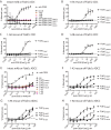
ELISA detection of AHAs binding to F(ab′)2 fragments or peptide analogues of the human IgG1 hinge region lower hinge region. F(ab′)2 MMP3 fragments (A) or F(ab′)2 MMP7 fragments (B) were coated on a 96-well plate, and AHAs were serially diluted. AHAs were detected with an HRP-conjugated anti-Fc reagent. Data are representative of three independent experiments. C, hinge analogue peptides were biotinylated 14-mers where the free C termini is indicated on the x axis. SCR represents a scrambled peptide. D5933 serum represents ELISA reactivity of the serum from the donor from which the AHAs were derived. Determinations were performed in triplicate wells with error bars representing S.D. Data are representative of three independent experiments.
Figure 4.
Figure 4.
ELISA detection of AHAs binding to F(ab′)2 fragments and Fab′ fragments to assess linear versus conformational epitope-binding dependence. F(ab′)2 MMP3 fragments (A) or Fab′MMP3 fragments (B) were coated on a 96-well plate, and AHAs were serially diluted. AHAs were detected with an HRP-conjugated anti-Fc reagent. F(ab′)2 MMP7 fragments (C) or Fab′MMP7 fragments (D) were coated on a 96-well plate, and AHAs were serially diluted. AHAs were detected with an HRP-conjugated anti-Fc reagent. Data are representative of two independent experiments.
Figure 5.
Figure 5.
Comparison of SPR sensorgrams for the interaction between protease-cleaved F(ab′)2 fragments and immobilized AHAs. Sensorgrams represent real-time binding interactions as a change in refractive index plotted as response units against time in seconds. Monovalent binding was achieved using an IgG capture format. A concentration series of each F(ab′)2 from 2 to 500 nm was flowed as mobile analyte, and binding was analyzed using a Langmuir 1:1 model. Affinity values are reported as nanomolar dissociation constants (KD) ± S.D. n = 3. n.m. refers to no measured specific binding.
Figure 6.
Figure 6.
Comparison of SPRi sensorgrams for the interaction between AHAs and immobilized protease-cleaved F(ab′)2 fragments. Sensorgrams represent real-time binding interactions as a change in refractive index plotted as response units against time in seconds. Avidity-based binding was achieved using a F(ab′)2 fragments capture format. A concentration series of each AHAs from 33 to 900 nm was flowed as mobile analyte, and binding was analyzed using a Langmuir 1:1 model. Affinity values are reported as nanomolar dissociation constants (KD) ± S.D. n = 3. n.m. refers to no measured specific binding.
Figure 7.
Figure 7.
Cell-based CDC and ADCC assays of AHAs on anti-CD20 F(ab′)2 fragments generated with the proteases MMP3, GluV8, MMP7, and IdeS. CDC assays were performed with WIL-2 cells as described under the “Materials and methods.” A, CDC with intact anti-CD20, AHAs 1-A3, 1-A6, and 1-A8 as well as anti-CD20 F(ab′)2 MMP3, F(ab′)2 GluV8, F(ab′)2 MMP7, and F(ab′)2 IdeS. B–D, CDC with 1-A3 (B), 1-A6 (C), and 1-A8 (D) against anti-CD20 F(ab′)2 MMP3, F(ab′)2 GluV8, F(ab′)2 MMP7, and F(ab′)2 IdeS. CDC assays were performed in triplicate wells, and error bars represent S.D. Data are representative of three independent experiments. ADCC assays were performed with WIL-2 cells and PBMC effector cells as described under “Materials and methods.” E, ADCC with intact anti-CD20, AHAs 1-A3, 1-A6, and 1-A8 as well as anti-CD20 F(ab′)2 MMP3, F(ab′)2 GluV8, F(ab′)2 MMP7, and F(ab′)2 IdeS. F–H, CDC with 1-A3 (F), 1-A6 (G), and 1-A8 (H) against anti-CD20 F(ab′)2 MMP3, F(ab′)2 GluV8, F(ab′)2 MMP7, and F(ab′)2 IdeS are shown. ADCC assays were performed in triplicate wells, and error bars represent S.D. Data are representative of three independent experiments.
Figure 8.
Figure 8.
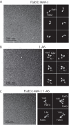
Architecture of F(ab′)2 MMP-3 and human anti-hinge antibodies 1-A6. Raw images for each of these specimens (left) and reference free 2D classes (right) are shown. A, raw images of F(ab′)2 MMP3 molecules appear to be monodisperse. Reference free classification indicates that the two Fab arms can orient at different angles, in respect to each other, suggesting an intrinsic flexibility of the hinge region. B, raw images and 2D classes of full-length AHA 1-A6 clone show that this sample is also homogeneous and presents the typical Y-shape antibody architecture with the CH2 domains oriented at 45° from the Fc region. C, images and classes obtained from the F(ab′)2 MMP3–1-A6 complex indicate that the full-length AHA 1-A6 can use its Fab fragments region to bind to either one or two copies of F(ab′)2 MMP3.
Figure 9.
Figure 9.
Schematic of proposed mechanism for how anti-hinge–specific B cells receive T cell help. A, anti-hinge–specific B cell binds to a cleaved IgG that is opsonized on a pathogen. PAMPs from the pathogen provide co-stimulation to the B cell. The B cell then internalizes and processes and presents antigen, including the pathogen antigen even though the BCR bound to cleaved IgG. B, B cell presentation of pathogen antigen on MHCII elicits cognate T cell help and further co-stimulation (e.g. through CD40–CD40L interactions) resulting in CSR and affinity maturation of the anti-hinge specific B cell. C, legend for the symbols depicted in A and B.

References

    1. Brezski R. J., and Monroe J. G. (2008) B-cell receptor. Adv. Exp. Med. Biol. 640, 12–21 10.1007/978-0-387-09789-3_2 - DOI - PubMed
    1. McHeyzer-Williams L. J., and McHeyzer-Williams M. G. (2005) Antigen-specific memory B cell development. Annu. Rev. Immunol. 23, 487–513 10.1146/annurev.immunol.23.021704.115732 - DOI - PubMed
    1. Klein U., Rajewsky K., and Küppers R. (1998) Human immunoglobulin (Ig)M+IgD+ peripheral blood B cells expressing the CD27 cell surface antigen carry somatically mutated variable region genes: CD27 as a general marker for somatically mutated (memory) B cells. J. Exp. Med. 188, 1679–1689 10.1084/jem.188.9.1679 - DOI - PMC - PubMed
    1. Wardemann H., Yurasov S., Schaefer A., Young J. W., Meffre E., and Nussenzweig M. C. (2003) Predominant autoantibody production by early human B cell precursors. Science 301, 1374–1377 10.1126/science.1086907 - DOI - PubMed
    1. van Schie K. A., Wolbink G. J., and Rispens T. (2015) Cross-reactive and pre-existing antibodies to therapeutic antibodies–effects on treatment and immunogenicity. MAbs 7, 662–671 10.1080/19420862.2015.1048411 - DOI - PMC - PubMed

Publication types

MeSH terms