

Review
doi: 10.1104/pp.17.01519.
Epub 2017 Nov 30.
The Actin Cytoskeleton: Functional Arrays for Cytoplasmic Organization and Cell Shape Control
Affiliations
- PMID: 29192029
- PMCID: PMC5761824
- DOI: 10.1104/pp.17.01519
Item in Clipboard
Review
The Actin Cytoskeleton: Functional Arrays for Cytoplasmic Organization and Cell Shape Control
Plant Physiol.
2018 Jan.
Abstract
Functionally distinct actin filament arrays cluster organelles and define cellular scale flow patterns for secretion.
Figures
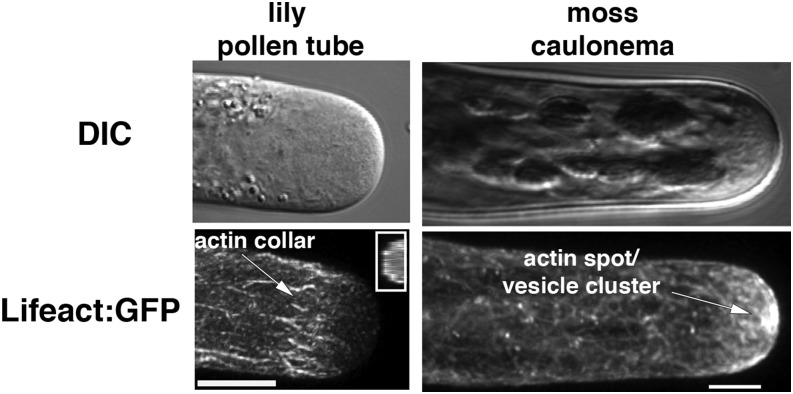
Actin organization in living tip-growing lily pollen tubes (left) and moss caulonema (right) detected with the Lifeact:GFP actin-binding probe. Top images are differential interference contrast (DIC) images of the cell and the bottom images are the same cell imaged for Lifeact:GFP fluorescence. The cortical actin collar or fringe is indicated with an arrow; a resliced view showing its cortical location is shown in the inset. The filaments in the cortical actin collar are predicted to have their plus ends oriented toward the apex. Actin bundles in the core cytoplasm in the shank are oriented with their plus ends away from the cell tip. The dense meshwork of actin filaments focused into an actin spot at the apex of moss caulonema is labeled in the lower right image. Bars = 5 μm. Figure is adapted with permission from Vidali et al. (2009a).
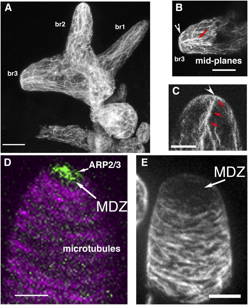
Actin and microtubule organization in Arabidopsis leaf hairs and cotton fibers during the process of cell elongation and tapering. A, Whole mounted trichome with branches that are becoming progressively tapered (defined as stage 4). The cell is labeled with an anti-actin antibody using the freeze-shattering technique. Note prominent tip actin in branches 2 (br2) and 3 (br3). B, Midplanes of br3 in A showing cortical actin at the apex and cytoplasmic bundles that are oriented toward the apical meshwork. C, Whole mounted trichome labeled with phalloidin. Apical actin meshwork and cytoplasmic bundles are labeled as in B. D, Live-cell image of GFP-tagged ARP2/3 complex (green) and microtubules (magenta). ARP2/3 localizes within the apical microtubule-depletion zone (MDZ) and is required to polymerize the apical actin meshwork. E, Whole mounted 1-DPA cotton fiber in the process of cell tapering labeled with an anti-α-Tubulin antibody. Bars = 5 μm.
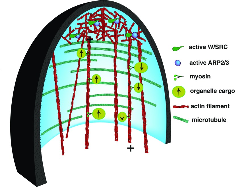
ARP2/3-dependent cytoplasmic organization in a developing trichome branch. The cartoon is a view of the cytoplasm from a medial longitudinal section through the young branch. In response to ROP-GTP signals, the W/SRC complex (green) physically interacts with and activates ARP2/3 (blue ellipse). ARP2/3 generates branched networks of actin filaments (red). ARP2/3 activation is restricted to an apical zone that has a reduced density of cortical microtubules. Aligned microtubules (cyan) along the cell flanks promote highly anisotropic cell elongation. Actin bundles orient longitudinally in the core cytoplasm and terminate at or near the apical actin meshwork. A subset of the actin bundles is composed of parallel actin filaments with their plus ends (marked with +) oriented either toward or away from the cell apex. These bundles act as roadways for directional actomyosin transport. The symbols representing the myosin motors, the organelle cargo, and their direction of movement are defined in the figure. The plasma membrane is shaded light blue, and the cell wall thickness and its longitudinal thickness gradient are represented in black.
References
-
- Augustine RC, Vidali L, Kleinman KP, Bezanilla M (2008) Actin depolymerizing factor is essential for viability in plants, and its phosphoregulation is important for tip growth. Plant J 54: 863–875 - PubMed
-
- Baskin TI. (2005) Anisotropic expansion of the plant cell wall. Annu Rev Cell Dev Biol 21: 203–222 - PubMed
Publication types
MeSH terms
Substances
LinkOut - more resources
Full Text Sources
Other Literature Sources
